
Hemodynamic Disorders - PowerPoint PPT Presentation
1 / 57
Title:
Hemodynamic Disorders
Description:
xmlns:stRef='http://ns.adobe.com/xap/1.0/sType/ResourceRef ... xmlns:tiff='http://ns.adobe.com/tiff/1.0/' tiff:Orientation 1 /tiff:Orientation ... – PowerPoint PPT presentation
Number of Views:3134
Avg rating:3.0/5.0
Title: Hemodynamic Disorders
1
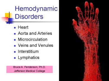
Hemodynamic Disorders
- Heart
- Aorta and Arteries
- Microcirculation
- Veins and Venules
- Interstitium
- Lymphatics
Bruce A. Fenderson, Ph.D. Jefferson Medical
College
2
Microcirculation - site of gas and nutrient/waste
exchange with tissues of the body
3
Hemodynamic Disorders
- Hyperemia
- Active
- Passive (chronic passive congestion)
- Hemorrhage
- Thrombosis
- Embolism and Infarction
- Edema
- Shock
4
Hyperemia
- Hyperemia is defined as excess amount of blood in
an organ. - Active hyperemia is an augmented supply of blood
to an organ usually as a response to increased
functional demand (eg inflammation). - Passive hyperemia (congestion) refers to
engorgement of an organ with venous blood due to
an impediment to venous return.
5
Passive Congestion of Liver
With the hepatic veins emptying into the vena
cava immediately inferior to the heart, the liver
is vulnerable to chronic passive congestion. The
changes are referred to as nutmeg liver.
6
Passive Congestion of Lungs (CHF)
- Increased pressure in the pulmonary alveolar
capillaries has 4 major consequences - Microhemorrhages release erythrocytes into the
alveolar spaces (iron-laden macrophages). - Pressure forces fluid into the alveolar spaces
(pulmonary edema). - Increased pressure stimulates fibrosis.
- Increased capillary pressure causes pulmonary
hypertension.
7
Heart Failure Cells(Iron-Laden Macrophages)
Prussian Blue Stain for Iron
8
Hemorrhage
- Hemorrhage is discharge of blood from the
vascular compartment to the exterior of the body
or into nonvascular body spaces. - A person may hemorrhage into an internal cavity
- bleeding peptic ulcer (arterial hemorrhage)
- esophageal varicosity (venous hemorrhage).
- Bleeding into an open (serous) cavity can result
in the accumulation of a large amount of blood to
the point of exsanguination.
9
Examples of Hemorrhage
- Hematoma - hemorrhage into soft tissue
- Hemoperitoneum - bleeding into peritoneum
- Hemarthrosis - bleeding into joint space
- Purpura - diffuse superficial hemorrhage
- Ecchymosis - larger superficial hemorrhage
- Petechia - pinpoint hemorrhage
10
Petechia (pin-point hemorrhages appear as red
dots)
11
Woman with Vaginal Bleeding
- Squamous Cell Carcinoma of the Uterine Cervix.
12
Man with Bleeding Peptic Ulcer
13
Blunt Trauma to the Head
Epidural Hematoma
14
Rupture of Heart MuscleHemopericardium - Cardiac
Tamponade
15
Thrombosis
- Thrombosis refers to the formation within a
vascular lumen of a thrombus, defined as an
aggregate of coagulated blood containing
platelets and fibrin. - A thrombus is by definition adherent to the
vascular endothelium and should be distinguished
from a simple blood clot. - The most common cause of arterial thrombosis is
atherosclerosis.
16
Platelet Activation
- Pathogenesis of Thrombosis
- Damage to the endothelium
- Alterations in blood flow
- Increased coagulability of blood
- Thrombin converts fibrinogen to fibrin.
17
Venous Thrombosis
Thrombi are adherent to the vessel wall. They
are composed of fibrin, platelets, and blood
cells..
18
Deep Venous Thrombosis
- Large majority (gt90) of venous thromboses occur
in the deep veins of the legs (DVT). - Occlusive thrombosis of the femoral or iliac
veins leads to severe congestion, edema, and
cyanosis. - DVT is treated with systemic anti-coagulants.
- Conditions that favor the development of deep
venous thrombosis include - Stasis of blood flow
- Injury Inflammation (phlebitis)
- Hypercoagulability of blood
- Advanced age
19
Arterial Thrombosis
?
Coronary artery
20
Infarction is a Complication of Arterial
Thrombosis
- Arterial thrombosis is the most common cause of
death in Western industrialized countries. - Thrombosis of a coronary or cerebral artery
results in myocardial infarct (heart attack) or
cerebral infarct (stroke).
21
Mural (Heart Wall) Thrombosis
- Myocardial infarction
- Atrial fibrillation
- Cardiomyopathy
- Endocarditis
22
Arterial Thrombi Are Laminated
Note laminations of platelets and fibrin (lines
of Zahn)
23
Fate of Thrombi
- Lysis
- Propagation
- Organization (in-growth of connective tissue)
- Canalization
- Detachment and Embolization
24
VenousEmboli
- Sources of venous emboli shown.
- Venous emboli travel through the heart to the
lungs.
25
Pulmonary Embolism
- Embolism is passage through the venous or
arterial circulation of any material capable of
lodging in a blood vessel. - Pulmonary embolism remains an important
diagnostic and therapeutic challenge. Pulmonary
thromboemboli are reported in more than half of
all autopsies. - This complication occurs in 1 to 2 of
post-operative patients over the age of 40.
26
Pulmonary Saddle Embolism
27
Amniotic and Fat Emboli
- Amniotic fluid embolism refers to the entry of
amniotic fluid containing fetal cells and debris
into the maternal circulation through the open
uterine and cervical veins. - Fat embolism describes the release of emboli of
fatty marrow into damaged blood vessels following
severe trauma to fat-containing tissue,
particularly accompanying bone fractures.
28
Fat Embolism
29
Fat Embolism
30
Arterial Embolization
- The heart is the most common source of arterial
emboli, which usually arise from mural thrombi or
diseased heart valves. - The major complication of thrombi in the heart is
detachment of fragments and transport to distant
sites (arterial thromboembolism). - Organs that suffer the most from arterial
embolism (ie undergo infarction) include - Brain
- Lower extremity
- Kidney
- Heart
31
Sources
Sources of arterial emboli..
32
Infarction
Common sites of infarction from arterial emboli.
33
Infarction
- Infarction is defined as the process by which
coagulative necrosis develops in an area distal
to the occlusion of an end-artery. The necrotic
zone is termed an infarct. - Pale infarcts are typical in the heart, kidneys,
brain, and spleen. - Red infarcts, which may result from either
arterial or venous occlusion, are also
characterized by coagulative necrosis but are
distinguished by bleeding.
34
Arterial Embolism Infarction
35
Multiple Splenic Infarcts
36
Cystic Brain Infarct
37
(No Transcript)
38
Myocardial Infarct (Pale)
39
Pulmonary Infarct (Red)
40
Extravascular Fluid and Edema
- Edema refers to the presence of excess fluid in
the interstitial spaces of the body. Edema may
be generalized or local. Local edema is often
associated with acute inflammation. - Non-inflammatory edema is due to either a
decrease in blood oncotic pressure or an
increase in blood hydrostatic pressure. - Control of extracellular fluid volume depends
largely on the regulation of renal sodium
excretion, which is influence by - Atrial natriuretic factor
- Renin-angiotensin system
- Sympathic nervous system activity
41
Summary
42
Examples of Edema
- Congestive heart failure (gthydrostatic pressure)
- Cirrhosis of the liver (ltoncotic pressure)
- Nephrotic syndrome (ltoncotic pressure)
- Cerebral edema
- Pulmonary edema
- Fluid in body cavities (effusions)
- Edema due to lymphatic obstruction
43
Congestive Heart Failure (CHF)
- CHF describes the consequences of inadequate
cardiac output relative to the needs of the body.
- It is estimated that two to three million people
in the United States have congestive heart
failure and 15 die annually. - Failure of the left ventricle is associated
principally with passive congestion of the lungs
and pulmonary edema.
44
Pulmonary Edema in CHF
CHF leads to increased capillary hydrostatic
pressure. Venous engorgement of the lungs leads
to the accumulation of a transudate in the
alveoli, a condition termed pulmonary edema.
45
Clinical Features of Pulmonary Edema in CHF
- Pulmonary edema refers to increased fluid in the
alveolar spaces and interstitium of lungs. - The patient becomes acutely short of breath and
bubbly rales are heard. In extreme cases, frothy
fluid is coughed up or wells up out of the
trachea. - Pulmonary fluid accumulation may go unnoticed
initially, but eventually dyspnea and coughing
become prominent. - Hypoxemia is manifested as cyanosis.
46
CHF
Complications of Congestive Heart Failure.
47
Pitting Edema in CHF
48
LymphEdema
Is this elephantiasis or elephantitis?
49
Cardiac Tamponade
- Pericardial fluid (effusion) may accumulate
rapidly, particularly with hemorrhage caused by a
ruptured myocardial infarct, dissecting aortic
aneurysm, or trauma. - In this circumstance, the pressure in the
pericardial cavity rises to exceed the filing
pressure of the heart, a condition termed cardiac
tamponade.
50
Fluid Loss and Overload
- Dehydration
- Over-hydration
51
Shock
- Shock is a condition of profound hemodynamic and
metabolic disturbance characterized by failure of
the circulatory systems to maintain an
appropriate blood supply to the microcirculation,
with inadequate perfusion of the vital organs. - Shock is not synonymous with low blood pressure,
although hypotension is commonly a part of the
shock syndrome.
52
Cardiogenic and Hypovolemic Shock
- Cardiogenic shock is usually caused by myocardial
infarction and less commonly by myocarditis. - Hypovolemic shock is secondary to a pronounced
disease in blood volume, caused by the loss of
fluid from the vascular compartment (eg,
diarrhea, excessive urine formation, and
perspiration are the major mechanisms of external
fluid loss.
53
Septic Shock
- Pathogenesis of septic shock involves
- Release of TNF by activated macrophages
- Injury to endothelial cells
- Increased vascular permiability
54
Hemorrhagic Infarction of Adrenal Glands in
Septic Shock
Normal Gland
55
Organ Pathology of Shock
- Heart - Petechial hemorrhages
- Kidney - Acute tubular necrosis
- Lung - Adult respiratory distress syndrome
- Gastrointestinal tract - Erosive gastritis
- Liver - Centrilobular congestion and necrosis
- Pancreas - Acute hemorrhagic pancreatitis
- Adrenals - Necrosis and insufficiency
56
Complications of Shock
57
Path Key Words
- Active hyperemia
- Air embolism
- Cardiogenic shock
- Chronic passive congestion
- Dissecting aneurysm
- Fat embolism
- Hematoma
- Hemopericardium
- Hypovolemic shock
- Petechia
- Pulmonary edema
- Thromboembolism
Recommended
CrystalGraphics Presentations


