
Proteomics: fundaments and applications - PowerPoint PPT Presentation
1 / 54
Title:
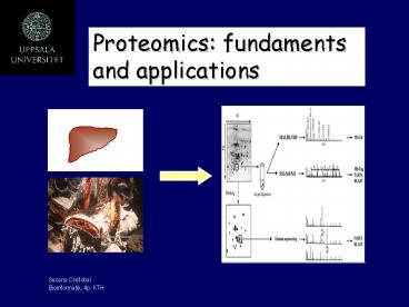
Proteomics: fundaments and applications
Description:
Protein world: study of less abundant proteins ... Passive elution of proteins. Analyze in a linear MALDI-TOF MS. Peptide mass FINGERPRINT: ... – PowerPoint PPT presentation
Number of Views:1480
Avg rating:3.0/5.0
Title: Proteomics: fundaments and applications
1
Proteomics fundaments and applications
Susana Cristobal Bioinformatik, 4p. KTH
2
0utline
- The virtue of proteomics
- Two dimensional gel electrophoresis
- Detection technology
- Identification methods
- Application of proteomics
Susana Cristobal Bioinformatik, 4p. KTH
3
The virtue of proteomics
- Proteome, the end product of the genome.
- Proteome dynamic entity.
- Protein world study of less abundant proteins
- Transcriptomics insufficient short-cut to study
most functional aspects of genomics
Susana Cristobal Bioinformatik, 4p. KTH
4
Sampling biological material
- Factors
- In cell cultures growth phase, culture
conditions,strain employed - Cell from multicellular organisms stage of
differentiation - Tissues from biopsy isolation of homogenous
cell populations
Susana Cristobal Bioinformatik, 4p. KTH
5
Phases of a large scale analytical process
- 1. Separation of biomolecules of interest
- Extraction of protein sample
- Cell culture
- Organelle isolation
- Two dimensional electrophoresis
- 2. Molecular characterization
- Detection technology
- Identification of proteins
- Differential expression profiles
Susana Cristobal Bioinformatik, 4p. KTH
6
Number of proteins in one cell
- High expression 105-106
- Moderate expression 103-104
- Low expression 101-102
Susana Cristobal Bioinformatik, 4p. KTH
7
Different strategies for proteome purification
and protein separation for identification by MS
- A. Separation of individual proteins by 2-DE.
- B. Separation of protein complexes by
non-denaturing 2-DE (BN-PAGE) - C. Purification of protein complexes by
immuno-affinity chromatography and SDS-PAGE. - D. Multidimensional chromatography.
- E. Organic solvent fractionation for separation
of complex protein mixtures of hydrobhobic
membrane proteins.
(van Wijk, 2001, Plant Physiology 126, 501-508)
Susana Cristobal Bioinformatik, 4p. KTH
8
Organelle can be separated by differential
velocity centrifugation
- Rupture plasma membrane to prepare tissue/ cell
homogenates - high speed blender
- Sonication
- tissue homogenize
- osmotic shock
( Molecular cell biology. Lodish. Fig 5-23)
Susana Cristobal Bioinformatik, 4p. KTH
9
Partially purified organelles can be better
separated by equilibrium density gradient
centrifugation
- How can you assess purity?
- Organelle-specific markers
- Cytchrome c, mitochondria
- Catalase, peroxisome
- Ribosome, rough ER
- Esterase, microsomes
Susana Cristobal Bioinformatik, 4p. KTH
(Lodish fig 5-24)
10
Organelle-specific antibodies are useful in
preparing highly purified organelles
Protein A o G is a bacterial molecule that
selectively binds Igs. Protein AAbAg complex
collected and dissociated to release organelle.
Susana Cristobal Bioinformatik, 4p. KTH
(Lodish fig 5-26)
11
Two-dimensional gel electrophoresis
- Solubilization of proteins in 2D electrophoresis
- Two dimensional electrophoresis with immobilized
pH gradients - Detection of proteins on 2DE
Susana Cristobal Bioinformatik, 4p. KTH
12
Solubilization of proteins in two dimensional
electrophoresis
- Goals
- Breaking macromolecular interaction (disulfide
bonds). - Preventing any artefactual modification of
polypeptides in the solubilization medium. - Removal of substances that may interfere with
2DE. - Keeping proteins in solution during 2DE process.
- There is no universal solubilization protocol.
- urea-reducer-detergent mixtures usually achieve
disruption of disulfide bonds and non-covalent
interactions.
Susana Cristobal Bioinformatik, 4p. KTH
13
Sample buffer
- Chaotropes
- 8M Urea
- 2M Thiourea/ 7M Urea
- Surfactants
- 4 CHAPS
- 2 CHAPS / 2 SB-14
- Reducing agents
- 65 mM DTE (dithioerythritol)
- 100 mM DTT ( dithiothreitol)
- 2 mM tributyl phosphine
- Ampholytes 2
Susana Cristobal Bioinformatik, 4p. KTH
14
How many quantities of samples can be loaded in
one IPG strip?
Identification of membrane proteins
Susana Cristobal Bioinformatik, 4p. KTH
(Govorun, 2002)
15
Two-dimensional gel electrophoresis
Internet-sites http//www.weihenstephan.de/blm/de
g/manual/manualwork2html02testp6 htm and
http//www.expasy.ch/ch2d/protocols.
Susana Cristobal Bioinformatik, 4p. KTH
16
First dimension IEFImmobilized pH gradients
(IPGs)
- IPG principle
- pH gradient is generated by a limited number
(6-8) of well defined chemicals (immobilines)
which are co-polymerized with the acrylamide
matrix. - IPG allows the generation of pH gradients of any
desired range ( broad, narrow, ultra-narrow)
between pH 3 and 12. - sample loading capacity is much higher.
- This is the method of choice for micropreparative
separation or spot identification.
Susana Cristobal Bioinformatik, 4p. KTH
17
First dimension IEFProcedure
- Rehydratation of IPGs dry strips
- Applying the sample
- in gel hydratation
- load coupling
- Running IPG strips
Susana Cristobal Bioinformatik, 4p. KTH
18
How many quantities of samples can be loaded in
one IPG strip?
Susana Cristobal Bioinformatik, 4p. KTH
19
How many quantities of samples can be loaded in
one IPG strip?
(18 cm) Analytical run 50-100 mg
Micropreparative runs 0.5-10 mg
Susana Cristobal Bioinformatik, 4p. KTH
20
Two dimensional electrophoresis with immobilized
pH gradient(Görg , 2000 Proteome research ,
chapter 4. Springer)
Susana Cristobal Bioinformatik, 4p. KTH
21
Two dimensional electrophoresis Running conditions
SampleCaenorhabditis elegans
- IEF dry strips pH 4-7
- Hydratation conditionsurea, thiourea, CHAPS,
DTT, ampholytes, iodoacetamine. - Passive 15h.
- Isoelectrofocussing
- 200v 1h
- 500v 1h
- 1000v 1h
- 5000v 3h
- SDS-PAGE 12
- Silver staining
Susana Cristobal Bioinformatik, 4p. KTH
22
Detection technologies in proteome analysis
- General detection methods.
- Differential display proteomics.
- Specific detection methods for post-translational
modifications.
Susana Cristobal Bioinformatik, 4p. KTH
23
General detection methods
- Organic dye- and silver-based methods
- Coomassie blue (R and G)
- Silver
- Radiactive labeling methods
- Reverse stain methods
- Flourescence methods
Susana Cristobal Bioinformatik, 4p. KTH
24
Differential display proteomics
- Detection techniques
- Difference gel electrophoresis (DIGE).
- Multiplexed proteomics (MP)
- Isotope-coded affinity tagging (ICAT)
- Differential gel exposure.
Susana Cristobal Bioinformatik, 4p. KTH
25
Summary of protein expression profile analysis
Susana Cristobal Bioinformatik, 4p. KTH
26
Difference gel electrophoresis (DIGE)
(Unlu, 1997, electrophoresis 18, 2071)
Susana Cristobal Bioinformatik, 4p. KTH
27
Multiplexed proteomics (MP) technology platform
(Steinberg, 2001, Proteomics 1,841, 2071)
Susana Cristobal Bioinformatik, 4p. KTH
28
Isotope-coded affinity tagging (ICAT) technology
platform
Very successful technique for identification of
integral membrane proteins
Susana Cristobal Bioinformatik, 4p. KTH
(Smolka, 2002, Mol Cell Proteomics 1, 19-29)
29
Differential gel exposure
(Monribot-Espagne, 2002, Proteomics 2, 229-240)
- Coelectrophoresis on 2DE of two protein samples.
- In vivo labelling, using 14C and 3H -isotopes.
- 2DE separation.
- Transfer on a PVDF membrane.
- 3H /14C ratio by exposure to two types of
imaging plates. - Investigate changes in the rate of synthesis of
individual proteins.
Susana Cristobal Bioinformatik, 4p. KTH
30
Image analysis
- Software commomly used to manipulate the gel
images - Imagemaster TM
- Melanie III TM
- Other functions
- Quantification
- Alignment
- Comparison
- Matching
- Synthetic image from the image of the sample
Susana Cristobal Bioinformatik, 4p. KTH
31
Example of data from differential display
proteomics
Susana Cristobal Bioinformatik, 4p. KTH
(Chevatier, 2000, Eur.J. Biochem. 267, 4624-4634)
32
Protein profiling in response to various
treatments at two different time-points
Susana Cristobal Bioinformatik, 4p. KTH
(Chevatier, 2000, Eur.J. Biochem. 267, 4624-4634)
33
General scheme of proteomic analysis
Susana Cristobal Bioinformatik, 4p. KTH
34
Pick the protein gel spot from the gel
- Pick up the protein gel spot from gel
- Manual
- Automatic
- In-gel digestion
- Washing process
- Dehydratation and drying
- Trypsin digestion (50 ng trypsin, 37C 16h)
- Extraction
- Desalt and concentrate the peptide
Susana Cristobal Bioinformatik, 4p. KTH
35
Identification methods
- Identification of proteins by mass spectrometry
- Identification of proteins by amino acid
composition after acid hydrolysis - Identification of proteins by amino acid
sequencing
Susana Cristobal Bioinformatik, 4p. KTH
36
Flow chart for the analysis of proteomes by MS
(van Wijk, 2001, Plant Physiology 126, 501-508)
Susana Cristobal Bioinformatik, 4p. KTH
37
Identification of eluted protein spots by
different MS approaches
- Extraction of intact protein (single peak)
- MALDI-TOF LINEAR mode
- Passive elution of proteins.
- Analyze in a linear MALDI-TOF MS.
- Peptide mass FINGERPRINT
- MALDI-TOF REFLECTRON mode
- In situ tryptic digestion of spots.
- Analyze in reflectron MALDI-TOF MS.
- Fragments, SEQUENCE
- LC-ESI MS/MS
- Separation in a C18 column.
- MS/MS analysis in a Q-TOF.
Susana Cristobal Bioinformatik, 4p. KTH
38
Comparison of MALDI-TOF and ESI-MS-MS approaches
to protein identification
- MALDI-TOF MS
- Sample on a a slide (crystalline matrix).
- Spectra indicate masses of the peptide ions.
- Protein identification by peptide mass
fingerprinting.
- ESI-MS-MS
- Sample in solution (high performance liquid
chromatography). - MS-MS spectra reveal fragmentation patterns.
- Protein identification by cross-correlation
algorithms.
Susana Cristobal Bioinformatik, 4p. KTH
39
Schematic of the MALDI quadrupole time of flight
instrument
- Advantages
- Mixture are analysed easily.
- It is highly tolerant to contaminants.
- High sensitivity. (picomol range)
- Good accuracy in mass determination.
- Quick and not expensive analysis.
- Disadvantages
- Low reproducibility and repeatability of single
shot spectra. (Averaging ) - Low resolution.
- Matrix ions interfere in the low max range.
Susana Cristobal Bioinformatik, 4p. KTH
40
Comparison of MALDI-TOF and ESI-MS-MS approaches
to protein identification
- MALDI-TOF spectrum
Susana Cristobal Bioinformatik, 4p. KTH
41
Correlating peptide masses to protein
sequence Positive identification
Susana Cristobal Bioinformatik, 4p. KTH
42
Peptides that span exon splices will be missed
when matching uninterpreted MS-MS data to genomic
DNA
(Jyoti, 2001, Trends 19, supp )
Susana Cristobal Bioinformatik, 4p. KTH
43
- ESI-MS-MS
- Peptide sequencing by nano-electrospray MS
Susana Cristobal Bioinformatik, 4p. KTH
44
Theoretical fragmentation of peptide
Susana Cristobal Bioinformatik, 4p. KTH
45
Peptide identification using mapping fingerprint
information
Experimental proteolytic peptides
Experimental MS
Intact protein
2DE gel
COMPUTER SEARCH
Theorectical MS
Theoretical proteolytic peptides
Protein sequence database
DNA sequence database
Susana Cristobal Bioinformatik, 4p. KTH
46
Databases of 2D-electrophoretic maps
Susana Cristobal Bioinformatik, 4p. KTH
(Govorum,2002)
47
Programs for comparison of 2D-proteomic maps
- Algorithms for gel matching
- algorithms based on characteristics of spot image
on gel - PDQuest (http//www.proteomeworks.bio-rad.com/inde
x.htm), - Phoretix 2D (http//www.phoretix.com/products/2d
products.htm), Melanie (http//www.expasy.ch/melan
ie) - 2) algorithms based on direct comparison of
images by distribution of intensity. - Z3 (http//www.2dgels.com),
- MIR (http//www.doc.ic.ac.uk/gzy).
Susana Cristobal Bioinformatik, 4p. KTH
48
Identification of mass-spectra Databases available
SWISS-PROT is a database of annotated protein
sequences it also contains additional
information on function of the protein, its
domain structure, posttranslational
modification(s), etc. - TrEMBL is a supplement
to SWISS-PROT, which contains all protein
sequences, translated from nucleotide sequences
of the EMBL database - PIR-International
(Protein Identification Resource, National
Biomedical Research Foundation, Washington, USA)
is also an annotated database of protein
sequences - NCBInr (National Center of
Biotechnological Information) is a database
containing sequences translated from DNA
sequences of GenBank and also sequences from PDB,
SWISS-PROT, and PIR databases - ESTdb
(Expressed Sequence Tags database, NCBI, NIH). -
programs operating with MS/MS only (SEQUEST,
PepFrag, MS-Tag, Sherpa).
Susana Cristobal Bioinformatik, 4p. KTH
49
Identification of mass-spectra Algorithms and
programs
Three main groups - programs using proteolytic
peptide fingerprint for protein identification
(PeptIdent, MultiIdent, ProFound) - programs
additionally operating with MS/MS spectra
(PepSea, MASCOT, MS-Fit, MOWSE) or with MS/MS
only - programs operating with MS/MS only
(SEQUEST, PepFrag, MS-Tag, Sherpa).
Susana Cristobal Bioinformatik, 4p. KTH
50
Identification of mass-spectra Algorithms and
programs
More perfect algorithms use additional
information such as isoelectric point of protein,
its molecular mass, amino acid composition, etc.
(PeptIdent, MultiIdent) The algorithm MOWSE is
more selective and sensitive than other
algorithms calculating only number of matching
peptides.
Susana Cristobal Bioinformatik, 4p. KTH
51
Alternatives to 2DE/MS. MudPIT
(Multidimensional protein identification
technology)
Susana Cristobal Bioinformatik, 4p. KTH
(Wolters, 2001, Anal.Chem 73, 5683-90)
52
Applications ?
- Cancer proteomics
- Peptidomics for profiling small proteins in
the human fluids - Neuroscience
- Toxicoproteomics a new preclinical tool to
revolutionary drug target discovery - Environmental pollution assessment
Susana Cristobal Bioinformatik, 4p. KTH
53
Schedule of a proteomics experiment
Day 1 Sample preparation and IEF 1. Load
protein sample onto IPG strip (IEF) 2. Run the
IEF (about 24 hours) 3. Polyacrylamide gel
casting
Day 2 Equilibrium IPG strip and running
SDS-PAGE 1. Remove IPG strip from IEF
machine 2. Equilibrium IPG strip 3. Put IPG
strip onto SDS-PAGE 4. Run the SDS-PAGE
(overnight)
Susana Cristobal Bioinformatik, 4p. KTH
54
Day 3 Staining, image scanning and image
analysis 1. Remove the gel from the cassette 2.
Stain the gel by SYPRO Ruby or silver 3. Scan
the gel image 4. Image analysis
Day 4 In-gel digestion, MALDI-TOF and database
search 1. Pick the protein gel spot from gel 2.
In-gel digestion 3. Spot the sample onto MALDI
chip 4. MALDI-TOF analysis 5. Database search
Susana Cristobal Bioinformatik, 4p. KTH
Recommended
CrystalGraphics Presentations


