
Mixed fat-dense stroma - PowerPoint PPT Presentation
1 / 111
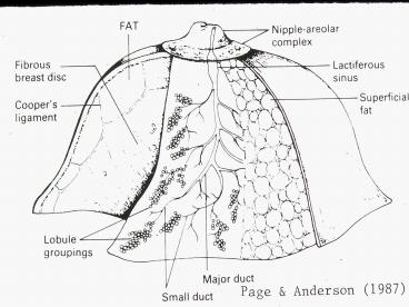
Title: Mixed fat-dense stroma
1
(No Transcript)
2
(No Transcript)
3
(No Transcript)
4
Mixed fat-dense stroma
5
Predominant dense stroma
6
Breast Density JAMA 276 33, 1996
7
Normal mammogram 68 yr old
8
Normal mammogram mixed dense stroma 52 yrs old
9
41 yr menstruating BRCA1 female palpable
lesion at marker
10
After 50 lb weight gain
11
HRT BEFORE HRT
12
61 yr female with lucent breast region of ductal
carcinoma-in-situ and a focus of invasive breast
cancer
13
Spiculated invasive breast cancer age 58
14
(No Transcript)
15
(No Transcript)
16
(No Transcript)
17
Interval cancer
- 8.18 / 10,000
- 22 false negative on screening
- 45 architectural distortion
- 100 associated with dense breast
- 67 invasive lobular carcinoma
- Rad 199 811, 1996
18
Suggestions for imaging of dense breast
- Ultrasound
- Digital mammography
- MRI
- CT
- Thallium, Tc99
- PET glucose, estrogen
- Rad. 188 297, 1993
19
Dense breastmammogram MRI T2 multiple cysts
20
Enhanced T1 MRI Mammogram 37 yr,
invasive ductal AdCa BRCA1
interpretation
dense breast no
identifiable pathology
21
Mammogram dense breast architectural distortion
?
Gadolinium enhanced MRI invasive AdCa
22
Silicone implant
23
(No Transcript)
24
ACS recommendations for MRI screening as adjunct
to mammography
- Recommend annual MRI screening
- Patients with BRCA mutations
- First degree relative of BRCA carrier
- Lifetime risk 2025
- Chest radiation _at_ ages 10-30 yrs
- Cowden (tricholemmomas) Bannayan-Riley-Ruvalcaba
(juvenile polyposis macrocephaly) syndromes
25
MRI guidance on surgical management of newly
diagnosed breast cancer
- 155 women diagnosed with invasive breast cancer
via needle biopsy following physical or
mammographic findings - Changes in surgical management
- Lumpectomy converted to mastectomy 10 cases
- Wider excision 21 cases
- Contralateral surgery 5 cases
- Arch
Surg 142 441, 2007
26
Negative mammogram contralateral breast
MRI gadolinium-enhanced mass, contralateral
breast
27
MCP 80 24, 2005
99Tc sestamibi scintiscanning
28
(No Transcript)
29
(No Transcript)
30
Irregular shaped density biopsy sclerosing
adenosis
31
Microcalcification within site of adenosis
32
Adenosis with microcalcification- calcium
phosphate
33
Calcium oxalate crystals
34
(No Transcript)
35
Lobular arrangement of calcium densities,
consistent with adenosis
36
Sclerosing adenosis with radial scar
37
Kopans (1998)
38
Radial scar 47 yr F
39
Radial scar of beastAJR 174 1075, 2000
- 12 patients mean age 61 years
- 12/12 discovered on mammogram mean size 22 mm
including spicules no calcifications - 12/12 not palpable
- 12/12 not associated with cancer
40
Microcystic disease
41
(No Transcript)
42
Large cyst age 51
43
Air infused into cystsee prior slide
44
Circumscribed cyst grayish fluid aspirated with
collapse
45
Some irregularity of margins fluid aspiration
with collapse- diagnosis cyst
46
Ultrasound CYST
47
Multiple cysts
48
Mass density with cystic component
associated vascular calcification. Dx
intracystic papillary carcinoma
49
Curved meniscal calcium densities
associated with cystic disease milk of calcium
50
Cyst with calcification
51
Calcium densities within cystic spaces
52
Calcification associated with multifocal regions
of fat necrosis
53
Focal breast fibrosisAJR 173 1657, 1999
- Duke University experience
- 80/894 (9) of imaging-guided biopsies
- 75/80 mammographically detected
- 43 Oval mass
- 39 Asymmetric density
- 9 Calcifications
- 5 Lobular mass
- 4 Irregular mass
54
Mastopathy in insulin-dependent diabetes Ann.
Surg. 205 529, 1987
- 8 patients M. age 33.9 yrs.
- 5/8 retinopathy
- Px hard, palpable mass
- Rx irregular mass
- Bx cellular fibrosis perivascular lymphocytic
infiltration. - ? Glycosylation of collagen
55
(No Transcript)
56
Fibroadenoma age 24
57
Solitary mass fibroadenoma
58
Ultrasound fibroadenoma
59
AJR 190 1219, 2008
60
Acceptable rates of growth for breast
fibroadenomas, diagnosed by FNA
Rad 229 233, 2003
- Solid breast masses interpreted as BIRADS 2
probably benign with FNA confirmation of
fibroadenoma may be safely followed if - Volume growth lt16 month in patients lt50yrs and
lt13 in patients gt50yrs
61
Fibroadenoma 28 yr old with palpable mass
62
Popcorn calcification, fibroadenoma
63
Solid calcification, fibroadenoma 64 yr old
female
64
Mammographic Findings in Phyllodes Tumor
- 10 cases
- 9/10 sharply defined, lobulated or round
- 5/10 halo sign
- 0/10 calcifications
- A.J.R. 157
716, 1991
65
Phyllodes tumor
66
Phyllodes tumor 52 yr F
67
Malignant phyllodes tumor
68
Phyllodes tumor, benign
69
Malignant phylloides tumor
70
Pseudoangiomatous stromal hyperplasiaMammography
11/22 hyperdense 10/22irregular borders Mod
Path 21 207, 2008
71
Bloody nipple discharge as a sign of breast
carcinoma
- Associated with cancer in 4 of cases
- When no other signs (palpatory mass, mammography,
skin changes) evident, nipple discharge is due to
cancer in only 8 of cases - Donegan
(2002)
72
Ductal papilloma
73
Solitary breast papilloma
- 21/24 nipple discharge
- 22/24 normal screening gram
- 11/19 normal detail gram
- 5/19 dilated duct (s)
- 2/19 nodules
- 13/15 abnormal galactogram
- 12/13 filling defect ductal
- dilation
- AJR 159 487, 1992
74
(No Transcript)
75
Atypical ductal hyperplasia
76
DCIScomedo type
77
DCIS__comedo type
78
(No Transcript)
79
(No Transcript)
80
DCIScomedo type with dystrophic calcification
81
alphabet calcification consistent with DCIS
82
alphabet calcification consistent with DCIS
83
Benign ectatic collecting duct
84
(No Transcript)
85
Cribiform ductal carcinoma-in-situ with
micro- calcifications
86
?
Cribriform DCIS with calcification
87
(No Transcript)
88
(No Transcript)
89
(No Transcript)
90
Microcalcification after lumpectomy AJR 188
393, 2007
- 68/402 patients
- 24 months median rate of development post
lumpectomy - 93 occurred in same quadrant
- 9/68 (9) BIRADS 4 or 5 6/9 malignant
91
Suture calcification
92
Lobular carcinoma in-situ
93
Mammographic findings in LCIS AJR
157 257, 1991
- 32/73 No radiologic abnormalities
- 11/73 Mass not suggestive of malignancy
- 10/73 Microcalcifications
- 6/73 Mass suggestive of malignancy
- 5/73 Asymmetry
94
Kopans (1998)
95
Kopans (1998)
96
Mammographic criteria I AJR
155 977, 1990
- Normal- BIRADS 1
- Benign-BIRADS 2
- Circumscribed, low density mass
- lt5 round uniform dense microcalcifications
- Probably benign-BIRADS 3
- Low density mass with partial loss of border
sharpness - lt15 round uniform dense microcalcifications
97
Mammographic criteria II AJR 155
977, 1990
- Suspicious for malignancy- BIRADS 4
- Low density mass with architectural distortion
- Circumscribed high density mass
- Small stellate mass without architectural
distortion - Microcalcifications are irregular in shape and
density
98
Mammographic criteria III AJR
155 977, 1990
- Malignant BIRADS- 5
- Circumscribed high density mass
- Stellate, spiculated mass with architectural
distortion - Pleomorphic or heterogeneous granular
calcifications, usually lt 5mm - Fine or branching, calcifications lt1mm in width
99
Malignant spiculation
100
Adencarcinoma with desmoplasia
101
Spiculated invasive tracts of ductal
adenocarcinoma
102
Spiculated mass, suspicious for malignancy
103
Grade 3 ductal adenocarcinoma small calcific
density fibroadenoma
104
Positive predictive values of various types of
mass densities Rad
165 101, 1987
- 0.00 opacity with regular borders
- 0.02 moderately suspicious
- microcalcifications
- 0.11 parenchymal distortion
- 0.35 opacity with poorly defined
- borders
- 0.56 strongly suspicious
- microcalcifications
- 0.75 stellate opacity
105
- as to tumors of the breastyou will be
abundantly convinced how many different species
existwhich do not emulate the nature of cancer. - Morgagni (1761)
106
Medullary carcinoma
107
Medullary carcinoma, breast
108
9 mo. progression medullary
carcinoma radiologist stated on 1st
report that the lesion was
representative of a fibroadenoma
109
Circumscribed breast cancers
- Variants of typical invasive ductal
adenocarcinoma - Medullary carcinoma
- Mucinous carcinoma
- Papillary carcinoma
- Phyllodes tumor
- Adenoid cystic carcinoma
-
Rad 242 683, 2007
110
Mucinous carcinoma
111
Mucinous carcinoma