
Evaluation of Thyroid Nodules - PowerPoint PPT Presentation
1 / 27
Title:
Evaluation of Thyroid Nodules
Description:
Evaluation of Thyroid Nodules Michael L. Tuggy, MD Swedish Family Medicine, Seattle, WA Case 1 42 y.o. male with no active medical problems. During your routine ... – PowerPoint PPT presentation
Number of Views:1290
Avg rating:3.0/5.0
Title: Evaluation of Thyroid Nodules
1
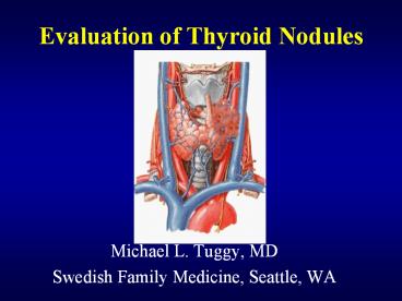
Evaluation of Thyroid Nodules
- Michael L. Tuggy, MD
- Swedish Family Medicine, Seattle, WA
2
Case 1
- 42 y.o. male with no active medical problems.
During your routine physical, note a thyroid
nodule. Told by ENT last year not to worry about
it. - PE 1 x 2cm R lower pole nodule.
- What information do you want from the patient?
3
Age as a Risk Factor
- Age
- young patients (lt20 years of age)
- thyroid nodules are much more likely to be
malignant (40-50). - elderly (gt60 years of age) -higher risk,
especially of more aggressive thyroid tumors.
4
Gender and Thyroid Nodules
- Gender
- male -higher risk if nodule present
- females
- have many more nodules
- less likely to be malignant.
- still have majority of thyroid cancers
5
Other major risks
- Radiation to head and neck.
- 40 risk of thyroid cancer usually 25 years
later. - Exposed populations- Polynesian studies
- Family History of MEN II, Gardners Syndrome,
Cowdens disease.
6
Historical Red Flags
- Recent growth
- Soft tissue swelling
- Vocal changes
- Dysphagia
- Signs of thyroid dysfunction
7
Case 2
- 26 y.o. Eritrean female with a 2-3 year history
of goiter. No symptoms but noted enlargement on
right for 1 year. - P.E. 3x4 cm Right sided thyroid mass, firm,
adherent to soft tissue. - What physical findings are worrisome?
- How can you best clarify the nature of the nodule?
8
Thyroid Exam
9
Physical Exam of the Thyroid
- Use both hands simultaneously to evaluate for
symmetry - Patient upright - screening exam
- Patient supine with neck in extension- detailed
exam. Swallowing assists in elevating gland. - Evaluation of other neck structures.
- Voice changes (recurrent laryngeal nerve).
10
(No Transcript)
11
Thyroid Scans
- Purpose
- Determine function of the gland and/or a nodule
within the gland - Hot nodules - usually independently functioning
nodules - Rarely, rarely malignant
- Cold nodules - either adenoma or maligancy
- 15 chance of malignancy in adults.
12
Thyroid Ultrasound
- Can identify presence of nodules.
- May be able to characterize follicular vs. solid.
- Not able to rule our malignant nodule
- Aid in biopsy.
Thyroid
13
Case 3
- 30 y.o. WF with enlarging cold benign thyroid
adenoma (diagnosis from previous FNA biopsy). - PE 4 x 5 cm mass on Right
- What do you do now?
14
Fine-Needle Aspiration
- Best tool for determining pathology other than
surgical excision. - Can be as high as 80 sensitive and 95
specific. - Operator dependent in obtaining adequate amount
of tissue. 25 gauge needle is optimal. - Should not be relied on if negative in patient
with previous neck irradiation. - Multifocal tumors common.
15
Interpreting the Biopsy Report
- What you get
- benign
- indeterminate
- suspicious
- inadequate specimen
- What it means
- benign - 90-95 likelihood it is benign
- indeterminate- who knows?
- suspicious- its malignant.
- inadequate specimen - do it again (and again)
16
Thyroid Malignancies- Papillary
- Most common
- 30 have node metastasis at diagnosis
- Radiation related
- Histologically, psammoma bodies distinguish from
benign adenoma.
17
Thyroid Malignancies-Follicular
- 20 of malignancies
- Distinguished from normal follicular adenomas by
invasion of capsule or blood vessels. - May be difficult to determine on FNA
18
Thyroid Malignancies- Medullary
- 5-10 of cases
- arise from the C cells which produce calcitonin
- diagnosis based on elevated thyrocalcitonin
levels and thyroid nodule (cold)
19
Thyroid Malignancies- Anaplastic
- lt 10
- Highly aggressive with local extension at time of
diagnosis. - No suitable therapy
- Prognosis lt 1 yr from diagnosis
20
Treatment
- For all malignancies, excision of the the lobe
(or if post-radiation the entire gland). - XRT- very specific and well tolerated- I131
therapy. - Anaplastic tumors - palliative radiation and XRT.
21
What about those benign nodules?
- No specific treatment is needed.
- Thyroid suppression may shrink size of adenomas
- Not proven to be effective or necessary
- May hide malignancies - ? Periodic biopsies or
scans.
22
Case 4 - This weeks puzzler!
- 40 y.o. WF s/p I131 ablation for Graves Dz. 6
years ago. - Persistant R thyroid nodule 2 x 1.5 cm in size.
- What is the likely diagnosis?
23
Outcomes
- Case 1. - Papillary cancer - 3 () nodes
- no metastasis at 1 year.
- Case 2. - Follicular cancer - 5 () nodes
- no metastasis at 1.5 years
- Case 3. - Large adenoma with incidental 1 cm
papillary carcinoma superior to nodule. - No recurrence at 5 years.
- Case 4. - Non-functional adenoma
24
Modified from Castro, MR, Gharib, H. Endocr
Pract 2003 9128.
25
SummarySolitary Nodule Evaluation
- TSH if low scan if hot nodule, then
observe. - Normal TSH - Do I scan first or FNA first?-
- high risk - scan and FNA
- Is the nodule cold or hot?
- Cold - FNA biopsy
- low risk - FNA
- if indeterminate- scan and re-FNA or excisional
biopsy. - Anti-perioxidase Antibody helpful if low- TSH
to diagnose thyroiditis.
26
(No Transcript)
27
Never assume a solitary thyroid nodule is benign.
Prove it.