
Chest Trauma - PowerPoint PPT Presentation
1 / 48
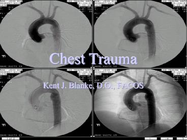
Title: Chest Trauma
1
Chest Trauma
- Kent J. Blanke, D.O., FACOS
2
Introduction
- Trauma 3rd leading cause of death in the U.S.
- Trauma is the leading cause of death in those
under 40 y.o.a. - There are 100,000 accidental deaths/yr and
9,000,000 disabling injuries yearly in the U.S. - 25 of deaths from blunt trauma are due solely to
chest injuries
3
Thoracic Trauma
4
Penetrating Chest Injuries
- Majority are stab wounds or gunshot wounds (GSW)
- Lower mortality rates--less likely to include
multiorgan injury - 85 of penetrating chest wounds can be treated
with tube thoracostomy and supportive measures
5
Penetrating Chest Injuries
- 25,000 deaths per year in the U.S. due to GSWs to
the chest
6
Penetrating Chest Trauma
- Wounds that enter or exit inferior to the nipple
or the posterior tip of scapula may perforate the
dome of the diaphragm. - Any penetrating wound such as this should be
considered to have an abdominal component until
proven otherwise.
7
Penetrating Chest Trauma Treatment
- ATLS protocol A,B,C,D,Es
- Emergency management
- Needle thoracentesis
- Tube thoracostomy
- Subxiphoid pericardotomy
- Video assisted thoracic surgery (VATS)
8
Work-up of Penetrating Chest Trauma
- Physical examination
- Look, Listen, Feel
- Contusions, diminished or absent breath sounds,
SQ emphysema can readily be found - CXR- best, least expensive and fastest initial
evaluation - Ultrasound-may soon replace CXR as initial
radiographic study in chest trauma - Angiography- to look for great vessel injuries
- CT Scan for better evaluation of chest wall and
parenchyma - Transesophogeal Echocardiography
9
Penetrating Chest Injuries
- Operative intervention required for
- Massive or persistent bleeding
- Massive air leak
- Tracheobronchial injuries
- Esophageal perforation
- Cardiac or great vessel injuries
- Post-traumatic empyema
10
Penetrating Chest Trauma
- Wounds that enter or exit inferior to the nipple
or the posterior tip of scapula may perforate the
dome or the diaphragm. - Any penetrating wound such as this should be
considered to have an abdominal component until
proven otherwise.
11
Penetrating Chest TraumaIndications for
Mechanical Ventilation
12
Intrapulmonary Foreign Bodies
- Bullets, fragments indications for removal
- Greater than 1.5 cm
- centrally located
- irregularly shaped
- sharp edged fragments
- FBs associated with gross contamination should
be removed
13
Intrapulmonary Foreign Bodies
- When left in lung
- 20 developed into chronic bronchitis
- 6 lung abscess
- 10 bronchopleural fistula
- 5 Empyema
14
Pulmonary Parenchymal Laceration
- Massive air leaks and hemorrhage require
immediate operation
15
High Velocity Missile Injuries
- Wounds due to high velocity missiles that travel
gt 25,000 ft/s are being seen with ever-increasing
frequency - Military and civilian
16
High Velocity Missile Injuries
- Cavitation phenomenon causes damage to
structures distal to the path of the missile. - Striking and shattering bone and other tissue may
add to the damage - Associated injuries to the large vessels and
bronchi is common - Severe pulmonary contusion
- Vietnam experience
17
Blunt Chest Trauma
- Higher mortality than penetrating trauma
- More frequent simultaneous injuries of multiple
organs - MVA leading cause of chest trauma with 50,000
deaths and 2 million disabling injuries/year
18
Categories of chest wall injuries
- Open pneumothorax
- Contusion and Hematoma
- Sternal fractures
- Scapular fractures
- Flail chest
- Intercostal vessel injury
19
Categories of Intra-thoracic Injuries
- Pulmonary
- Pneumothorax, hemothorax
- Pulmonary contusion
- Pulmonary laceration
- Vascular
- Great vessel disruption (Ao dissection, pulmonary
vasculature) - Cardiac
- Blunt Cardiac Injury, Penetrating injury
20
Work-up of Blunt Chest Trauma
- Physical examination
- Look, Listen, Feel
- Contusions, diminished or absent breath sounds,
SQ emphysema can readily be found - CXR- best, least expensive and fastest initial
evaluation - Ultrasound-may soon replace CXR as initial
radiographic study in chest trauma - Angiography- to look for great vessel injuries
- CT Scan for better evaluation of chest wall and
parenchyma - Transesophogeal Echocardiography
21
Categories of chest wall injuries
- Contusion and hematoma
- Rich vascular network established by intercostal
arteries w/ each rib - Internal mammary arteries on each side of sternum
- Rib fx bleed from raw surface exposure of the
bone and muscle tears
22
Categories of chest wall injuries
- Open pneumothorax
- When the defect in the chest wall is larger than
the trachea, pt is unable to ventilate. - Apply occlusive dressing on 3 sides
- Air cannot enter, but can exit through the flap
- Prevents pneumothorax
- Definitive management
23
Categories of chest wall injuries
- Pneumothorax
- Needle thoracentesis
- Chest tube
24
Operative Intervention for Hemothorax
- As noted previously
- Hemothorax massive initial drainage more than
1,000 cc or - Continuous bleeding of 200 cc/hr for 2 hrs
25
Fractured Ribs Chest Wall Trauma
- 70 of chest wall trauma is caused by MVAs
- 15 secondary to falls
- Blunt chest trauma accounts from 81 of thoracic
injuries in children, 78 in the elderly - Children are more likely to be injured as
pedestrians (35 vs 11 in the elderly)
26
Fractured Ribs Chest Wall Trauma
- The presence of 3 or more fx ribs on x-ray is an
indication for the need of tertiary care - Pts with rib fx are more likely to require
thoracotomy and laparotomy - The likelihood of splenic and hepatic injury is
increased by 1.7 and 1.4 times, respectively
27
Fractured Ribs Chest Wall Trauma
- Rib fxs are found in 52 of patients with
documented cardiac contusion - Mortality doubles with there are 3 or more ribs
- Blunt trauma with chest injury increases
mortality rate by 27 than without chest
injuries. Associated risk for death increases - Pneumo by 38
- Hemothorax by 42
- Pulmonary contusion by 56
- Flail chest by 69
28
Blunt Cardiac Injury
29
Blunt Cardiac Injury
- EKG (for any blunt chest injury, persistent
tachycardia, ST-T changes or ectopy) - Cardiac enzymes (CPK, CK-MB and Troponin I) see
EAST guideline - Echocardiography (TEE)
30
Categories of chest wall injuries
- Sternal fractures
- 80 associated with steering wheel impact
- 62 have blunt cardiac injury (BCI)
31
Categories of chest wall injuries
- Scapular fractures
- 3 of blunt trauma cases
- 54 have pulmonary contusions
- 11 have associated ipsilateral subclavian,
axillary or brachial artery injury - Over 1/3 are missed on initial evaluation
32
Categories of chest wall injuries
- Flail chest
- Fx of at least 4 consecutive ribs in 2 or more
places - Incompetent segment of chest wall large enough to
impair respirations - Paradoxic motion hinders creation of the expected
ipsilateral negative inspiratory force
33
Categories of chest wall injuries
- Flail chest
- Combination of pulmonary contusion and flail
chest has a mortality of 42 - Pulmonary contusion with flail chest 75 require
ventilation - Flail chest ALONE 48 require ventilation tx
- Aggressive respiratory txs and IS with pain
control
34
Categories of chest wall injuries
- Flail chest
- Internal splinting of mechanical ventilation
until fibrous stabilization of the chest wall is
apparent - Usually heavy sedation
- SIMV with PS
- PEEP or CPAP
- Sandbagging
- DO NOT use rib belts
- Surgery Staples, Kirschner wires and plates
- Analgesia
35
Pulmonary Contusion
- Pulmonary contusions are not innocuous injuries
- 11 of pts with isolated pulmonary contusion die
- ARDS develops in nearly 20 ARDS carries a 50
mortality
36
Intra-thoracic TraumaPulmonary Contusion
- Occurs in nearly 50 of all chest trauma
- Injury occurs to
- Alveolar-capillary walls
- Intra-alveolar hemorrhage
- Interstitial edema
- Increased tissue wt, airway and arterial
resistance, decreased compliance, decreased
surfactant content, decreased blood flow
37
Pulmonary Contusion
- Increase in pulmonary vascular resistance and
A-aO2 difference - Diagnosis
- Dyspnea
- Tachypnea
- Hemoptysis
- Cyanosis
- Hypotension
38
Pulmonary Contusion
- Physical signs
- Inspiratory rales, decreased Vt
- Patchy alveolar infiltrates due to intra-alveolar
hemorrhage - Intrapulmonary bleeding reaches maximal extent
within 6 hrs - Progression of a pulmonary contusion on X-ray
after 48 hrs should raise suspicion that
aspiration, bacterial pneumonitis or ARDS has
developed
39
Pulmonary Contusion
- Treatment
- Oxygen to maintain PaO2 above 60 mmHg
- Vigorous chest physiotherapy
- Use colloids instead of crystalloids when rapid
volume replacement is needed - Place PA catheter when large or rapid volume
replacement is needed - Use of steroids and antibiotics are controversial
40
Intra-thoracic Trauma Great Vessel and
Mediastinal Trauma
- Aorta
- Pulmonary vessels
- Tracheobronchial lacerations
- Esophageal lacerations
41
Intra-thoracic Trauma Great Vessel and
Mediastinal TraumaWork-up
- Plain CXR to identify thoracic aorta injuries
- Look for air in the mediastinum
- Persistent airleak should cue into
- Bronchopulmonary or tracheobronchial injury
- Mediastinitis, tube feedings in chest tube or
saliva in chest tube should cue into - Esophageal injury
42
Intra-thoracic Trauma Great Vessel and
Mediastinal TraumaWork-up
- Bronchoscopy
- Esophagoscopy
- CT
- Serial CXR
43
Initial CXR of Concern
44
Indications for Angiography
- Lateral deviation of the NGT in esophagus
- Widened mediastinum (gt8cm)
- Loss of visualization of the aortic knob
- Hematoma of the Left cervical pleura (pleural
cap) - Depressed left main stem bronchus
- Rt lateral deviation of the trachea
45
Indications for Angiography
- Widened mediastinum (gt8cm)
46
Indications for Angiography
- Forward displacement of the trachea on the
lateral CXR - Fx of the 1st or 2nd rib
- Massive chest trauma w/ multiple rib fx
- Fx or dislocation of the thoracic spine
- Major deceleration injury
47
Complete Aortogram
48
(No Transcript)