
Bacterial Morphology Arrangement - PowerPoint PPT Presentation
Title:
Bacterial Morphology Arrangement
Description:
Bacterial Morphology Arrangement Robert Hooke (1635-1703) English Scientist First to use the microscope to observe cells Coined the term cell Anton van ... – PowerPoint PPT presentation
Number of Views:482
Avg rating:3.0/5.0
Title: Bacterial Morphology Arrangement
1
Bacterial Morphology Arrangement
2
Robert Hooke (1635-1703)
- English Scientist
- First to use the microscope to observe cells
- Coined the term cell
3
Anton van Leeuwenhoek1632-1723
- Dutch scientist
- Invented the first compound microscope
- First to observe LIVING cells
- Blood cells and protists
4
Robert Brown1773-1858
- Scottish botanist
- In 1831 he was the first person to observe the
nucleus of a cell
5
Schleiden Schwann1804-1881 1810-1882
6
Developing Cell Theory 1838
- Schleiden
- Said all plants are made up of cells
- Schwann
- Said all animals are made up of cells
7
Cell Theory Overview
- All organisms are made of one or more cells
Unicellular or Multicellular. - All cells carry on life activities.
- New cells arise only from other living cells.
8
Prokaryotic vs Eukaryotic
- PROKARYOTIC
- Simplest form
- Lack membrane bound structures
- Lack true nucleus
- Example bacteria and cyanobacteria
- EUKARYOTIC
- Most common
- Possess membrane bound structures and a nucleus
- Found in most living things
9
Sizes of Cells
- Eukaryotic are usually larger than prokaryotic
- Both nutrients and wastes are constantly entering
and exiting cells - Vary in size and shape
10
Size relationships among prokaryotes
11
Bacterial Morphology Arrangement
- 1. Rod or Bacilli
- a.Streptobacilli
- b. Bacilli
- 2. Cocci
- a. Cocci
- b. Diplococci ( e.g. Neisseria meningitidis)
- c. Streptococci ( e.g. Streptococcus pyogenes)
- d. Staphylococci (e.g. Staphylococcus aureus)
- e. Sarcina
- f. tetrads ( Micrococcus species)
12
Bacterial Shapes, Arrangements, and Sizes
- Variety in shape, size, and arrangement but
typically described by one of three basic shapes - coccus - spherical
- bacillus rod
- coccobacillus very short and plump ( Brucella
abortus) - Streptobacilli ( Bacillus subtilus)
- diplobacilli
- spirillum - helical, comma, twisted rod,
- spirochete spring-like- flexible ( Treponema
pallidum) - vibrio gently curved ( Vibrio cholera)
- Spirilla- rigid ( Borrelia species)
- Pleomorphic variable in shape ( Corynebacterium)
12
13
13
14
Bacterial Shapes, Arrangements, and Sizes
- Arrangement of cells is dependent on pattern of
division and how cells remain attached after
division - cocci
- singles
- diplococci in pairs
- tetrads groups of four
- irregular clusters
- chains
- cubical packets
- bacilli
- chains
- palisades
14
15
15
16
Streptococcus sp.
17
Bacterial morphologies (1)
18
Bacterial morphologies (2)
19
Bacterial morphologies (3)
20
Bacterial Morphology Arrangement
- 3 Spirl
- a. Vibrio
- b. Spirillum
- c. Spirochete
21
(No Transcript)
22
Bacterial morphologies (4)
23
Borrelia (spirochete)
24
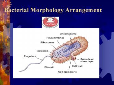
- Bacterial Cell Structures Functions
Pili
25
Bacterial Cell Structure
- Appendages - flagella, pili or fimbriae
- Surface layers - capsule, cell wall, cell
membrane - Cytoplasm - nuclear material, ribosome, mesosome,
inclusions etc. - Special structure - endospore
26
- Appendages
- 1. flagella
- Some rods and spiral form have this.
- a). function motility
- b). origin cell membrane flagella attach to
the cell by hook and basal body which consists
of set(s) of rings and rods - Gram - 2 sets of ring and rods, L, P,
S, M rings and rods . e.g. E. coli - Gram S, M rings and rods .e.g. B.
megaterium
27
Flagella
- Motility - movement
- Swarming occurs with some bacteria
- Spread across Petri Dish
- Proteus species most evident
- Arrangement basis for classification
- Monotrichous 1 flagella
- Lophotrichous tuft at one end
- Kophotrichous tuft at both ends
- Amphitrichous both ends
- Peritrichous all around bacteria
28
(No Transcript)
29
Structure of the flagellum
30
- c).Origin (continued)
- The structure of the bacterial flagella allows it
to spin like a propeller and thereby propel the
bacterial cell clockwise or counter clockwise
wave like motion. - Bacterial flagella provides the bacterium with
mechanism for swimming toward or away from
chemical stimuli, a behavior is knows as
CHEMOTAXIX, chemosenors in the cell envelope can
detect certain chemicals and signal the flagella
to respond. - d). structure
- protein in nature subunit flagellin (
globular protein)
31
Flagella movement(1)
32
Flagella movement(2)
33
2. Fimbriae and Pili Fimbriae Shorter than
flagella and straighter , smaller, hairlike
appendages . Only on some gram- bacteria. a).
function adhere. Not involve in motility. One
of the invasive mechanism on bacteria. Some
pathogens cause diseases due to this (Antigenic
characteristic). Prevent phagocytosis.
34
pili - sex factor. If they make pili, they are
or donors of F factor. It is necessary for
bacterial conjugation resulting in the transfer
of DNA from one cell to another. It have been
implicated in the ability of bacteria to
recognize specific receptor sites on the host
cell membrane.
35
Conjugation in E. coli
36
- b). Origin Cell membrane
- c). Position common pili , numerous over the
cell, usually called sex pile, 1-4/cell - d). Structure composed of proteins which can
be dissociated into smaller unit Pilin . It
belongs to a class of protein Lectin which bond
to cell surface polysaccharide.
37
- II. CELL SURFACE LAYER
- 1. Glycocalyx Capsule or slime layer
- Many bacteria are able to secrete material that
adheres to the bacterial cell but is actually
external to the cell. - It consists of polypeptide and polysaccharide on
bacilli. Most of them have only polysaccharide.
It is a protective layer that resists host
phagocytosis. Medically important (
Streptococcus pneumonia).
38
Capsule and Slime layer
- The layer is well organized and not easily washed
off, it is capsule - Slime layer, unorganized material that is easily
removed. - They give mucoid growth on agar plate
- B. anthracis has a capsule of poly-D-glutamic
acid, while S. pyogenes made of Hyaluronic acid. - Function Resistant phagocytosis, Protect against
desiccation, Attachment to surface of solid
objects.
39
Axial Filaments
- Present in spirochetes ( Treponema pallidum cause
syphilis) - Function is motility gliding motility
- Bundles of fibrils that arise at the ends of the
cell
40
Spirochetes
- Axial filament
- Structurally similar to flagella
- Unique location under an outer membrane
41
2. Bacterial Cell Wall General structure
mucopolysaccharide i.e. peptidoglycan. It is
made by N-acetylglucosamine and N-acetylmuramic
acid. tetrapeptide ( L-alanine-
isoglutamine-lysine-alanine) is attached. The
entire cell wall structure is cross linked by
covalent bonds. This provide the rigidity
necessary to maintain the integrity of the
cell. N-acetylmuramic acid is unique to
prokaryotic cell.
42
Cell walls of bacteria(2)
43
Cell walls of bacteria(3)
44
Cell walls of bacteria(4)
45
Cell walls of bacteria(1)
46
Structure of peptidoglycan(1)
47
Structure of peptidoglycan(2)
48
- a). Gram positive bacterial cell wall
- Thick peptidoglycan layer
- pentaglycin cross linkage.
- Teichoic acid (TA) Polymer of ribitol
glycerol joined by phosphate groups - Some have peptioglycan teichoic acid.
- All have lipoteichoic acid.
49
Function of Teichoic acids Antigenic
determinant Participate in the supply of Mg to
the cell by binding Mg regulate normal cell
division. For most part, protein is not
found as a constituent of the G cell wall
except M protein on group streptococci
50
Structure of the Gram-positive Cell Wall
51
- (b) Gram negative bacterial cell wall
- Thin peptidoglycan
- Tetrapeptide cross linkage
- A second membrane structure protein and
lipopolysaccharide (LPS). - Toxicity endotoxin on lipid A of LPS.
glucosamine- glucosamine-long - polysaccharide- repeated sequences of a few
sugars (e.g. gal- mann-rham) n10-20 O antigen
52
Structure of peptidoglycan(3)
53
Toxicity endotoxin on lipid A of
lipopolysaccharide. glucosamine-
glucosamine-long FA FA FA FA
polysaccharide- repeated sequences of a few
sugars (e.g. gal- mann-rham) n10-20 O antigen
54
Chemistry of LPS
55
The Gram-negative outer membrane(1)
56
The Gram-negative outer membrane(2)
57
(No Transcript)
58
Atypical Cell Walls
- Some bacterial groups lack typical cell wall
structure i.e. Mycobacterium and Nocardia - Gram-positive cell wall structure with lipid
mycolic acid (cord factor) - pathogenicity and high degree of resistance to
certain chemicals and dyes - basis for acid-fast stain used for diagnosis of
infections caused by these microorganisms - Some have no cell wall i.e. Mycoplasma
- cell wall is stabilized by sterols
- pleomorphic
58
59
- 2. Cell Membrane
- Function
- a. control permeability
- b. transportes and protons for cellular
metabolism - c. contain enzymes to synthesis and transport
- cell wall substance and for metabolism
- d. secret hydrolytic enzymes
- e. regulate cell division.
- Fluid mosaic model. phospholipid bilayer
and protein (structure and enzymatic function).
Similar to eukaryotic cell membrane but some
differs. e.g. sterols such as cholesterol in Euk
not in Prok.
60
60
61
Functions of the cytoplasmic membrane(1)
62
Functions of the cytoplasmic membrane(2)
63
Transport proteins
64
Classes of membrane transporting systems(1)
65
Classes of membrane transporting systems(2)
66
Bacterial Internal Structures
- Cell cytoplasm
- dense gelatinous solution of sugars, amino acids,
and salts - 70-80 water
- serves as solvent for materials used in all cell
functions
66
67
Bacterial Internal Structures
- Chromosome
- single, circular, double-stranded DNA molecule
that contains all the genetic information
required by a cell - DNA is tightly coiled around a protein,
aggregated in a dense area called the nucleoid.
67
68
The bacterial chromosome and supercoiling
69
Bacterial Internal Structures
- Plasmids
- small circular, double-stranded DNA
- free or integrated into the chromosome
- duplicated and passed on to offspring
- not essential to bacterial growth and metabolism
- may encode antibiotic resistance, tolerance to
toxic metals, enzymes and toxins - used in genetic engineering- readily manipulated
and transferred from cell to cell
69
70
Bacterial Internal Structures
- Ribosomes (70 S)
- made of 60 ribosomal RNA and 40 protein
- consist of two subunits large and small
- procaryotic differ from eucaryotic ribosomes in
size and number of proteins - site of protein synthesis
- present in all cells
70
71
71
72
- 3. Mesosomes ( mostly in Gram ve)
- A large invaginations of the plasma membrane,
irregular in shape. - a. increase in membrane surface, which may be
useful as a site for enzyme activity in
respiration and transport. - b. may participate in cell replication by serving
as a place of attachment for the bacterial
chromosome.
73
- 4. Inclusions
- Not separate by a membrane but distinct.
- Granules of various kinds
- glycogen ( used as carbon source),
- polyhydroxybutyric acid droplets (PHB)
- i.e. fat droplets and have Lipid inclusion
- inorganic metaphosphate (metachromatic
granules or Volutin granules) - in general,
starvation of cell for almost any nutrients leads
to the formation of this to serve as an
intracellular phosphate reservoir (
Corynebacterium).
74
PHB
75
- 5. Chromatophores
- Only in photosynthetic bacteria and blue green
algae. Prok. no chloroplast, pigment found in
lamellae located beneath the cell membrane. - Sulfur Granules Mainly in Thiobacillus, convert
H2S to S
76
76
77
- IV. Special Structure
- Endospores
- Spore former Sporobactobacilli and
Sporosarcinae (Gram cocci)- no medical
importance. - Bacillus and Clostridium ( Gram Rod) have
medical importance. Coxiella ( Gram ve Rod)
cause Q fever. - Position median, sub-terminal and terminal
have small water, high calcium content and
dipicolinic acid (calcium dipicolinate) - Extremely resistant to heat, UV, chemicals etc.
may be due to many S containing A.A for disulfide
groups.
78
The process of endospore formation
- After the active growth period approaching the
stationary growth phase, a structure called
forespore develops within the cells. - It consists of coat, cortex and nuclear
structure.
79
(No Transcript)
80
Negatively Stained Bacillus (A) Vegetative Cell
(B) Endospore
81
Dipicolinic acid
82
82
83
Detailed stepsin endospore formation(1)
84
Detailed stepsin endospore formation(2)
85
Detailed stepsin endospore formation(3)
86
(No Transcript)
87
PROCARYOTIC vs. EUCARYOTIC CELLS
88
PROCARYOTIC vs. EUCARYOTIC CELLS
89
PROCARYOTIC vs. EUCARYOTIC CELLS
90
PROCARYOTIC vs. EUCARYOTIC CELLS
Recommended
CrystalGraphics Presentations


