
1985 Nobel Laureates - PowerPoint PPT Presentation
1 / 30
Title:
1985 Nobel Laureates
Description:
Cholic acid and bile salt biosynthesis. Steroid hormone and vitamin D biosynthesis. Membrane permeability and fluidity (dampens phase transitions) ... – PowerPoint PPT presentation
Number of Views:90
Avg rating:3.0/5.0
Title: 1985 Nobel Laureates
1
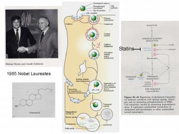
Statins
1985 Nobel Laureates
2
Physiological and Cellular Roles for
Cholesterol Cholic acid and bile salt
biosynthesis Steroid hormone and vitamin D
biosynthesis Membrane permeability and fluidity
(dampens phase transitions) Membrane
microdomains for protein sorting and signal
transduction Pathological Consequences of
Dysfunctional Cholesterol Regulation Atherosclero
sis Hardening of the arteries Plaque formation
in coronary arteries caused by LDL
deposition Hypercholesterolemia (genetic defect
in LDL receptor) 600 1000 mg cholesterol /
dL, normal is 150 mg/dL Regulation of
Cholesterol Levels Biosynthetic HMG-CoA
reductase phosphorylation and feedback
inhibition Transcriptional regulation of
biosynthetic enzymes Sterol Regulatory Element
Binding Protein (SREBP) Uptake from serum
lipoprotein particles via LDL and HDL
receptors Transcriptional regulation of the LDL
receptor Sterol Regulatory Element Binding
Protein (SREBP)
3
Transcriptional Regulation of Cellular
Cholesterol Levels
Cellular Cholesterol is too low Cellular
Cholesterol is too high Increased Txn of
HMG-CoA Decreased Txn of HMG-CoA reductase and
the LDL reductase and the LDL receptor
genes receptor genes Cholesterol levels
rise Cholsterol levels decrease
Txn
No Txn
SREBP
Gene
Gene
SRE
SRE
How do the cells know if they have enough
cholesterol? How does cholesterol influence the
amount of SREBP in the nucleus?
4
SRE Sterol Regulatory Element DNA sequence
required for transcriptional regulation of genes
in response to cholesterol SREBP Sterol
Regulatory Element Binding Protein Transcription
factor that binds SRE to induce
transcription SCAP SREBP Cleavage Activating
Protein Somehow activates SREBP cleavage when
cells are low on cholesterol S1P Site 1
Protease Cleaves SREBP in cytosolic loop S2P
Site 2 Protease Cleaves SREBP within the first
transmembrane domain
5
A SCAP mutation that causes a loss of cholesterol
regulation is found in a TMD
6
The transmembrane region of SCAP is similar to
other proteins that interact with
cholesterol. SCAP is the best candidate for
being the cholesterol sensor. How does SCAP
control the cleavage of SREBP by Site 1 Protease?
7
Conclusions
8
Localization of SCAP Do SCAP N-linked oligos
acquire Golgi modifications?
These data suggest that SCAP remains in the ER of
cells grown with sterols but moves to the Golgi
in the absence of sterols. SCAP N-linked
oligosaccharides must be trimmed by Mann II to
become EndoH resistant. Additional controls
indicate that other ER resident glycoproteins do
not become Endo H resistant in cholesterol
depleted cells. Western blot probed with a
monoclonal Ab to SCAP.
Nohturfft PNAS Fig 1
9
Subcellular Fractionation by Differential
Centrifugation
Golgi
10
Subcellular Fractionation by Equilibrium Density
Gradient Centrifugation
5-24
Assay marker enzyme to detect presence of
organelle. A marker enzyme is a known resident of
an organelle. The distribution of an enzyme whose
localization is unknown can be compared to the
distribution of marker enzymes.
11
Localization of SCAP by differential
centrifugation and density gradient
MannII is a Golgi marker Calnexin is an
ER marker
SCAP from cells without sterols has
Golgi-modified N-linked oligosaccharides but is
physically located in the ER
Nohturfft PNAS Fig 2
12
Localization of SCAP GFP-SCAP is functional in
vivo
CHO cell line that is SCAP deficient
Without SCAP, SREBP is unstable and so the SCAP
deficient cells also have less SREBP
Introduction of GFP-SCAP into these cells allows
stable expression of SREBP, and its sterol
regulated cleavage.
Nohturfft Cell, Fig 1
13
The amount of GFP-SCAP in the Golgi complex
increases when sterols are depleted
In this experiment, cells were fixed and stained
with anti-mannosidase primary Ab followed by a
secondary Ab coupled to a red flourescent
molecule. The autofluorescence of GFP survives
this treatment. Sterols were more efficiently
removed in these experiments, as compared to the
fractionation experiment, by using HPCD, which
will extract choleserol from the plasma membrane.
Nohturfft Cell, Fig 2
14
GFP-SCAP after sterol deprivation
GFP-SCAP was visualized in living cells over time
after sterol deprivation. Movement of SCAP to
the Golgi complex, along with SREBP that is
tightly associated with SCAP, correlates with
cleavage of SREBP by site 1 protease. Where is
site 1 protease?
Nohturfft Cell, Fig 3
15
Site-1 Protease Localizes to the Golgi complex
(S1P-)
WGA Wheat Germ Agglutinin. This is a lectin
that binds specifically to GlcNAc at the end of
an oligosaccharide. Later compartments of the
Golgi complex contain the GlcNAc transferase and
are therefore rich in oligosaccharides with
terminal GlcNAcs. WGA is covalently coupled to a
red fluorescent molecule and used like an Ab.
SIP is detected by IF.
DeBose-Boyd, Cell Fig 3
16
How does cholesterol regulate these events? Is
sterol-regulated transport of SCAP-SREBP the
primary mode of regulation, or does cholesterol
depletion also activate site 1 protease? Is it
absolutely necessary for SCAP-SREBP to go to the
Golgi to get cleaved by S1P? What happens if we
bring S1P back to the ER? Will we bypass sterol
regulation?
17
Influence of brefeldin A (BFA) on the Golgi
complex
Cell expressing a GFP tagged Golgi protein is
treated with BFA
18
BFA induces Endo H resistance of SCAP in the
presence of sterols
BFA causes the Golgi to fuse with the ER.
Nohturfft PNAS Fig 4
This suggests that enzymes in the Golgi complex
are moving back to the ER of BFA treated cells.
Does this cause processing of SREBP?
19
Western blot probed for SREBP
BFA bypasses the sterol-mediated suppression of
SREBP cleavage.
DeBose-Boyd, Cell Fig 1
20
BFA can bypass the requirement for SCAP, but not
S1P
The mutant cells indicated were transformed with
an HSV epitope-tagged form of SREBP
DeBose-Boyd, Cell Fig 2
21
BFA relocates S1P from the Golgi to the ER
Late Golgi compartments do not fuse back to the
ER of cells teated with BFA
DeBose-Boyd, Cell Fig 4
22
All Golgi proteins are initially translocated
into the ER before moving to the Golgi. Why
doesnt the newly synthesized S1P cleave SREBP as
it is passing through the ER?
preproSite 1 Protease
TMD
Site 1 protease is a Type I integral membrane
protein It is synthesized in the ER as a high
molecular weight precursor (S1P-A) that is
enzymatically inactive. When S1P reaches the
Golgi, it cleaves itself to generate the S1P-C
form, which is enzymatically active.
DeBose-Boyd, Cell Fig 5
23
The BFA experiments suffer from the fact that
nearly all Golgi proteins are returned to the ER,
including any site 2 protease that is in the
Golgi. Is the return of S1P alone sufficient to
bypass sterol regulations of SREBP cleavage? To
address this question, the S1P TMD was replaced
with a KDEL sequence, which will cause retrieval
of S1P back to the ER.
DeBose-Boyd, Cell Fig 6
24
S1P-KDEL can bypass sterol regulation and the
SCAP requirement for SREBP cleavage
DeBose-Boyd, Cell Fig 7
25
DeBose-Boyd, Cell Fig 8
How does sterol deprivation cause the movement of
SREBP-SCAP to the Golgi? Do sterols regulate the
incorporation of SREBP-SCAP into COPII vesicles
budding from the ER?
26
In vitro COPII vesicle budding assay using
sterol-depleted cells
p58 known COPII cargo
grp94 ER resident
grp78 ER resident
Ribophorin 1 ER membrane resident
ER microsomes were incubated with ATP, GTP and
cytosol (containing Sar1 and COPII components).
Vesicles and microsomes are separated and Western
blotted for proteins shown above.
Nohturfft Cell, Fig 4
27
HPCD depletes cells of cholesterol. The 0 minute
time point are cells with cholesterol and you see
very little SCAP in the vesicle fraction. As
cholesterol is depleted, more and more SCAP is
incorporated into the COPII-coated
vesicles. This event precedes the appearance of
cleaved SREBP in the nucleus.
SCAP
SCAP
Nohturfft Cell, Fig 5
28
(A) Cholesterol is added back to cells lacking
cholesterol. This rapidly prevents SCAP
inclusion into vesicles. (B) Important control
showing cholesterol (25-HC) specifically prevents
SCAP inclusion into vesicles, but not other cargo
such as VSVG-T7 or p58.
Nohturfft Cell, Fig 6
29
SCAP is required for inclusion of SREBP into
COPII vesicles
SCAP minus cells with
Nohturfft Cell, Fig 8
30
Model
When bound to cholesterol, SCAP is is in a
conformation that somehow prevents inclusion of
SREBP-SCAP into COPII vesicles. When
cholesterol levels drop, SCAP is in its
cholesterol-free conformation that somehow allows
inclusion of SREBP-SCAP into COPII vesicles. The
question remains as to how SCAP-SREBP is sorted
into COPII vesicles, and how cholesterol prevents
this soring event.
COPII
DeBose-Boyd, Cell Fig 8
Recommended
CrystalGraphics Presentations


