
peadiatric radiology for medical student - PowerPoint PPT Presentation
Title: peadiatric radiology for medical student
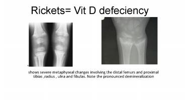
1
Rickets Vit D defeciency
- shows severe metaphyseal changes involving the
distal femurs and proximal tibias ,radius , ulna
and fibulas. Note the pronounced demineralization
2
The anterior ends of the ribs become enlarged and
cupped (arrows), so-called "rickety rosary".
3
Osteogenesis imperfecta (OI) is a rare genetic
disorder of the synthesis of collagen that
affects bone and connective tissue that can also
be referred to as brittle bone disease
4
Congenital diaphragmatic hernia Chest X-ray
(A.P.) displacement of right lung and mediastinum
towards left and resultant right lung collapse.
5
Congenital diaphragmatic hernia Chest X-ray (A.P.
view) of 1 hr old newborn baby herniation of
bowel contents into left thoracic cavity through
diaphragm with displacement of left lung and
mediastinum towards right and resultant right
lung collapse.
6
Congenital diaphragmatic hernialeft sided
confirm with oral contrast
7
cardiomegaly
8
Boat shape heart Fallots tetrology oligemia,
increased lung lucency
9
(No Transcript)
10
Cystic fibrosis (CF) is an inherited disease that
affects the lungs, digestive system, sweat
glands, and male fertilityThick walled
bronchiectasis Bronchiectasis. Hyperinflation.
lobar collapse.
11
Hyaline membrane disease also known as neonatal
respiratory distress syndrome
Diffuse ground-glass appearance to both lungs and
hypoaeration
12
Positive ventilator respiration
13
Hydrocephalus CT of the brain of a neonate. The
lateral ventricles (V) are grossly dilated. A
ventriculoperitoneal shunt tip is in the right
lateral ventricle (arrow)
14
Hydrocephalus If CSF is obstructed within the
ventricular system it is labelled
non-communicating Hyd.If the obstruction at
surface pathways communicating Hyd.
- Hydrocephalus refers increase in cerebrospinal
fluid volume associated with ventricular
dilatation and IVP. - Impaired absorption of CSF is usually due to some
degree of obstruction along CSF pathways.
15
Perthe's diseasePerthe's disease (avascular
necrosis of the capital femoral epiphysis) occurs
in the age range 2 - 12 years (majority 4 - 8
yrs.).
16
Oteosarcoma Typically it produces periosteal new
bone formation and sclerosis. Destruction with
spiculated periosteal reaction ("sunray
appearance") Codman's triangle.Osteomyelitis
can also produce an aggressive appearance.
17
Following plain films, The following imaging
techniques are used to evaluate the disease
- Radionuclide bone scan to look for synchronous
lesions or bone metastases.
18
CT of the chest to look for lung metastases is a
next step.
19
MRI of the lesion to look for Marrow extent and
soft tissue component,As well as extension
across the physis or joint.
20
TRUMA Acute blood is white on CT. Intracerebral
areas of haemorrhage are secondary to contusion
(axonal injury). Blood is also seen in the
subdural space (arrow) outlining the superior
sagittal sinus.
21
CT scan Shows the bone windows of the same image
reveal multiple fractures of the skull (arrows)
and angulation of the coronal and lambdoid
sutures (arrowheads).
22
Pediatric kind of fractures
- Torus or buckle fracture, Green stick fracture,
Todler fracture, Salter Harris fracture ,
23
Torus or buckle fracture, TODLER Results from
compression of a bone and is often seen in the
distal radial metaphysis.
24
Green stick A fracture occurs only on one side
of the cortex of the bone whilst the other side
bends so that a cortical injury can only be seen
along one surface.
25
Spiral tibial fractures Toddler fractures
26
SALTER
- 1 S slipped
- 2 Aabove
- 3 L lower
- 4 TeTogether
- 5 R Rund
27
Salter s Harris type 1 fracture.
Salter s Harris type 2 fracture.
28
Salter s Harris type 4 fracture
29
A mass encasing the vessels Neuroblastoma
30
A mass not encasing the vessels Wilms tumor (
peak sign with kidney).
31
Wilm's tumor metas to lung L to L Neuroblastoma
metas bone b to b
32
Pneumoperitonium, Riglers sign
Recommended
CrystalGraphics Presentations


