
Fluorescence Image Slide Scanner-OptraSCAN - PowerPoint PPT Presentation
Title:
Fluorescence Image Slide Scanner-OptraSCAN
Description:
OptraSCAN offers best Fluorescence Image Slide Scanner no matter the size of the pathology lab. Tel : +1-408-524-5300 Contact us at- info@optrascan.com Visit- – PowerPoint PPT presentation
Number of Views:30
Title: Fluorescence Image Slide Scanner-OptraSCAN
1
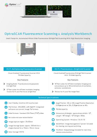
OptraSCAN Fluorescence Scanning Analysis
Workbench Small Footprint, Automated Whole Slide
Fluorescence Bright?eld Scanning With High
Resolution Imaging
OS-FLi Fluorescence Bright?eld Scanner
OS-FL Multiplexing Fluorescence Scanner
Cloud-Enabled Fluorescence Bright?eld Scanner
With 15-Slide Capacity
Cloud-Enabled Fluorescence Scanner With 15-Slide
Capacity
Key Features
Key Features
14 ?lter cubes for ef?cient multiplex imaging and
can produce up to 30 combinations of excitation,
emission and dichroic
Imaging Mode Fluorescence and grayscale in
bright?eld
6 ?lter cubes for ef?cient multiplex imaging. 5
slots for FL and 1 for mono bright?eld
14 slots for FL and 1 for bright?eld
Technical Speci?cations
Magni?cation 20x or 40x magni?cation
Resolution 0.50 µm/pixel at 20x, 0.25 µm/pixel
at 40x
User friendly, Intuitive LED touchscreen
File Format JPEG2000, .otiff, BigTIFF,
integrated software can convert image ?le formats
15 slide capacity
Slide Formats Standard 25x75mm (1"x3") slides
Dimensions Weight Approximate Width- 12",
Length- 16", Height- 12", Weight- 60lbs
Bar code and case reconciliation
Operating System Windows 7, 8, 8.1, 10
Image capture region 25x50mm
IMAGEPath Image Management System included for
viewing, storing and archiving
Image Storage Space approx. 300 MB for a single
channel for a 15mm x 15 mm tissue
TELEPath Telepathology included for real-time,
remote consultations
Data Storage 1-10 TB
2
FL Viewer IHC Multiplex Software
Key Features
Large image support
Comprehensive feature extraction
Illumination correction, vignetting correction
3D reconstruction
Image manipulations Brightness Contrast and
opacity
Photobleaching correction
Pixel to pixel spatial registration
Custom channel naming
Individual signal optimization
Layer blending
Spectral unmixing
Image operations
Multi-level cell segmentation
Atlas mapping
Gating to construct cells from segmented
cellular parts
Pan-and-zoom functionality for high resolution
images
Drawing importing of user-de?ned regions of
interest
Robust quantitative analysis for each imaged
channel
Technical Speci?cations
Supports CZI, BigTIFF, JP-2000 and standard TIFF
with no restriction on image size number of
channels
Features associated with each cellular object is
computed available for viewing and analysis
Software is natively compatible and seamlessly
integrated to support end- to-end image data
processing and analysis multiplexed ?uorescent
images
Segmented cells are displayed in a cell tray
Operating system Windows 7, 8, 8.1, 10
Software provides precise pixel-to-pixel spatial
registration for all imaged channels per
specimen, including those sequentially acquired
after repeated antibody stripping, restaining and
reimaging
3
Multi-Level Cell Segmentation Detection
algorithms to identify and classify cellular
entities Algorithms can be ?ne tuned by user
3D Re-Construction Selection of multiple
sections Fetching of composite segmented cell and
process objects that need to be reconstructed 3
Dimensional (3-D) visualization
Gating Module Morphological operations between
segmented objects in different channels to
reconstruct cells Addition and subtraction of
segmented objects between two or more channels
supported
Data Export In FCS ICE Software supports FCS
and ICE export ?le formats compatible with 3rd
party ?ow cytometry and image cytometry softwares
Pan-And-Zoom Functionality For High Resolution
Images Real-time pan and zoom Software supports
functionality of drawing user adjustable ROIs
dropdown/ select the ROI option for selecting a
particular area in the input image The software
supports adding annotations for the color
channels (square, rectangle, circle, ellipsoid,
polygonal, freeform) to classify and compare the
data across multiple areas of interest The
software supports functionality to save the ROIs
drawn on the image
info_at_optrascan.com
OptraSCAN is an ISO13485 certi?ed company.
www.optrascan.com
OptraSCAN whole slide scanners are CE marked for
IVD use.
OptraSCAN Systems are for research use only in
North America.
_at_optrascan
Recommended
CrystalGraphics Presentations


