
Biophotonics - PowerPoint PPT Presentation
1 / 14
Title:
Biophotonics
Description:
Biomedical Engineering and Engineering Healthcare Cluster ... histopathology. LEBS can detect alterations in. histologically normal-appearing cells due to the ... – PowerPoint PPT presentation
Number of Views:1013
Avg rating:3.0/5.0
Title: Biophotonics
1
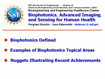
NSF Directorate for Engineering Division
of Chemical, Bioengineering, Environmental, and
Transport Systems (CBET) Bioengineering and
Engineering Healthcare Cluster Biophotonics,
Advanced Imaging, and Sensing for Human
Health Program Director - Leon Esterowitz -
lesterow _at_ nsf.gov
- ? Biophotonics Defined
- ? Examples of Biophotonics Topical Areas
- ? Nuggets Illustrating Recent Achievements
2
Biophotonics
? Photonics is the technology of generating
and harnessing light and other forms of
radiant energy whose quantum unit is the
photon ? Biophotonics applies photonics to
the fields of medicine, biology and
biotechnology
2
3
Examples of Biophotonic Topical Areas
Slide 1 of 2
- ? CONTRAST AGENTS - New classes of photonic
- probes and contrast agents to label
structures - and push the envelope of optical sensing to
the - limits of detection, resolution, and
identification - ? MOLECULAR IMAGING - Image and data fusion
- between optical imaging, spectroscopic
- techniques, and conventional imaging
modalities - for imaging diseases at the molecular and
- cellular level
3
4
Examples of Biophotonic Topical Areas
Slide 2 of 2
- ? NEUROPHOTONICS - Development and
- application of photonic tools such as large
scale - parallel interfaces and interconnects for
study - and control of neural systems
- ? MICRO- and NANO-PHOTONICS - Development
- and application of nanoparticle fluorescent
- quantum-dots sensitive, multiplexed, high-
- throughput characterization of
macromolecular - properties of cells nanomaterials and
- nanodevices for biomedicine
4
5
SGER Advances in Biophotonics to Enable
Pancreatic Cancer Screening
Vadim Backman - Northwestern University
Of all major types of cancer, pancreatic cancer
is the most lethal. The disease carries a dismal
five-year survival rate below 5. The major
reason is that no currently available techniques
allow diagnosis of pancreatic cancer at a stage
when a tumor is amenable to surgical
resection. Sponsored by NSF, this group
invented and developed a novel optical
technology, low-coherence enhanced
backscattering (LEBS), which senses subtle
changes in tissue nanoarchitecture otherwise
undetectable by histopathology. LEBS can detect
alterations in histologically normal-appearing
cells due to the presence of precancer in a
different part of an organ. This group showed
that LEBS-derived optical markers from
normal-appearing periampullary duodenal mucosa
can discriminate between pancreatic cancer
patients and normal controls with 95
sensitivity and 91 specificity. Moreover, the
diagnostic performance of these optical markers
was not compromised by confounding factors such
as tumor location and stage. Thus, these data
provide the first evidence that optical analysis
of histologically normal duodenal mucosa can
predict the presence of pancreatic cancer without
direct visualization of the pancreas.
Low-coherence Enhanced Backscattering (LEBS)
signal from duodenal mucosa. It is signals like
this one that contain information about tissue
nano/microarchitecture and whose alterations in
otherwise histologically normal-appearing tissue
are diagnostic for the presence of pancreatic
cancer. Credit Vadim Backman Young Kim,
Northwestern University
CBET-0733868
5
6
An Ultrafast Micro-Scalpel with Vision
Adela BenYakar - Northwestern University
The Ben-Yakar group has developed a unique
miniaturized probe that combines
femtosecond-laser microsurgery (FLMS) with
two-photon microscopy (TPM). The successful
development of the probe has been achieved due
to a novel optical design and photonics devices
such as photonic crystal fibers and MEMS
scanning mirrors. Using this probe, the
Ben-Yakar group has demonstrated
three-dimensional (3D) imaging of live cancer
cells in tissue phantoms, which are 3D cell
cultures engineered to mimic the optical
properties of natural biological tissue. In
addition, selective ablation of individual
cells was demonstrated with high precision.
Such a device constitutes a novel all-optical
seek-and-treat tool, capable of diagnostics as
well as microsurgery with unrivaled precision.
This combined FLMS/TPM device would be valuable
in a variety of medical applications, from early
cancer detection and removal, to dermatology.
An Ultrafast Micro-Scalpel with Vision. A
three-dimensional rendering of the combined
femtosecond laser microsurgery and two-photon
imaging probe designed by the Ben-Yakar Group.
SEM micrographs (inset) of (1) the air-core
photonic crystal fiber and (2) the MEMS scanning
mirror design are shown. Credit Adela
Ben-Yakar, University of Texas at Austin
6
7
A High Resolution, Lensless On-chip Microscope
System
Changhuei Yang - California Institute of
Technology
The Yang group developed a high resolution
on-chip microscope design that results in a
microscope that is roughly the size of
Washingtons nose on a quarter. This device
abandons the conventional microscope design and
instead uses a novel array in conjunction with
microfluidic flow to perform high resolution
imaging. This design, termed Optofluidic
Microscopy (OFM), uniquely combines the strength
of optics and microfluidics. The device does
not contain any lenses or other optical elements
and it can be implemented using existing
semiconductor and microfluidic technologies.
The Yang group employed this fully operational
on-chip Optofluidic Microscope system and used
it to image C. elegans a popular animal model.
The team showed that, despite the fact that it is
108 times smaller than a conventional
microscope, the image quality of the device is
comparable to a conventional 20x microscope.
The Optofluidic Microscope operates without
optical elements that are generally associated
with a conventional microscope. There are no eye
pieces, no sample stages, and no microscope
objectives. Instead the imaging principle is
based on a novel aperture array arrangement that
is emplaced directly onto a linear sensor
array. Credits Xiquan Cui and Changhuei Yang,
California Tech
On-chip microscope. The microscope itself is
about the size of Washington's nose on a
quarter.
7
BES-0547657
8
Functional Imaging with Diffuse Optical
Wavefields
Eric L. Miller - Northeastern University
Aims of Project ? Develop reduced order
non-linear inversion schemes that exploit
MRI-based structural information ? Develop
new methods for processing DOT data over time
? Develop fast forward model for DOT brain
imaging
Diffuse optical tomography of brain
function Passive movement of the right arm of a
premature baby stimulated brain activation as
indicated by increased blood flow causing an
increase in blood volume and hemoglobin oxygen
saturation.
8
9
Large-Vertical-Displacement (LVD) Microactuator
MEMS- based Micromirrors and Microlenses for
Biomedical Imaging
Huikai Xie - University of Florida
4. Fabricated Devices
- 1. Motivation
- ? High mortality of cancers is due to lack of
early detection modalities. - ? Commonly used biopsy is risky and has low early
detection rate. - ? Optical coherence tomography (OCT) is a
non-invasive high-resolution imaging technique,
but conventional OCT is bulky and not suitable
for in vivo internal organ imaging and OCT has
poor lateral resolution. - 2. Objective
- ? Design MEMS actuators for large
- vertical displacement of
- micromirrors and microlenses, for
- phase-only scanning, and
- focusing, respectively
- ? The micromirror can be used for
- axial scanning in interferometry,
- while the tunable microlens can
3. Design Concept
0.2mm vertical displacement at 6V DC, scan rate
of 2kHz
- 5. Research Plan
- ? Integrate microlens on a LVD device using
polymer droplets - ? Integrate capacitive vibration sensors for
position control - ? Develop MEMS-based confocal imaging probes for
in vivo imaging of internal organs
The basic idea is to use an oppositely tilted
bimorph beam to compensate the tilted mirror, and
thus the mirror surface will move vertically when
a current is applied to both bimorph actuators.
9
10
Multimodal Miniature Microscope for Early Cancer
Detection
Slide 1 of 2
M. Descour - University of Arizona
10
11
Multimodal Miniature Microscope for Early Cancer
Detection
Slide 2 of 2
M. Descour - University of Arizona
Ultra-compact microscope developed by M. Descour
of the University of Arizona, together with the
use of contrast agents, demonstrates the clear
distinction between benign and early cancerous
lesions. A pen-sized, battery-powered
multi-modal miniature microscope, designed to
specifically image microscopic and molecular
features of pre-cancer, is the goal of this
research.
11
12
Genetic Optimization of Ultrabright Ag Nanodot
Biolabels
Robert Dickson - Georgia Tech Yih-Ling Tzeng
- Emory University
- Dendrimer encapsulated Ag nanodots Idealized
single biolabels - ? Emission from sub-nm,
- 2-8 atom Ag nanocluster
- ? Water soluble due to
- protective
- poly(amidoamine)
- dendrimer
- encapsulation
- ? Greatly reduced blinking
- on single molecule level
- ? Individual nanodots easily observed with weak
Hg lamp excitation - (gt20x brighter than organic dyes)
- ? Multicolored and incredibly photostable
outstanding single molecule - labeling potential
12
13
Photoactivated Coupling of Nano-particle
Multilayers and Nerve Cells
Nicholas Kotov - Oklahoma State University
Massoud Motamedi - University of Texas-Galveston
13
14
Artificial Retina Concept
Mark Humayan University of Southern
California
14
Recommended
CrystalGraphics Presentations


