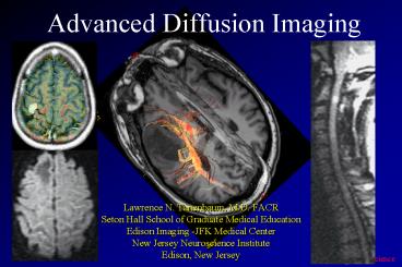
Advanced Diffusion Imaging - PowerPoint PPT Presentation
1 / 170
Title: Advanced Diffusion Imaging
1
Advanced Diffusion Imaging
- Lawrence N. Tanenbaum, M.D. FACR
- Seton Hall School of Graduate Medical Education
- Edison Imaging -JFK Medical Center
- New Jersey Neuroscience Institute
- Edison, New Jersey
2
Advanced Diffusion Imaging
- Spine and spinal cord
- High B values
- Diffusion tensor imaging and tractography
3
Diffusion imagingspine indications
- cord lesions
- demyelinating disease
- infarction
Pusan 2000
4
Diffusion imagingspine indications
- cord lesions
- demyelinating disease
- infarction
- vertebral lesion characterization
Pusan 2000
5
Kuala Lumpur 2000
6
(No Transcript)
7
(No Transcript)
8
(No Transcript)
9
(No Transcript)
10
(No Transcript)
11
(No Transcript)
12
(No Transcript)
13
(No Transcript)
14
(No Transcript)
15
(No Transcript)
16
(No Transcript)
17
(No Transcript)
18
(No Transcript)
19
(No Transcript)
20
Third generation gradients
- shorter TE per B value
- higher SNR, reduced susceptibility artifact
EchoSpeed 23 mT/m
EchoSpeed Plus 33 mT/m
B1000
TE95ms
TE75ms
21
TwinSpeed
B 1200 TE 69.9
B 2000 TE 79.1
B 3000 TE 87.6
B 4000 TE 94.4
B 6000 TE 105.2
B 5000 TE 100.2
B 7000 TE 109.7
22
EchoSpeed plus
B 1500
B800
23
NVi
B800
B1900
24
NVi
B800
B1900
25
B 1200
B 10000
26
B 1900
High B DWI
NVi/ CVi
white matter shine through
B 800
27
1000
28
(No Transcript)
29
NVi
B800
B1900
30
(No Transcript)
31
B 800
B 2000
32
(No Transcript)
33
6 55 directions
34
High SNR DWI 1.5 T
35
Diffusion tensor imaging
- quantitative evaluation of the rate and direction
of water motion - rate of diffusion measured by ADC
- directionality measured by relative or fractional
anisotropy
36
Diffusion tensor
- matrix of numbers derived from diffusion
measurements in several different directions (at
least 6) - mathematical model of diffusion in 3D space
- diffusivity in any arbitrary direction
- direction of maximum diffusivity
37
Diffusion tensor imaging
- fiber tracts impose directionality (anisotropy)
- direction of maximum diffusivity coincides with
WM tract orientation - anisotropy is the extent to which the ellipsoid
deviates from that of a sphere
38
Diffusion tensor imaging
- tensor matrix (water diffusivity) visualized as
an ellipsoid - diameter in any direction estimates diffusivity
in that direction - principle axis oriented in direction of maximum
diffusivity - orientation characterized by 3 eigenvectors
- shape determined by 3 eigenvalues
39
fractional anisotropy
Diffusion Tensor Imaging of Cerebral White
Matter A Pictorial Review of Physics, Fiber
Tract Anatomy, and Tumor Imaging Patterns Brian
J. Jellison, et.al. American Journal of
Neuroradiology 25356-369, March 2004
40
fractional anisotropy
41
Diffusion anisotropy imaging
- tractography
- operative planning
- sensorimotor localization
- eloquent tract localization
- imaging occult disease
- neuropsychiatric applications
- TBI
- schizophrenia
- dementia (MID vs. DAT)
- epilepsy
- brain maturation
42
real-time fMRI sensorimotor Cortex 35
seconds NVi / CVi
43
Tensor imaging anisotropy study -- 55 directions
44
relative anisotropy
Real-time FMRI
45
Real-time FMRI
46
cavernous malformation
47
(No Transcript)
48
(No Transcript)
49
(No Transcript)
50
RT F M R I
51
tensor DWI fractional anisotropy
52
tensor DWI fractional anisotropy
53
(No Transcript)
54
3T
55
metastatic disease
FMRI 3T
56
FA
RTIP
FA
FMRI 3T
avg
glioma
57
3 mm diffusion tractography 3T
58
Derek Jones, Institute of Psychiatry, UK
59
Superior Longitudinal Fasciculus
Anterior Commissure
Cingulum
Uncinate Fasciculus
Inferior Fronto-Occipital Fasciculus
Fornix
Derek Jones, Institute of Psychiatry, UK
60
WM fiber tracts
- association fibers
- interconnect cortical areas in each hemisphere
- projection fibers
- interconnect cortical areas with deep nuclei,
brain stem, cerebellum and spinal cord - efferent (corticofugal) and afferent
(corticopetal) - commissural fibers
- interconnect similar cortical areas between
hemispheres
61
association fibers
Cingulum
Superior Longitudinal Fasciculus
- cingulum
- superior occipitofrontal fasciculus
- inferior occipitofrontal fasciculus
- uncinate fasciculus
- superior longitudinal (arcuate) fasciculus
- inferior longitudinal (occipitotemporal)
fasciculus
Uncinate Fasciculus
62
cingulum
- begins in paraolfactory area of the cortex below
the rostrum of the CC - courses within the cingulate gyrus and arches
around the CC - extends forward into the parahippocampal gyrus
and uncus
63
Diffusion Tensor Imaging of Cerebral White
Matter A Pictorial Review of Physics, Fiber
Tract Anatomy, and Tumor Imaging Patterns Brian
J. Jellison, et.al. American Journal of
Neuroradiology 25356-369, March 2004
64
Inferior occipitofrontal fasciculus
cingulum
Diffusion Tensor Imaging of Cerebral White
Matter A Pictorial Review of Physics, Fiber
Tract Anatomy, and Tumor Imaging Patterns Brian
J. Jellison, et.al. American Journal of
Neuroradiology 25356-369, March 2004
65
(No Transcript)
66
superior occipitofrontal f.
- lies beneath the superior aspect of the CC
- connects occipital and frontal lobes
- extends posteriorly along the dorsal border of
the caudate nucleus - separated from superior longitudinal f. by corona
radiata and internal capsule
Diffusion Tensor Imaging of Cerebral White
Matter A Pictorial Review of Physics, Fiber
Tract Anatomy, and Tumor Imaging Patterns Brian
J. Jellison, et.al. American Journal of
Neuroradiology 25356-369, March 2004
67
Diffusion Tensor Imaging of Cerebral White
Matter A Pictorial Review of Physics, Fiber
Tract Anatomy, and Tumor Imaging Patterns Brian
J. Jellison, et.al. American Journal of
Neuroradiology 25356-369, March 2004
68
inferior occipitofrontal fascic.
- connects occipital and frontal lobes
- far inferior to superior occipitofrontal f.
- extends along inferolateral edge of the claustrum
below the insula - mid portion bundled with the mid portion of the
uncinate f.
Diffusion Tensor Imaging of Cerebral White
Matter A Pictorial Review of Physics, Fiber
Tract Anatomy, and Tumor Imaging Patterns Brian
J. Jellison, et.al. American Journal of
Neuroradiology 25356-369, March 2004
69
inferior occipitofrontalfasciculus
- posteriorly joins the inf longitudinal f., the
descending portion of the sup longitudinal f. and
portions of the geniculocalcarine t. to form much
of the sagittal stratum - connects the occipital lobe to the rest of the
brain
Diffusion Tensor Imaging of Cerebral White
Matter A Pictorial Review of Physics, Fiber
Tract Anatomy, and Tumor Imaging Patterns Brian
J. Jellison, et.al. American Journal of
Neuroradiology 25356-369, March 2004o
70
inferior occipitofrontal fasciculus
Inferior occipitofrontal fasciculus
cingulum
Inferior longitudinal fasciculus
Diffusion Tensor Imaging of Cerebral White
Matter A Pictorial Review of Physics, Fiber
Tract Anatomy, and Tumor Imaging Patterns Brian
J. Jellison, et.al. American Journal of
Neuroradiology 25356-369, March 2004
71
uncinate fasciculus (UF)
- hooks around the lateral fissure
- connects the orbital and inferior frontal gyri of
the frontal lobe to the anterior temporal lobe - anteriorly it is parallel and inferomedial to and
in midportion joins the inferior occipitofrontal
f. before heading inferolaterally into the
anterior temporal lobe
Diffusion Tensor Imaging of Cerebral White
Matter A Pictorial Review of Physics, Fiber
Tract Anatomy, and Tumor Imaging Patterns Brian
J. Jellison, et.al. American Journal of
Neuroradiology 25356-369, March 2004
72
Diffusion Tensor Imaging of Cerebral White
Matter A Pictorial Review of Physics, Fiber
Tract Anatomy, and Tumor Imaging Patterns Brian
J. Jellison, et.al. American Journal of
Neuroradiology 25356-369, March 2004
73
superior longitudinal f. (SLF)
- massive (largest) fiber bundle that sweeps along
the superior margin of the insula - arcuate fasciculus
- connects frontal lobe cortex to parietal,
temporal and occipital lobe cortices
Diffusion Tensor Imaging of Cerebral White
Matter A Pictorial Review of Physics, Fiber
Tract Anatomy, and Tumor Imaging Patterns Brian
J. Jellison, et.al. American Journal of
Neuroradiology 25356-369, March 2004
74
Diffusion Tensor Imaging of Cerebral White
Matter A Pictorial Review of Physics, Fiber
Tract Anatomy, and Tumor Imaging Patterns Brian
J. Jellison, et.al. American Journal of
Neuroradiology 25356-369, March 2004
75
superior longitudinal f. (SLF)
76
inferior longitudinal f. (ILF)
- connects temporal and occipital lobe cortices
- traverses the length of the occipital lobe
- joins the IOFF, the inferior aspect of the SLF
and the optic radiations to form much of the
sagittal stratum traversing the occipital lobe
Diffusion Tensor Imaging of Cerebral White
Matter A Pictorial Review of Physics, Fiber
Tract Anatomy, and Tumor Imaging Patterns Brian
J. Jellison, et.al. American Journal of
Neuroradiology 25356-369, March 2004
77
Diffusion Tensor Imaging of Cerebral White
Matter A Pictorial Review of Physics, Fiber
Tract Anatomy, and Tumor Imaging Patterns Brian
J. Jellison, et.al. American Journal of
Neuroradiology 25356-369, March 2004
78
inferior longitudinal f. (ILF)
Inferior occipitofrontal fasciculus
cingulum
Inferior longitudinal fasciculus
Diffusion Tensor Imaging of Cerebral White
Matter A Pictorial Review of Physics, Fiber
Tract Anatomy, and Tumor Imaging Patterns Brian
J. Jellison, et.al. American Journal of
Neuroradiology 25356-369, March 2004
79
projection fibers
- corticospinal tracts
- corticobulbar tracts
- corticopontine tracts
- geniculocalcarine tracts (optic radiations)
80
Corticospinal, pontine and bulbar tracts
- major efferent fibers connecting motor cortex to
the brain stem and spinal cord - cant be parsed with tractography techniques
corticospinal
frontopontine
temporoparieto occipito pontine
Diffusion Tensor MR Imaging of the Brain and
White Matter Tractography Elias R. Melhem1,2,
Susumu Mori1,2, Govind Mukundan1, Michael A.
Kraut1,2, Martin G. Pomper1 and Peter C. M. van
Zijl1,2 AJR 2002 1783-16
81
corticospinal
frontopontine
temporo parieto occipito pontine
Diffusion Tensor MR Imaging of the Brain and
White Matter Tractography Elias R. Melhem1,2,
Susumu Mori1,2, Govind Mukundan1, Michael A.
Kraut1,2, Martin G. Pomper1 and Peter C. M. van
Zijl1,2 AJR 2002 1783-16
82
corticospinal tracts
- fibers converge in CR
- continue through posterior limb internal capsule
- turn medially to the cerebral peduncle then
vertically on the way to the lateral funiculus of
the cord
Diffusion Tensor Imaging of Cerebral White
Matter A Pictorial Review of Physics, Fiber
Tract Anatomy, and Tumor Imaging Patterns Brian
J. Jellison, et.al. American Journal of
Neuroradiology 25356-369, March 2004
83
corticospinal tracts
- fibers converge in CR
- continue through posterior limb internal capsule
- turn medially to the cerebral peduncle then
vertically on the way to the lateral funiculus of
the cord
Diffusion Tensor Imaging of Cerebral White
Matter A Pictorial Review of Physics, Fiber
Tract Anatomy, and Tumor Imaging Patterns Brian
J. Jellison, et.al. American Journal of
Neuroradiology 25356-369, March 2004
84
cortico spinal fibers
peduncle
b stem
Diffusion Tensor Imaging of Cerebral White
Matter A Pictorial Review of Physics, Fiber
Tract Anatomy, and Tumor Imaging Patterns Brian
J. Jellison, et.al. American Journal of
Neuroradiology 25356-369, March 2004
85
corticobulbar tracts
- fibers converge in CR
- continue through genu internal capsule
- turn medially to the cerebral peduncle (medial
and dorsal to corticospinal fibers) - predominantly terminate at CN nuclei
Diffusion Tensor Imaging of Cerebral White
Matter A Pictorial Review of Physics, Fiber
Tract Anatomy, and Tumor Imaging Patterns Brian
J. Jellison, et.al. American Journal of
Neuroradiology 25356-369, March 2004
86
internal capsule
- conduit of fibers to and from the cerebral cortex
- anterior limb
- primarily anteroposteriorly directed projection
fibers to and from the thalamus (thalamocortical
projections) as well as frontopontine tracts - posterior limb
- superoinferiorly oriented fibers of the CS, CB
and CP tracts
Diffusion Tensor Imaging of Cerebral White
Matter A Pictorial Review of Physics, Fiber
Tract Anatomy, and Tumor Imaging Patterns Brian
J. Jellison, et.al. American Journal of
Neuroradiology 25356-369, March 2004
87
ext capsule
opt rad
redL-R greenA-P blueS-I
Diffusion Tensor Imaging of Cerebral White
Matter A Pictorial Review of Physics, Fiber
Tract Anatomy, and Tumor Imaging Patterns Brian
J. Jellison, et.al. American Journal of
Neuroradiology 25356-369, March 2004
88
geniculocalcarine tracts
- optic radiation connects the lateral geniculate
nucleus to occipital cortex - inferior fibers sweep around the posterior horns
of the lateral ventricles and terminate in the
calcarine cortex - superior fibers take more direct route
89
geniculocalcarine tracts
- optic radiation connects the lateral geniculate
nucleus to occipital cortex - inferior fibers sweep around the posterior horns
of the lateral ventricles and terminate in the
calcarine cortex - superior fibers take more direct route
90
redL-R greenA-P blueS-I
optic rad
Diffusion Tensor Imaging of Cerebral White
Matter A Pictorial Review of Physics, Fiber
Tract Anatomy, and Tumor Imaging Patterns Brian
J. Jellison, et.al. American Journal of
Neuroradiology 25356-369, March 2004
91
commissural fibers
- corpus callosum
- anterior commissure
Anterior Commissure
92
corpus callosum
- massive accumulation of fibers connecting
corresponding areas of cortex between the
hemispheres
93
corpus callosum
- body fibers are transversely oriented
- genu and splenium fibers course anteriorly and
posteriorly to reach the poles of the hemispheres - near mid-sagittal plane fibers are left-right
oriented
Diffusion Tensor Imaging of Cerebral White
Matter A Pictorial Review of Physics, Fiber
Tract Anatomy, and Tumor Imaging Patterns Brian
J. Jellison, et.al. American Journal of
Neuroradiology 25356-369, March 2004
94
redL-R greenA-P blueS-I
Diffusion Tensor Imaging of Cerebral White
Matter A Pictorial Review of Physics, Fiber
Tract Anatomy, and Tumor Imaging Patterns Brian
J. Jellison, et.al. American Journal of
Neuroradiology 25356-369, March 2004
95
redL-R greenA-P blueS-I
Diffusion Tensor Imaging of Cerebral White
Matter A Pictorial Review of Physics, Fiber
Tract Anatomy, and Tumor Imaging Patterns Brian
J. Jellison, et.al. American Journal of
Neuroradiology 25356-369, March 2004
96
anterior commissure
- crosses through the lamina terminalis
- anterior fibers connect the olfactory bulbs and
nuclei - posterior fibers connect middle and inferior
temporal gyri
97
cingulum
sup longitudinal f
sup occipitofrontal f.
inf occipitofrontal f.
ant commissure
Diffusion Tensor Imaging of Cerebral White
Matter A Pictorial Review of Physics, Fiber
Tract Anatomy, and Tumor Imaging Patterns Brian
J. Jellison, et.al. American Journal of
Neuroradiology 25356-369, March 2004
98
brain stem
Diffusion Tensor Imaging of Cerebral White
Matter A Pictorial Review of Physics, Fiber
Tract Anatomy, and Tumor Imaging Patterns Brian
J. Jellison, et.al. American Journal of
Neuroradiology 25356-369, March 2004
99
brain stem
Diffusion Tensor Imaging of Cerebral White
Matter A Pictorial Review of Physics, Fiber
Tract Anatomy, and Tumor Imaging Patterns Brian
J. Jellison, et.al. American Journal of
Neuroradiology 25356-369, March 2004
100
brain stem
Diffusion Tensor Imaging of Cerebral White
Matter A Pictorial Review of Physics, Fiber
Tract Anatomy, and Tumor Imaging Patterns Brian
J. Jellison, et.al. American Journal of
Neuroradiology 25356-369, March 2004
101
(No Transcript)
102
Diffusion Tensor MR Imaging of the Brain and
White Matter Tractography Elias R. Melhem1,2,
Susumu Mori1,2, Govind Mukundan1, Michael A.
Kraut1,2, Martin G. Pomper1 and Peter C. M. van
Zijl1,2 AJR 2002 1783-16
103
(No Transcript)
104
Jellison, et.al. AJNR 25356-369, March 2004
105
Diffusion Tensor Imaging of Cerebral White
Matter A Pictorial Review of Physics, Fiber
Tract Anatomy, and Tumor Imaging Patterns Brian
J. Jellison, et.al. American Journal of
Neuroradiology 25356-369, March 2004
106
(No Transcript)
107
(No Transcript)
108
(No Transcript)
109
(No Transcript)
110
(No Transcript)
111
HD MRI 3T
.21 mm in plane
112
(No Transcript)
113
HD Prop MRI 3T
.21 mm in plane
114
(No Transcript)
115
(No Transcript)
116
(No Transcript)
117
(No Transcript)
118
Residual glioma
Perfusion
DWI B1000
119
(No Transcript)
120
(No Transcript)
121
(No Transcript)
122
Diffusion Tensor Imaging of Cerebral White
Matter A Pictorial Review of Physics, Fiber
Tract Anatomy, and Tumor Imaging Patterns Brian
J. Jellison, et.al. American Journal of
Neuroradiology 25356-369, March 2004
123
(No Transcript)
124
(No Transcript)
125
(No Transcript)
126
(No Transcript)
127
redL-R greenA-P blueS-I
128
anterior limb int capsule
posterior limb int capsule
129
(No Transcript)
130
cortico spinal fibers
sup long fasciculus
inf long fasciculus
post limb int cap
cortico spinal fibers
post limb int cap
post limb int cap
131
(No Transcript)
132
(No Transcript)
133
(No Transcript)
134
(No Transcript)
135
(No Transcript)
136
(No Transcript)
137
(No Transcript)
138
(No Transcript)
139
(No Transcript)
140
(No Transcript)
141
(No Transcript)
142
(No Transcript)
143
(No Transcript)
144
(No Transcript)
145
(No Transcript)
146
(No Transcript)
147
(No Transcript)
148
(No Transcript)
149
(No Transcript)
150
(No Transcript)
151
Glioma
Perfusion
DWI B1000
152
(No Transcript)
153
(No Transcript)
154
(No Transcript)
155
(No Transcript)
156
cingulum
Inf longitudinal f.
optic radiations
157
(No Transcript)
158
(No Transcript)
159
(No Transcript)
160
(No Transcript)
161
(No Transcript)
162
(No Transcript)
163
(No Transcript)
164
(No Transcript)
165
U fibers
166
(No Transcript)
167
optic radiations and inferior longitudinal f.
168
Tensor Diffusion
2
Scott Atlas, M.D.
169
(No Transcript)
170
(No Transcript)
171
(No Transcript)
172
Mesial temporal sclerosis 3.0 T
K. Thulborn MD
173
(No Transcript)
174
(No Transcript)
175
(No Transcript)
176
Hippocampal sclerosis
177
Hippocampal sclerosis
3 mm 55 dirns 5 min
178
(No Transcript)
179
Alzheimer disease
- loss of tract count and reduced FA within counts
in fronto-occipital and thalamo-frontal tracts in
early AD - corresponds to quantitative EEG findings of a
loss of coherence between the occipital and
frontal lobes - reduced FA in corpus callosum
180
www. drtmasters .com
JFK Medical Center
Mardi Gras NO 2001
181
www.drtmasters.com
182
For further information contact the CMRS at
888-350-CMRS or visit www.cmrs.org.
183
www.drtmasters.com
184
(No Transcript)
185
(No Transcript)
186
(No Transcript)
187
(No Transcript)
188
(No Transcript)
189
(No Transcript)
190
(No Transcript)
191
(No Transcript)
192
(No Transcript)
193
(No Transcript)
194
(No Transcript)
195
(No Transcript)
196
(No Transcript)
197
(No Transcript)
198
(No Transcript)
199
(No Transcript)
200
(No Transcript)


