
1' Receptor superfamilies - PowerPoint PPT Presentation
1 / 37
Title:
1' Receptor superfamilies
Description:
Ion channel receptors (Ligand gated ion channels) Five glycoprotein subunits. traversing cell membrane. Messenger. Receptor. INDUCED. FIT. GATING' (ion channel. opens) ... – PowerPoint PPT presentation
Number of Views:244
Avg rating:3.0/5.0
Title: 1' Receptor superfamilies
1
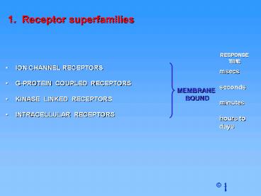
1. Receptor superfamilies
- ION CHANNEL RECEPTORS
- G-PROTEIN COUPLED RECEPTORS
- KINASE LINKED RECEPTORS
- INTRACELLULAR RECEPTORS
2
Ion channel receptors (Ligand gated ion channels)
Five glycoprotein subunits traversing cell
membrane
Cationic ion channels for K, Na, Ca2
excitatory Anionic ion channels for Cl-
inhibitory Ex cationic nAChR, iGluR, 5HT3, ATP,
anionic GABAA, Gly
3
What determines selectivity of each ion
channel? 1. hydrated size of the ion Li 0.6
A Na 0.95A K 1.33A Rb 1.48A 2.
nature of sidechains lining pore of
channel/selectivity filter Ex K channel
has 8 oxygens Great review article Science
310, 2005, 1461.
4
Ion channel receptors (Ligand gated ion channels)
nAChR 255 kDa, non-selective cation channel
Na, K, Ca2
2a, b, g, d subunits
Each subunit highly homologous (40)
5
Ion channel receptors (Ligand gated ion channels)
glycine receptor
3a, 2b subunits
anion channels Cl-, SCN-, I-, Br-, NO3-
most relevant physiologically
6
Ion channel receptors (Ligand gated ion channels)
Structure of protein subunits (4-TM receptor
subunits)
4 Transmembrane (TM) regions (hydrophobic)
Ex nAChR, 5HT3, glycine, GABAA
7
Ion channel receptors (Ligand gated ion channels)
Cartoon of ion channel
Note TM2 of each protein subunit lines the
central pore
8
Ion channel receptors (Ligand gated ion channels)
Gating
9
Extracellular
Ex iGluR
10
Extracellular
Ex ATP receptor (purinoceptors P2X1-P2X7)
11
Ion channel receptors (Ligand gated ion channels)
Gating
- Fast response measured in msec
- Ideal for transmission between nerves
- Binding of messenger leads directly to ion flows
across cell membrane 108 ions/sec - Ion flow secondary effect (signal transduction)
12
G-protein-coupled receptors (7-TM receptors)
Structure - Single protein with 7
transmembrane regions
13
G-protein-coupled receptors (7-TM receptors)
Ligands
- Monoamines e.g. dopamine, histamine,
noradrenaline, acetylcholine (muscarinic) - Nucleotides
- Lipids
- Hormones
- Glutamate
- Ca2
In general, mediate action of hormones and
slow-acting neurotransmitters
14
G-protein-coupled receptors (7-TM receptors)
Ligand binding site - varies depending on
receptor type
A) Monoamines, nucleotides, lipids - pocket in
TM helices B) Peptide hormones (Ex oxytocin,
glucagon, insulin) bind to top of TM helices
extracellular loops N-terminal chain C)
Hormones (Ex epinephrine, norepinephrine,
estrogen, vitamin D) bind to extracellular loops
N-terminal chain D) Glutamate, Ca2 -
N-terminal chain
15
G-protein-coupled receptors (7-TM receptors)
Bacteriorhodopsin rhodopsin family
- Rhodopsin visual receptor in retina
- Many common receptors belong to this same family
- Implications for drug selectivity depending on
similarity (evolution) - Membrane bound receptors difficult to crystallize
- X-Ray structure of bacteriorhodopsin solved -
bacterial protein similar to rhodopsin - Bacteriorhodopsin structure used as template
for other receptors - Construct model receptors based on template and
amino acid sequence - Leads to model binding sites for drug design
- Crystal structure for rhodopsin now solved -
better template
16
G-protein-coupled receptors (7-TM receptors)
Bacteriorhodopsin rhodopsin family
17
G-protein-coupled receptors (7-TM receptors)
Receptor types and subtypes
Reflects differences in receptors which recognize
the same ligand
18
G-protein-coupled receptors (7-TM receptors)
Receptor types and subtypes
- Receptor types and subtypes not equally
distributed amongst tissues. - Target selectivity leads to tissue selectivity
Heart muscle - b1 adrenergic receptors Fat
cells - b3 adrenergic receptors Bronchial
muscle - a1 b2 adrenergic receptors GI-tract
- a1 a2 b2 adrenergic receptors
19
Kinase linked receptors (1 TM)
- Bi-functional receptor / enzyme in 1 molecule
(activates enzyme directly- doesnt require G
protein as signaling molecule) - Activated by hormones (ie insulin), growth
factors, cytokines (growth factors that regulate
the differentiation, proliferation, and
activities of various types of blood cells, ie
interleukins) - Over-expression can result in cancer (drug
target) - Loss of function developmental defects or
hormone resistance
20
Tyrosine kinase linked receptors
Structure
Extracellular N-terminal chain
Intracellular C-terminal chain
21
Tyrosine kinase linked receptors
Reaction catalyzed by tyrosine kinase
22
Tyrosine kinase linked receptors
Epidermal growth factor receptor (EGF-
R) (EGF promotes growth and proliferation of
mesenchymal, glial, and epithelial cells)
Induced fit opens tyrosine kinase active sites
23
Tyrosine kinase linked receptors
Epidermal growth factor receptor (EGF- R)
- Active site on one half of dimer catalyses
phosphorylation of Tyr residues on other half - Dimerisation of receptor is crucial
- Phosphorylated regions act as binding sites for
further proteins and enzymes - Results in activation of signalling proteins and
enzymes - Message carried into cell
24
Tyrosine kinase linked receptors
Insulin receptor (tetrameric complex)
Kinase active site opened by induced fit
25
Tyrosine kinase linked receptors
Growth hormone receptor Tetrameric complex
constructed in presence of growth hormone
Kinase active site opened by induced fit
cytoplasmic
26
Tyrosine kinase linked receptors
Signalling pathways
27
Tyrosine kinase linked receptors
Signalling pathways
28
Tyrosine kinase linked receptors
Signalling pathways
29
Tyrosine kinase linked receptors
Signalling pathways
Ras member of small G-protein family similar to
a subunit of larger G protein family
30
Tyrosine kinase linked receptors
Signalling pathways
31
Intracellular receptors
- Chemical messengers must cross cell membrane
- Chemical messengers must be hydrophobic
- Example - steroids and steroid receptors
- 50 members important in directly regulating
gene - transcription. Called nuclear hormone
receptors or - nuclear transcription factors.
32
Intracellular receptors
Structure
CO2H
H2N
Zinc fingers contain cysteine residues (SH) and
histidine residues (NH) to allow for S-Zn and
N-Zn interactions
33
Intracellular receptors
Mechanism
1. Messenger crosses membrane
5. Complex binds to DNA
2. Binds to receptor
6. Transcription switched on or off
3. Receptor dimerization
7. Protein synthesis activated or inhibited
4. Binds co-activator protein
34
Intracellular receptors
Estrogen receptor
35
Intracellular receptors
Estrogen receptor
- Phenol and alcohol of oestradiol are important
hydrogen binding groups - Binding site is spacious and hydrophobic
- Phenol group of oestradiol positioned in narrow
slot - Orientates rest of molecule
36
Intracellular receptors
Estrogen receptor
- Raloxifene is an antagonist (anticancer agent)
- Phenol groups mimic phenol and alcohol of
estradiol - Interaction with Asp351 is important for
antagonist activity - Side chain prevents receptor helix H12 folding
over as lid - AF-2 binding region not revealed
- Co-activator cannot bind
37
Intracellular receptors
Estrogen receptor
Tamoxifen (Nolvadex) - anticancer agent which
targets Estrogen receptor