
Pubertal Neurogenesis - PowerPoint PPT Presentation
1 / 40
Title:
Pubertal Neurogenesis
Description:
Figure 2 is a nissl-stained photomicrograph showing the subnuclei of the pig PVN. Figure 3 is a photomicrograph of a Nissl-stained section of the PVN. ... – PowerPoint PPT presentation
Number of Views:180
Avg rating:3.0/5.0
Title: Pubertal Neurogenesis
1
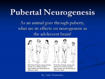
Pubertal Neurogenesis
- As an animal goes through puberty, what are its
effects on neurogenesis in the adolescent brain?
By Carly Christensen
2
Overview
- Part I What is Puberty?
- Looking at the neural basis of puberty and
adolescence - Pubertal hormones and behavioral maturation
- Part II Puberty and Neurogenesis
- What is neurogenesis?
- The areas of the brain that were examined
- Dentate Gyrus of the Hippocampus, and Forebrain
Subventricular Zone - Hypothalamus
- Part III Unanswered Questions and Future
Direction - What about the other structures involved in
reproduction? - What happens when neurogenesis is ablated?
3
Part I
What is Puberty?
4
What is Puberty and Adolescence?
According to Cheryl Sisk and Douglas Foster in
The Neural Basis of Puberty and Adolescence
- Puberty and adolescence mark the changing of a
child into an adult. - Puberty refers to the activation of the
hypothalamic-pituitary-gonadal axis that
cumulates in gonadal maturation. - Adolescence is the maturation of adult social and
cognitive behaviors. - The transition from puberty to adulthood involves
both gonadal and behavioral maturation.
5
Adolescent Maturation of Reproductive Behavior
Steroid hormones remodel and activate neural
circuits during adolescent brain development,
which leads to the development of sexual salience
of sensory stimuli, sexual motivation, and
expression of copulatory behaviors in certain
social contexts.
6
- Transient activation of the HPG axis during late
prenatal/ early postnatal life causes an increase
in gonadal steroids and in turn sexual
differentiation. GnRH secretion decreases after
HPG activation, and hormone pulse slows
throughout the prepubertal period. After this
period of quiescence, puberty begins when GnRH
secretion gradually increases and levels off to
stimulate gonadotropin and steroid hormone
secretion, resulting in complete gonadal
maturation and reproductive behavior. - Possible triggers that induce re-emergence of
GnRH secretion at puberty are melatonin, body
fat, and leptin. - Copulatory behavior can still be activated by a
dose of testosterone in castrated males during
adolescence, but not in prepubertal males. - Remodeling of the brain during adolescence
includes increased myelination and decreased gray
matter volume in cortical areas, synaptic
elaboration and pruning in striatum and PFC, cell
death in primary visual cortex, and changes in
connectivity in the amygdala and PFC. - These rearrangements are thought to be linked to
making decisions, planning, drug sensitivity, and
reward seeking behavior.
7
GnRH neurons are key to gonadal and behavioral
maturation
- GnRH is a decapeptide produced by specialized
neurons that secrete pulses of hormone from nerve
terminals in the median eminence of the basal
hypothalamus. - The brain initiates activation of the GnRH system
at puberty onset, leading to an increase in
steroid hormone production. - Steroid hormones in turn modulate GnRH secretion
in the brain, and organize and activate neural
circuits mediating reproductive behavior during
adolescence.
The pubertal increase in GnRH neuronal activity
and gonadotropin secretion is timed by a
developmental clock and fine-tuned by integration
of permissive signals
8
Pubertal Hormones Organize the Brain
- In an article by Schulz and Sisk et al., evidence
that adolescent brain development is a
prerequisite for the activation of reproductive
behavior was reported in the Syrian Hamster. - It was also found that pubertal gonadal hormones
shape adolescent brain development through their
organization of neural circuits.
Perinatal hormone secretions sexually
differentiate behavioral neural circuits and
pubertal hormone secretions improve and finish
these processes during adolescence to allow for
the display of sex-typical social behaviors in
adulthood
9
Adolescent Exposure to Testicular Hormones
- Figure 3 shows that testicular hormone
deprivation during puberty causes deficits in
masculine reproductive behavior. - Males deprived of testicular hormones during
puberty (No-T_at_P) exhibited diminished mounting
and T_at_P (with testicular hormones) displayed
significantly more mounts. - Figure 5 shows that adolescent testicular
hormones defeminize lordosis behavior. - Lordosis a posture assumed by some female
mammals during mating, in which the back arches
downward - Males exposed to testicular hormones during
pubertal development display longer lordosis
latencies than males deprived of pubertal
testicular hormones .
Figure 3
Figure 5
10
Figure 6
- Adolescent exposure to testicular hormones
organizes male agonistic (combative) behavior. - In Figure 6, adolescent testicular hormones
increase aggressive and decrease submissive
behaviors in adulthood. - Males exposed to testicular hormones during
puberty (T_at_P) displayed more attacks and fewer
escape dashes than males deprived (No-T_at_P)
regardless of testosterone treatment. - _______________________________________
- Adolescent exposure to ovarian hormones
defeminizes female reproductive behavior. - Figure 7 represents the finding that pubertal
ovarian hormones defeminize lordosis behavior. - Females exposed to ovarian hormones during
puberty (O_at_P) display longer lordosis latencies
than females deprived of pubertal ovarian
hormones (No-O_at_P).
Figure 7
11
Conclusions
- Gonadal steroid hormones are important at both
stages of behavioral development because of their
ability to influence cell survival, cell
phenotype, synaptic organization, and neural
circuitry. - During the adolescent period of development, both
ovarian and testicular secretions have organizing
actions, which cause the adult behaviors of
females and males to relate. - Support for adolescence being a sensitive period
- The fact that steroid hormones bring out
different behavioral responses before and after
adolescence. - The effects on behavior resulting from the
absence of hormones during adolescence are not
reversed by hormone replacement in adulthood.
12
Part II
Puberty and Neurogenesis
13
Defining characteristics of Adult Stem Cells
- Proliferate
- Multipotential
- Last life-time
- (extensive self-renewal)
Taken from Dr. Kippins slides
14
Neurogenesis
- The generation of new neurons after birth has
been identified in various structures of the
adult brain including the forebrain
subventricular zone (SVZ), hippocampal dentate
gyrus (DG), and paraventricular nucleus (PVN) of
the hypothalamus. - These areas were examined in the various papers
that will be discussed today.
15
The effect of Maturation on Neurogenesis
- According to He and Crews et al. adolescence is
marked by risk-taking, exploration, novelty
seeking, social interaction, activity, and play
along with changes in hormones and growth
factors. - It has been shown that overproduction of axons
and synapses occur during early puberty and rapid
pruning follows later in adolescence, while
neurogenesis in the SVZ and DG continues
throughout life in humans and rodents. - Jun He and Fulton Crews set out to determine if
neurogenesis changes during the transition from
adolescence to adulthood.
16
Subjects, Materials, Methods
- IGF-1 is a growth factor during postnatal
development known to enhance neurogenesis in
developing mice. - Transgenic mice that over-express IGF-1 show
overgrowth of the brain causing higher brain
weight compared to wildtype controls. - Transgenic mice that over-express IGF binding
protein-1 (IGFBP-1) causes a reduction in
neurogenesis resulting in brain growth
retardation (activation of IGFBP-1 inhibits IGF-1
bioactivity). - BrdU (exogenous mitotic marker bromodeoxyuridine)
and DCX (endogenous neuronal marker doublecortin)
were used to measure the level of neurogenesis in
this study. - BrdU Injections were given once a day 300
mg/kg/day for 2 days. Animals were killed 24h
after last injection.
17
Increased Proliferation in Adolescent DG
- Figure 1 represents the number of BrdU cells in
the hippocampus of adult and adolescent brains. - The number of BrdU cell/ DG section is
significantly higher in adolescent hippocampus
than adults in all genetic background of strains
(IGF-1, IGFBP-1, and WT). - Figure 2 represents BrdU cells in DG of
hippocampus. - AC) Adolescent hippocampus
- BD) Adult hippocampus.
Figure 1
Figure 2
18
Increased Differentiation of Neuroprogenitors in
Adolescent Hippocampus
- Figure 3 represents the expression of DCX
(doublecortin) in the hippocampus of adolescent
and adult brains. - The DCX immunoreactivity is significantly higher
in adolescent hippocampus than those of the
adults in all strands of mice. - Figure 4 represents DCX expression in the
hippocampus of adolescent and adult brains.
Adolescent mice were 30 days old, while adult
mice were 120 days old.
Figure 3
Figure 4
19
Increased Proliferation in SVZ of Adolescents
- Figure 5 shows enhanced BrdU-immunoreactivity in
the forebrain SVZ of the adolescents compared to
the adults. - BrdU-labeling is significantly higher in the SVZ
of the adolescents than in the adults. - On the right, adolescents are the 30 day olds and
adults are the 120 day olds.
Figure 5
20
Conclusions Further Implications
- Both forebrain and hippocampal neurogenesis is
significantly reduced during brain maturation
from adolescence to adults. - Their results suggest that the influence of IGF-1
on neurogenesis most likely occurs before the
animals reach adolescence, since the IGF-1
transgenic mice didnt contribute to the decrease
in neurogenesis. - The high level of neurogenesis in adolescent
brain found in this study could be due to the
high level of neuroplasticity during adolescence.
- The significant decline of neurogenesis may
indicate a critical window of opportunity where
the neuronal circuitry is still modifiable for
further adaptation. - Any manipulation during this critical period
could lead to more damage in adulthood (i.e.
alcohol-induced reduction of neurogenesis). - Therefore, the development of the adolescent
brain is a period of susceptibility and prospect.
21
Leptin Promotes Adult Neurogenesis in the
Hippocampus
- Leptin is an adipose-derived hormone encoded by
the obese (ob) gene that is linked to various
physiological processes within the hippocampus. - Leptin is known for its role in the control of
food intake and body weight, which is believed to
be mediated by interaction with LepRb in the
hippocampus. - It has also been found that leptin facilitates
spatial learning and memory and produces
anti-depressant effects. - Adult neurogenesis has been thought to mediate
hippocampal-dependent learning and therapeutic
actions of anti-depressants. - In the study by Garza et al., the impact of
leptin on cell proliferation, differentiation,
and survival in the DG of adult mice was
examined.
22
Effect of Leptin on Cell Proliferation
- Figure 1 shows the effects of leptin
administration on cell proliferation in the DG of
adult mice - BrdU was used to label cell proliferation
- Mice were injected i.p. with leptin (1mg/kg) or
vehicle twice daily for 1,5, or 14 days followed
by BrdU labeling - A) Acute treatment (1d)
- B) Short-term treatment (5d)
- C) Chronic treatment (14d)
- D E) show BrdU-labeled cells in adult DG of
mice treated for 14 days - D1 E1) show BrdU-labeled cells in PV thalamus
treated for 14 days - F) shows high magnification of proliferating
cells
23
Effect of Leptin on Cell Differentiation
- Figure 2 shows the effect of leptin
administration on cell differentiation. - Mice were injected i.p. with leptin (1mg/kg) or
vehicle twice daily for 14 days followed by BrdU
labeling and were perfused 28 days later. - A) A significantly higher number of BrdU cells
remained in the leptin-treated group compared to
the control group 28d after BrdU - B) The effect of leptin on the of BrdU-labeled
cells double-labeled for NeuN or GFAP (higher for
leptin-treated groups, not significant) - C) Co-localization of BrdU with NeuN under
confocal microscope - D) Confocal microscope images show
co-localization of BrdU with GFAP
24
The Effect of Leptin on Proliferation
- Figure 4 shows the expression of the long form
leptin receptor (LepRb) in adult hippocampal
stem/progenitor cells - B) cells stained with Nestin (green) are for
leptin receptor (red) - Figure 5 shows the effects of leptin treatment on
proliferation of adult hippocampal
stem/progenitor cells - Cells were treated with various concentrations of
leptin (1-30nM) for 48h and labeled with BrdU
(10microM) in the last 4h of incubation - A) The number of BrdU-labeled cells was increased
by leptin treatment at concentrations of 1nM and
3nM compared to the control - B) Microscopic images represent BrdU-labeled
adult hippocampal progenitor cells
25
Effect of Leptin on Differentiation of Cultured
Cells
- Figure 6 shows the effects of leptin treatment on
differentiation of cultured adult hippocampal
stem/progenitor cells - Cells that were treated with leptin (1nM) for 48h
and labeled with BrdU (10microM) in the last 4h
of incubation were allowed to differentiate for 8
days before fixation - A) Microscopic images show that BrdU-labeled
cells differentiated into neuronal (TuJ1 in red)
or glial (GFAP in green) cells - B) The of BrdU-labeled cells that were TuJ1 or
GFAP was not altered by leptin treatment
26
Significance
- Leptin was demonstrated to promote adult
hippocampal neurogenesis both in vitro and in
vivo. - Leptin-stimulated neurogenesis resulted from
increased cell proliferation, as leptin showed no
significant effect on cell differentiation and
survival. - Cell proliferation in the DG was increased by
chronic, not short-term or acute, administration
of leptin. - Leptin is known to suppress appetite, increase
energy expenditure, and reduce body weight gain.
Since dietary restriction and physical activity
have been shown to increase hippocampal
neurogenesis, leptins effects on neurogenesis
may be induced by negative energy balance
following chronic administration. - LEPTIN INCREASES THE PRODUCTION OF NEW NEURONS IN
THE ADULT DENTATE GYRUS.
27
Neurogenesis in the PVN of the Pig Hypothalamus
- In the article by Raymond et al., there is
evidence suggesting that neurogenesis occurs in
the adult hypothalamus, including centers
containing oxytocin and vasopressin producing
neurons. - The pig hypothalamus contains nuclei that release
the hormones oxytocin (OT) and vasopressin (VP),
which include the supraoptic nucleus (SON),
vasopressin and oxytocin-containing nucleus
(VON), and PVN. - Oxytocin plays a role in stimulating prolactin
release from the anterior pituitary gland and
regulates ingestive behaviors. - Vasopressin is an anti-diuretic that has been
suggested to play a role in reproductive and
sexual behavior. - In a study by Rankin et al., it was hypothesized
that there is existence of proliferating neurons
in the VON, and the occurrence of neuronal
proliferation is greater in adolescent pigs than
in mature pigs. - The VON is located bilateral to the third
ventricle in the anterior of the pigs
hypothalamus and contains VP and OT neurons. - It increases in size, volume, and neuron number
during puberty (16-30 weeks) and continues to
grow into adulthood.
28
Visualizing Methods
- OT immunoreactivity was identified by the
presence of dark blue-black precipitate in the
cytoplasm of cells. These cells were identified
as neurons because of their morphology and
antigenic reaction to the functional OT marker. - PCNA immunoreactivity was identified by the
presence of a brown granular precipitate in the
nuclei of neurons and glia. - VP immunoreactivity was identified by the
presence of red granular precipitate in the
cytoplasm of neurons. - Figure 1 represents transverse sections through
the pig hypothalamus showing the rostrocaudal
progression of the PVN.
Figure 1
29
Neurogenesis of oxytocin-containing neurons in
the PVN
- Oxytocin-containing neurons were observed in the
SON and PVN. - Within the PVN, OT-containing neurons were
observed at the dorsal end of the PVMM
subnucleus. - PCNA cells occurred most frequently in the PVLM
subnucleus of the PVN and were more numerous than
OT-containing neurons. - Figure 2 is a nissl-stained photomicrograph
showing the subnuclei of the pig PVN. - Figure 3 is a photomicrograph of a Nissl-stained
section of the PVN. There is a high density of
large spindle-shaped neurons in the PVLM and
lower density of the medium-sized neurons in the
PVMM. The PVMP contains a low density of small
neurons.
Figure 2
Figure 3
30
- Neurons double-labeled for OT and PCNA were found
in the SON and PVN of all pigs. - They were identified as neurons based on their
size and morphology, thought to be neurons
producing a neurotransmitter such as vasopressin. - In the tissue stained for VP and OT, the density
of VP-containing neurons was highest in the PVLM
subnucleus. - There was a significantly higher number of OT
PCNA labeled neurons in the PVN on lactating sows
and adult gilts compared to puberty gilts.
Figure 4- Photomicrograph from the PVN of a
tissue section stained immunohistochemically for
OT, proliferating cell nuclear antigen (PCNA) and
counterstained with hematoxylin. The unlabeled
neuron (small arrowhead), glial cells (double
arrow), and neurons for OT (large arrow), PCNA
(large arrowhead) and OT PCNA (small arrow).
Figure 5- Comparison of mean OT, OTPCNA, PCNA,
unlabeled neurons and total counts in PVN of
puberty gilts, adult gilts, and lactating sows
31
Postnatal Neurogenesis in the VP and
OT-containing nucleus of the HypothalamusImmunohi
stochemistry
- Figure 1 represents photomicrographs of PCNA-VP
stains in two hypothalamic nuclei of the pig - Granular blue- VP immunohistochemistry
- Brown- PCNA immunohistochemistry
- A) VON of 249-week-old dry sow
- Densely packed
- B) VON of 23-week-old gilt (young female)
- Smaller and less dense than older sow
- C) Double-labeled (VP-PCNA) neuron in VON of
adolescent gilt (short arrow) - Long arrow- VP without PCNA
- D) SON of 23-week-old gilt (mostly VP neurons,
less PCNA) - E) Double labeled (PCNA-VP) neuron in SON of an
adolescent gilt
Figure 1
32
Quantitative Analysis
- Figure 2 shows the comparison of mean VON volume
between adolescent gilts and mature dry sows - VON volume is significantly larger in mature dry
sows than in adolescent gilts - Figure 3 shows the comparison of the mean VP-PCNA
neurons counts in VON of adolescent gilts and
mature dry sows - VP counts are significantly greater in mature
sows, while PCNA-VP counts are significantly
greater in adolescent gilts - PCNA-VP cells indicate the generation of a new
neuron
Figure 2
Figure 3
33
Discussion
- The first study showed that the PVN of the adult
female pig contains proliferating neurons (PCNA),
OT-containing neurons and newly generated
OT-containing neurons. - Because the proportion of double-labeled cells
was significantly higher in lactating sows than
in puberty gilts, but there was no overall
significant difference in the number of neurons
in the PVN, it can be considered that
neurogenesis may occur to replace neurons that
have been lost or damaged. - The replacement of these old neurons with new,
short-lived neurons may allow for constant
upgrade of brain circuits. - As pigs experience puberty and repeated estrous
cycles as they age, the prepubertal neurons may
be replaced with those needed for brain function
by a sexually mature adult mammal. - Age may be an influencing factor in neurogenesis
of adult pigs. The up-regulation of OT-containing
neurons may be correlated with age and driven by
sexual maturation. - The decrease in proportion of double-labeled
(PCNA-VP) neurons with age suggests a slowing of
the recruitment of neurons in the old dry sow.
34
Importance?
- The significant volume increase of the VON
between adolescence and maturity, increase in
number of neurons, and recently proliferated
neurosecretory cells suggests recruitment of new
neurons in the hypothalamus. - The identification of recently mitotic VP neuron
suggests that postnatal neurogenesis is used by
the VON to increase in size from adolescence to
maturity. It also shows that neurogenesis in the
hypothalamus is occurring into adulthood. - The number and proportion of double-labeled
(PCNA-VP) cells was significantly higher in
adolescent pigs, while the number of PCNA-OT
cells was greater in lactating sows and adult
gilts. - Since the hypothalamus is important to
reproductive regulation, neurogenesis could be
related to the reproductive requirements of the
animal.
35
What Have We Learned?
- The development of the adolescent brain is a
period of plasticity. - Both forebrain and hippocampal neurogenesis is
significantly reduced during brain maturation
from puberty to adulthood. - Neurogenesis has been shown in the hypothalamus,
hippocampus, and forebrain SVZ of the adolescent
brain. The decrease in neurogenesis during brain
maturation from adolescence to adulthood may be
due to rewiring and strengthening of synapses as
the brain matures. - Leptin was demonstrated to promote adult
hippocampal neurogenesis (DG), resulting from
cell proliferation. - Neurogenesis in the hypothalamus could be related
to the reproductive requirements of the animal. - Significantly increased numbers of proliferating
OT-neurons were found in the lactating and adult
sow, while there were greater amounts of
proliferating VP-neurons in the adolescent gilt.
Studies in several species have suggested various
factors that can stimulate neurogenesis in
different regions of the adult mammalian brain.
These include the environment, photoperiod,
growth factors, and hormones.
36
Part III
There is Still Ambiguity
37
Unanswered Questions
- There have only been a handful of studies on the
topic of pubertal neurogenesis. - What about the other areas of the brain that are
affected by puberty? - Are they affected by neurogenesis?
- What about the Olfactory Bulb? It is a major
location of neurogenesis as well as puberty,
since smell is an important aspect of rodents
every day lives. - In a study by Schreibman et al., a structural and
functional link between olfactory and
reproductive systems in platyfish was found. It
was demonstrated by the connection of receptors
in the nasal epithelium to a center in the brain
that has a role in the reproductive system. There
is large morphological increase in the nasal
epithelium during sexual maturation. - This needs to be studied further and related to
neurogenesis. - The two studies about the hypothalamus only used
pigs. What about other species? - What happens if neurogenesis is ablated? How
would it affect puberty?
38
The Sexual (Subcortex) Brain
- The medial preoptic area (MPOA) receives direct
and indirect input from brain areas that are
important for the assimilation of sexually
relevant information. - Olfactory stimulation is received by the
olfactory bulbs (OB), the OB project to the
medial amygdala (MeA), which relays information
to the bed nucleus of stria terminalis (BST) and
the MPOA. - Additionally, the MPOA and MeA receive
somatosensory input (from genitals) via the
central tegmental field (CTF). - In turn, the MPOA projects to the ventral
tegmental area (VTA) and the brain stem (BS)
which project to nucleus accumbens (NAc). - Lateral hypothalamus involved in inhibiting mPOA
and NAc.
Taken from Dr. Kippins slides
39
Where to go in the Future
- Look at the effects of ablation of neurogenesis
on puberty. - Using GCV-activated GFAP-TK or mitotic toxins
- Study circuits in the brain related to puberty,
and how they are affected by neurogenesis. - Look at other regions in the brain besides the
hypothalamus, hippocampus, and SVZ. - Research on humans?
40
References
- Garza, JC, Guo, M, Zhang, W, Lu, XY (2008).
Leptin promotes adult hippocampal neurogenesis in
vivo and in vitro. The Journal of biological
chemistry, - He , J., Crews, FT. (2007). Neurogenesis
decreases during brain maturation from
adolescence to adulthood. Pharmacology,
biochemistry, and behavior. 86(2), 327-33. - Rankin, SL, Partlow, GD, McCurdy, RD, Giles, ED,
Fisher, KR (2003). Postnatal neurogenesis in
the vasopressin and oxytocin-containing nucleus
of the pig hypothalamus. Brain research. 971(2),
189-96. - Raymond, AD, Kucherepa , NN, Fisher, KR, Halina,
WG, Partlow, GD (2006). Neurogenesis of
oxytocin-containing neurons in the
paraventricular nucleus (PVN) of the female pig
in 3 reproductive states puberty gilts, adult
gilts and lactating sows. Brain research.
1102(1), 44-51. - Schreibman, MP, Margolis-Kazan , H,
Halpern-Sebold , L, O'Neill , PA, Silverman, RC
(1984). Structural and functional links between
olfactory and reproductive systems
puberty-related changes in olfactory epithelium.
Brain research. 302(1), 180-3. - Schulz, KM, Sisk, CL (2006). Pubertal hormones,
the adolescent brain, and the maturation of
social behaviors Lessons from the Syrian
hamster. Molecular and cellular endocrinology.
254-255, 120-6. - Sisk, CL, Foster, DL (2004). The neural basis
of puberty and adolescence. Nature neuroscience.
7(10), 1040-7.