
Diffuse Lymphatic Tissue' This is a section of the ileum illustrating diffuse lymphatic tissue' Diff - PowerPoint PPT Presentation
1 / 20
Title:
Diffuse Lymphatic Tissue' This is a section of the ileum illustrating diffuse lymphatic tissue' Diff
Description:
This is a section of the jejunum illustrating a dense diffuse lymphatic tissue ... This is a section of palatine tonsil containing several secondary nodules ... – PowerPoint PPT presentation
Number of Views:607
Avg rating:3.0/5.0
Title: Diffuse Lymphatic Tissue' This is a section of the ileum illustrating diffuse lymphatic tissue' Diff
1
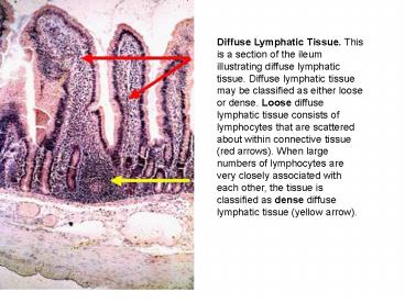
Diffuse Lymphatic Tissue. This is a section of
the ileum illustrating diffuse lymphatic tissue.
Diffuse lymphatic tissue may be classified as
either loose or dense. Loose diffuse lymphatic
tissue consists of lymphocytes that are scattered
about within connective tissue (red arrows). When
large numbers of lymphocytes are very closely
associated with each other, the tissue is
classified as dense diffuse lymphatic tissue
(yellow arrow).
2
Diffuse Lymphatic Tissue. This is a section of
the jejunum illustrating a dense diffuse
lymphatic tissue consisting primarily of large
numbers of closely associated lymphocytes giving
it a dark appearance (red arrows).
3
Nodular lymphatic tissue. This is a section of
appendix, which typically contains large numbers
of lymphatic nodules (red arrows). These nodules
are classified as secondary due to the presence
of germinal centers (yellow arrows). The green
arrow is indicating a patch of dense diffuse
lymphatic tissue.
4
Palatine Tonsil. This is a section of palatine
tonsil containing several secondary nodules
(yellow arrows) embedded in dense diffuse
lymphatic tissue (orange arrow). The surface of
the tonsil is lined by a non-keratinized
stratified squamous epithelium (red arrow). A
small amount of connective tissue lies just deep
to the epithelium.
5
Palatine Tonsil. This is a higher magnification
of the previous image illustrating the epithelial
lining (red arrow). A small amount of connective
tissue lies just deep to the epithelium (green
arrow). Nodular lymphatic tissue is located deep
to the connective tissue.
6
Palatine Tonsil. This image illustrates one of
several crypts typically found associated with
palatine tonsil (red arrow). Debris can collect
in these crypts, as there is no effective
flushing mechanism.
7
Palatine Tonsil - Capsule. Palatine tonsils are
classified (along with pharyngeal and lingual
tonsils) as partially encapsulated lymphatic
organs. Dense connective tissue forms a band
(partial capsule) around the deep portion of the
tonsil (green line).
8
Lymph Node. Lymph nodes are surrounded by a dense
connective tissue capsule (black arrows). Deep to
the capsule is the cortex of the lymph node (C).
This area contains lymphatic nodules (red arrows)
embedded in a dense diffuse lymphatic tissue. The
central portion of a lymph node is called the
medulla (M) and contains cords of lymphatic
tissue, but no nodules. The two areas are
separated by the red line in this illustration.
9
(No Transcript)
10
Lymph Node-Cortex. This is a high magnification
of the outer portion of a lymph node. The green
arrow is indicating the dense connective capsule.
Projecting off of the capsule are bands of
connective tissue, termed trabeculae (blue arrow)
that penetrate the cortex. Just deep to the
capsule is the subcapsular sinus (red arrow)
containing lymph that entered this region via
afferent lymphatic vessels. The lymph may
percolate through the nodular lymphatic tissue to
the medulla or pass to the medulla via trabecular
sinuses (yellow arrow) surrounding the trabeculae.
11
Lymph Node-Cortex. This is a higher magnification
of the outer portion of a lymph node illustrating
the capsule (green arrow), subcapsular sinus (red
arrow), and trabecular sinus (yellow arrow). The
cortical region of a secondary nodule, containing
small lymphocytes, is separated from the germinal
center (GC) by the red line. The germinal center
contains small and large lymphocytes,
macrophages, and plasma cells.
12
Lymph Node-Afferent Vessels. Lymph enters the
subcapsular sinuses via afferent lymphatic
vessels (red arrow). The lymph then passes to the
medullary sinuses via the trabecular sinuses
(fast route) or by percolating through the
cortical lymphatic tissue (slow route).
13
Spleen. The image on the left is a low
magnification of a spleen. A capsule of dense
connective tissue and smooth muscle surrounds
this organ. The bulk of the spleen contains red
pulp (black arrows), which consists of cords of
lymphatic tissue among blood vascular sinuses.
The white pulp (encircled in red) consists of
dense collections of lymphocytes surrounding
central arteries (yellow arrows). The image on
the right is a high magnification of the red pulp
illustrating several splenic cords or cords of
Billroth (red arrows).
14
Spleen. This is high magnification of the splenic
capsule (green arrow). This structure consists of
dense connective tissue and smooth muscle.
Numerous trabeculae (black arrow) project off of
the capsule and penetrate the substance of the
organ. White pulp is separated from the red pulp
by red lines. Yellow arrows point to the central
arteries.
15
Spleen. This is a high magnification of the
parenchyma of the spleen. Numerous areas of white
pulp are separated from the red pulp by red
lines.
16
Spleen-White Pulp. This is a high magnification
of white pulp (encircled in red). A central
artery is indicated by a black arrow. The red
pulp contains numerous blood vascular sinuses.
17
Thymus. The thymus is surrounded by a capsule of
connective tissue (not shown). Projections from
the capsule separate the organ into lobes and
lobules. The central portion of a lobule, the
medulla (M), is light staining and contains small
and large lymphocytes. The outer part of the
lobule is dark staining and termed the cortex and
contains small lymphocytes. The thymus lacks
lymphatic nodules. stroma is composed of
epithelial reticular cells.
18
(No Transcript)
19
Thymus. This image is a high magnification of the
approximate separation of the medulla and the
cortex (red line). The medulla contains unique
structures termed Hassalls (thymic) corpuscles
(red arrows). These whorl-shaped structures,
which increase with age, consist of concentric
layers of epithelial reticular cells.
20
Thymus-Medulla. The image on the left is a high
magnification of a thymic medulla illustrating
Hassalls corpuscles (red arrows). The image on
the right is a high magnification of a Hassalls
corpuscle (yellow arrows) demonstrating the
concentric arrangement of the epithelial
reticular cells. The central portion of the
corpuscle may contain keratin and occasionally,
calcification.
Recommended
CrystalGraphics Presentations


