
Four Basic Components of Signal Movement Through Neuron - PowerPoint PPT Presentation
1 / 30
Title:
Four Basic Components of Signal Movement Through Neuron
Description:
Integration of input signal at trigger zone ... Cholinergic neurons and receptors Nicotinic (agonistic) and muscarinic (antagonist) ... – PowerPoint PPT presentation
Number of Views:207
Avg rating:3.0/5.0
Title: Four Basic Components of Signal Movement Through Neuron
1
Four Basic Components of Signal Movement Through
Neuron
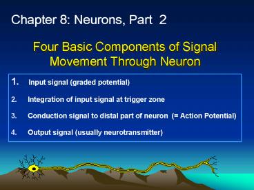
Chapter 8 Neurons, Part 2
- Input signal (graded potential)
- Integration of input signal at trigger zone
- Conduction signal to distal part of neuron (
Action Potential) - Output signal (usually neurotransmitter)
2
Review of Solute Distribution in Body Fluids
- The gradient of K is the main source of the
membrane potential - Change in permeability ot Na can allow influx of
Na - Depolarization
- Electric signal created
- Controlled by gated channels
3
Graded Potentials
- Trigger Zone
- Usually Axon Hillock
- and/or Initial segment of axon
- Many Na Channels
- Some stimuli may be inhibitory
- Hyperpolarizing effect
Fig 8-7
4
Graded Potentials
- Location Any receptor
- Strength ( amplitude) strength of triggering
event - Travel over short distances to trigger zone
- Amount of local current flow is variable
- Diminish in strength as they travel
- May be depolarizing (EPSP) or hyperpolarizing
(IPSP)
Fig 8-7
5
Subthreshold potential vs. Suprathreshold
potential
Graded potential starts here
Fig 8-8
AP
6
Conduction Signals Action Potentials (AP)
- Location ?
- Travel over long distances
- Do not lose strength as they travel
- Are all identical (all-or-none principle) 100mV
amplitude - Represent movement of Na and K across membrane
Ability to propagate the AP Excitability
7
Ion Movement across Cell Membrane During AP
- Sudden increase in Na permeability
- Na enters cell down electrochemical gradient (
feedback loop for .5 msec) - Influx causes depolarization of membrane
potential electrical signal - What stops feedback loop? The Na inactivation
gate closes.
8
Na Channels in Axon Have 2 Gates
- Activation gate and Inactivation gate
- Na entry based on pos. feedback loop ? needs
intervention to stop - Inactivation gates close in delayed response to
depolarization - ? stops escalating pos. feedback loop
Fig 8-10
9
Model of Activation and Inactivation Gates
10
AP-Graph
- has 3 phases
- 1. Rising (Na permeability ?)
- 2. Falling (K permeability ?)
- 3. Undershoot or Hyperpolarization
11
Graded potentials
- Produce an effect that increases with distance
from the point of stimulation - Produce an effect that spreads actively across
the entire membrane surface - May involve either depolarization or
hyperpolarization - Are all-or-none
- All of the above
12
The principal cause of early repolarization of a
nerve fiber after an adequate stimulus has been
applied is
- An increase in the diffusion of K into the
neuron - An increase in the diffusion of Na out of the
neuron - And increase in the diffusion of Na into the
neuron - And increase in the diffusion of K out of the
neuron - A decrease in the diffusion of Na into the neuron
13
Absolute Relative Refractory Periods
No movement of Na possible
- Na channels
- reset to resting
- state, K channels
- still open higher
- than normal
- Stimulus
- necessary
Fig 8-12
14
Refractory Periods
- Limit signal transmission rate (no summation!)
- Assure one way transmission!
- Remember that the Na and K concentration
gradients remain nearly unchanged!
Animation
Forward current excites, backward current does
NOT re-excite !
15
Conduction of AP
- Graded Potential
- Cytoplasmic flow
- AP starts at Axon Hillock
- Na gates open
- Na into axon
- K moves out
- Hyperpolarizes membrane briefly
- resets membrane for next AP
16
Conduction speed depends on . . . .
- Axon diameter (the larger the faster)
- Size constraints on axons become problem with
increasing organismal complexity - Membrane resistance
- High resistance of myelin sheath reduces leakage
of current (ion) flow between axon and ECF - Saltatory Conduction from node to node
Fig 8-17
Fig 8-18
17
1. Axon Diameter
Fig 8-17
18
2. Signal Transduction in Myelinated Axon
Fig. 8-18
Animation
Demyelination diseases (E.g. ?)
19
The primary problem in hypokalemia is that
- Neurons are harder to excite because their
resting potential is hyperpolarized - Neurons are hyper-excitable because their resting
potential is closer to threshold - Neurons respond too quickly to smaller graded
potentials - A and C
- B and C
20
The basis of neural integration is
- Addition of postsynaptic potentials overlapping
in time and space - Command signals from central pattern generators
- Spontaneous activity in pacemaker neurons
- The area under the curve of postsynaptic
potentials overlapping in time and space - All of the above
21
How would blocking the ability for retrograde
transport in an axon affect the activity of a
neuron?
- The neuron would not be able to produce NT
- The neuron would not be able to have APs
- The cell body would not be able to export
products to the axon terminal - The cell body would not be able to respond to
changes in the distal end of the axon - The neuron would be unable to depolarize when
stimulated.
22
Output Signal Communication at Synapses
Whats this?
- Synapse point where neuron meets target cell
(e.g. ?) - 2 types
- chemical
- electrical
- 3 components of chemical synapse
- presynaptic cell
- synaptic cleft
- postsynaptic cell
23
Chemical Synapses
- Majority of synapses
- Use neurotransmitters to carry info from cell to
cell - Axon terminals have mitochondria synaptic
vesicles containing neurotransmitter
24
Events at the Synapse
- AP reaches axon terminal
- Voltage-gated Ca2 channels open
- Ca2 entry
- Exocytosis of neurotransmitter containing vesicles
Ca2 Signal for Neurotransmitter Release
25
Synapse
Fig 8-21
26
3 Classes of Neurotransmitters (of 7)
Fig 8-22
- Acetyl Choline (ACh)
- Made from Acetyl CoA and choline
- Synthesized in axon terminal
- Quickly degraded by ACh-esterase
- Cholinergic neurons and receptors Nicotinic
(agonistic) and muscarinic (antagonist) - Amines
- Serotonin (tryptophane) and Histamine (histidine)
- SSRI antidepressants
- Dopamine and Norepinephrine (tyrosine)
- Widely used in brain, role in emotional behavior
(NE used in ANS) - Adrenergic neurons and receptors - ? and ?
- Gases
- NO (nitric oxide) and CO
- Others AA, (e.g., GABA), lipids, peptides,
purines
27
Synthesis and Recycling of ACh at Synapse
Fig 8-22
28
Postsynaptic Responses
- Can lead to either EPSP or IPSP (p.277)
- Any one synapse can only be either excitatory or
inhibitory - Fast synaptic potentials
- Opening of chemically gated ion channel
- Rapid of short duration
- Slow synaptic potentials
- Involve G-proteins and 2nd messengers
- Can open or close channels or change protein
composition of neuron
29
Integration of Neural Information Transfer
- Multiple graded potentials are integrated at axon
hillock to evaluate necessity of AP - 1. Spatial Summation stimuli from different
locations are added up - 2. Temporal Summation sequential stimuli added
up
Fig 8-26
Fig 8-25
30
1. Spatial Summation
31
2. Temporal Summation
32
Synapse most vulnerable step in signal
propagation
- Many disorders of synaptic transmission, e.g.
- Myasthenia gravis (PNS)
- Parkinsons (CNS)
- Schizophrenia (CNS)
- Depression (CNS)
- Many toxins
33
Chapter 9, The CNS
- Blood Brain Barrier
- Diencephalon (between-brain)
- Integration of sensory information
34
Blood Brain Barrier (p299)
- Allows careful selection of what substances can
cross to neurons - Capillary walls are different
- Fewer pores
- Tight junctions
- Special carriers
- Water soluble substances do not cross easily.
- Lipophilic molecules can cross
- Vomiting Center in medulla oblongata and
posterior pituitary have no BBB. Why??
35
Diencephalon (between-brain)
- Between brainstem and cortex
- Thalamus is a relay station
- Like spinal cord, can modify information
- Hypothalamus is center of maintenance
- Autonomic integration and output
- RH to anterior pituitary
36
Integration of sensory information
- Functional Areas (like compartmentation)
- Sensory (becomes perception)
- Motor
- Association (for integration)
- Both brain and spinal cord
- Modulation of Output
- Reticular formation (p 303)
- Group of nuclei in brain stem
- State of arousal
- Specific NT
37
The End
38
(No Transcript)
39
A(n) ________ functions to passively move ions
across a membrane against the direction of their
active transport.
- pump
- channel
- symporter
- antiporter
- exchanger
40
When it becomes harder for the neuron to fire, is
has become
- refracted
- polarized
- hyperpolarized
- depolarized
- repolarized
41
Starting with the arrival of the AP at the
terminal of a motor neuron and ending with the
beginning of an EPSP which of the following is a
correct temporal sequence?
- vesicle fusion ? inward Ca2 current ?
transmitter exocytosis ? synaptic delay ?
postsynaptic channel opens ? transmitter binds to
postsynaptic receptor - Inward Ca2 current ? vesicle fusion ?
postsynaptic channels open ? transmitter
exocytosis ? synaptic delay ? NT binds to
postsynaptic receptor - Inward Ca2 current ? vesicle fusion ?
transmitter exocytosis ? transmitter binds to
postsynaptic receptor ? postsynaptic channel
opens - transmitter binds to postsynaptic receptor ?
postsynaptic channel opens ? hydrolysis of
transmitter ? postsynaptic channel closes
42
When an adequate stimulus is applied to an axon
- The amplitude of the AP is directly proportional
to the strength of the applied stimulus - The amplitude of the AP is inversely proportional
to the strength of the applied stimulus - The speed of the nerve impulse conduction is
inversely proportional to the diameter of the
nerve fiber - The amplitude of the AP does not vary with the
strength of the stimulus - The first gate to open is the Na inactivation
gate
43
Toms father suffers a stroke that leaves him
partially paralyzed on his right side. What type
of glial cell would you expect to find in
increased numbers in the damaged area of the
brain that is affected by the stroke?
- Astrocytes
- Oligodendrocytes
- Schwann cells
- Ependymal cells
- Microglia
Recommended
CrystalGraphics Presentations


