
Simple Stains - PowerPoint PPT Presentation
1 / 6
Title:
Simple Stains
Description:
Simple Stains ... Different types of staining methods are used to make the cells and their ... Blot the with bibulous paper. Differential Staining ... – PowerPoint PPT presentation
Number of Views:422
Avg rating:3.0/5.0
Title: Simple Stains
1
Simple Stains
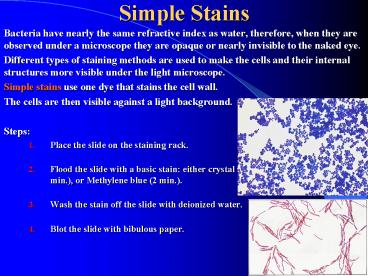
- Bacteria have nearly the same refractive index as
water, therefore, when they are observed under a
microscope they are opaque or nearly invisible to
the naked eye. - Different types of staining methods are used to
make the cells and their internal structures more
visible under the light microscope. - Simple stains use one dye that stains the cell
wall. - The cells are then visible against a light
background. - Steps
- Place the slide on the staining rack.
- Flood the slide with a basic stain either
crystal violet (1 min.), Safranin (2 min.), or
Methylene blue (2 min.). - Wash the stain off the slide with deionized
water. - Blot the slide with bibulous paper.
2
Differential Staining
- Differential Stains use two or more stains and
allow the cells to be categorized into various
groups or types. - Both techniques allow the observation of cell
morphology, or shape, but differential staining
usually provides more information about the
characteristics of the cell wall (Thickness). - The most common differential stain used in
microbiology is the Gram Stain. - Basic stains, due to their positive () charge
will bind electrostatically to negatively charged
molecules such as many polysaccharides, proteins
and nucleic acids. - Acid stains ( - ) bind to positively charged
molecules which are much less common, meaning
acidic stains are used only for special purposes.
- Some commonly encountered basic stains are
crystal violet, safranin (a red dye) and
methylene blue. - Basic stains may be used alone (a simple stain)
or in combination (differential stain) depending
on the experiment involved.
3
Gram Staining
- The Gram Stain is a differential stain.
- Four different reagents are used and the results
are based on differences in the bacterial cell
wall. - Gram Positive bacteria have a relatively thick
cell wall composed of a special carbohydrate
called Peptidoglycan. - Gram Negative bacteria have a much thinner cell
wall composed of the same carbohydrate,
Peptidoglycan, but with certain chemical
differences, such as the presence of
Lipopolysaccharides (LPS). - Notice that both Gram Positive and Gram Negative
bacteria have a cell wall composed primarily of
Peptidoglycan. - Gram Staining Steps
- Crystal violet acts as the primary stain. Crystal
violet may also be used as a simple stain because
it dyes the cell wall of any bacteria. - Grams iodine acts as a mordant (Helps to fix the
primary dye to the cell wall). - Decolorizer is used next to remove the primary
stain (crystal violet) from Gram Negative
bacteria (those with LPS imbedded in their cell
walls). Decolorizer is composed of an organic
solvent, such as, acetone or ethanol or a
combination of both.) - Finally, a counter stain (Safranin), is applied
to stain those cells (Gram Negative) that have
lost the primary stain as a result of
decolorization.
4
Gram Staining Procedure
5
Gram Staining Results
- When reporting the results of the Gram stain you
indicate the type of stain used, the reaction,
and the morphology of the cells observed. - Round (spherical), purple (or dark blue) cells
are reported as Gram positive cocci (GPC). - Rod-shaped, purple (or dark blue) cells are
reported as Gram positive bacilli (GPB). - The standard abbreviations for the four types of
Gram stain and morphology are - Gram Positive Cocci (GPC)
- Gram Positive Bacilli (GPB)
- Gram Negative Cocci (GNC)
- Gram Negative Bacilli (GNB)
- Notice that both Gram Positive and Gram Negative
bacteria have a cell wall composed primarily of
Peptidoglycan. - Spiral-shaped bacterial cells do not Gram stain
well and are usually observed using dark-field
microscopy. There are no standard abbreviations
for Gram stain reactions of the spirilla of
medical importance. - Certain other types of bacteria may not Gram
stain well, such as, Acid-fast Mycobacteria
(Mycobacterium tuberculosis).
6
Bacterial Cell Shapes
- Bacteria can have several different shapes, but
the primary shapes we will be observing are - Spherical or round cells cocci (plural) or
coccus (singular) - Rod shaped bacilli (plural) or bacillus
(singular) - Spiral shaped spirilla
- Some bacteria have characteristic clustering or
arrangements, usually due to how the cells divide
and whether they remain attached together when
they divide. - Diplococci divide in one plane and remain
attached together after cell division. - Streptococci divide in one plane and form long
chains of attached cells. - Staphylococci divide in many planes and remain
attached together forming a grape-like cluster