
Lab - PowerPoint PPT Presentation
1 / 36
Title:
Lab
Description:
Corniculate Cartilage. Cricoid Cartilage. Arytenoid Cartilages. Corniculate Cartilages. Vocal Cords in the Larynx. Trachea. w/ tracheal cartilage. Primary Bronchi ... – PowerPoint PPT presentation
Number of Views:37
Avg rating:3.0/5.0
Title: Lab
1
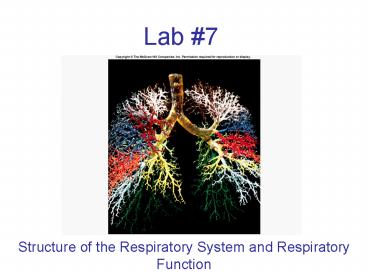
Lab 7
- Structure of the Respiratory System and
Respiratory Function
2
Respiratory System
- Provides O2 to the cells of the body and removes
CO2 - O2 is the terminal electron acceptor in cells
where ATP is made - CO2 is a waste product of cellular respiration
- Too much CO2 in blood increases the PH in the
body - Integrates the respiratory system with the other
systems in the body
3
Overview of Respiratory Anatomy
Nose
Nasal Cavity
Pharynx
Larynx
Trachea
Lungs
Bronchi
4
Bones of the Nasal Cavity
Perpendicular plate of Ethmoid Bone
Vomer
Nasal Cartilage
5
The Nose
Vestibule (stratified squamous epithelium)
Nasal Turbinates
Superior Middle Inferior
External Nares
Entrance protected by coarse guard hairs
Posterior Nares or Choanae
Nasal Cavity and mucous membrane (respiritory
epithelium)
Behind vestibule - Made of PSCCE and Goblet cells
6
PSCCE
Goblet Cells
Mucosal Lining
7
http//io.uwinnipeg.ca/simmons/15l4p26.htm
8
EM of Mucosal Lining
Cilia
Goblet Cell
9
The Pharynx
Nasopharynx
Uvula
Oropharynx
Epiglottis
Laryngopharynx
10
Cartilage of the Larynx and Trachea
Thyroid Cartilage (Adams apple)
Cricoid Cartilage
11
Tracheal Cartilages
Arytenoid Cartilages
Cricoid Cartilage
12
Vestibular folds
13
Epiglottis
Cuneiform Cartilage
Corniculate Cartilage
14
Corniculate Cartilages
Arytenoid Cartilages
Cricoid Cartilage
15
Vocal Cords in the Larynx
16
(No Transcript)
17
Trachea w/ tracheal cartilage
Primary Bronchi
Secondary Bronchi
18
Location of the Carina
Carina
19
Esophagus
Posterior Tracheal Membrane
Trachealis Muscle
Tracheal Ring (tracheal cartilage)
Respiratory epithelium
20
Respiritory epithelium (PSCCE)
Goblet Cells
21
Right Superior Lobe
Left Superior Lobe
Right Middle Lobe
Right Inferior Lobe
Left Inferior Lobe
Cardiac Notch
22
Human Lung
Left Lung
Right Lung
The lungs are located in pleural caveties -
parietal pleura is the outer membrane on the
chest cavity wall - visceral pleura adheres to
the surface of the lungs
23
Bronchi Bronchioles Respiratory
Bronchioles Alveolar Ducts Alveoli
Respiratory epithelium
Alveolar sacs
Simple squamous epithelium
24
http//science.nhmccd.edu/biol/respiratory/alveoli
.htm
25
http//science.nhmccd.edu/biol/respiratory/alveoli
2.htm
26
http//science.nhmccd.edu/biol/respiratory/alveoli
3.htm
27
Lung Histology
- The division of lung into many small sacs
increases surface area - Allows for rapid and extensive diffusion of O2
- Type II alveolar cells (septal cells) decrease
the surface tension in the lungs by secreting
surfactant - Alveolar macrophages phagocytize dust particles
in the lungs
28
http//www.wlap.org/wl-repository/umich/phys/satmo
rn/2003/20031108-annarbor-01-blumberg/real/sld016.
htm
29
http//www.metrohealth.org/Clinical/Pathology/Syll
abus/images/section1/mcr064a.jpg
30
Respiration
- O2 requirements vary with the bodys needs
- Variation in the loading of O2 or CO2 in the
lungs can be accomplished by - Increasing/decreasing the volume of air w/ each
inhalation - Adjusting breathing rate
- CO2 adjustment is important in maintaining
acid-base balance in the blood
31
Inspiration
- Diaphragm Contracts
- Volume of Thoracic Cavity Increases
- Decrease in Pressure
- Air Moves Into Lungs
Inspiration
32
Expiration
- Diaphragm Relaxes
- Volume of Thoracic Cavity Decreases
- Increase in Pressure
- Air Moves Out of Lungs
Expiration
33
Mechanics of Breathing
- Tidal Volume
- The normal volume of breath exhaled
- Expiratory Reserve Volume
- The maximal amount of air one can exhale after a
normal exhalation - Vital Capacity
- Total Volume of Air that can be forcefully
expelled from the lungs after maximal inhalation
34
Mechanics of Breathing
- Inspiratory Reserve Volume
- Maximum amount of air one can inspire after a
normal inhalation - Vital Capacity
- Air that can be exhaled with maximum effort after
max inhalation - Residual Volume
- Volume of remaining in lungs after exhaling vital
capacity - IRV ERV TV VC
35
Mechanics of Breathing
- Flow Pressure/Resistance
- Therefore, increase Resistance, decrease flow
- To overcome increased resistance, increase
Pressure to return Flow to normal - Emphysema
36
Mechanics of Breathing
- CO2 H2O ?? H HCO3-
- Catalyzed by Carbonic Anhydrase
- Increase CO2 drive towards products
- Increase H, decrease pH
- Decrease CO2 drive towards reactants
- Decrease H, increase pH
Recommended
CrystalGraphics Presentations


