Dichotomy key - PowerPoint PPT Presentation
Title:
Dichotomy key
Description:
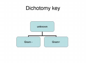
Dichotomy key unknown Gram - Gram+ Part 7 - Gram negative aerobic rods and cocci Part 8- Gram negative facultative rods and coccobacilli Part 8A- Gram negative ... – PowerPoint PPT presentation
Number of Views:186
Avg rating:3.0/5.0
Title: Dichotomy key
1
Dichotomy key
2
(No Transcript)
3
(No Transcript)
4
Part 7 - Gram negative aerobic rods and cocci
Gram negative aerobic rods / cocci
Fluorescent pigments on common media Pseudomonas
5
Part 8- Gram negative facultative rods and
coccobacilli
6
Part 8A- Gram negative facultative rods and
coccobacilli
Lactose fermented Within 48 h
Citrate utilized
Red methyl Positive Citrobacter
H2S produced Indole negative C. freundii
H2S not produced Indole variable C. intermedius
7
Part 8B- Gram negative facultative rods and
coccobacilli
Lactose not fermented Within 48 h
Growth well on common media Proteus
Growth very slow or non existent on common
media Yersinia
Urease positive
Urease Negative P. inconstans
Gelatin hydrolyzed
Gelatin not hydrolyzed
Maltose fermented P.vulgaris
Maltose not fermented P.mirabilis
Mannitol fermented P.rettgeri
Mannitol not fermented M. morganii
8
Part 14- Gram positive cocci
Gram positive cocci
Cells in irregular clusters Tetrads or packs of
8. Catalase positive
Cells in pairs or chains Catalase
negative Streptococcus
Glucose fermented Staphylococcus
9
Part 15- Endospores- forming rods and cocci
Endospores- forming rods and cocci
Cells are rod shaped Bacillus
Cells sperical, In sarcinal packs Sporosarcinal
urea
10
Part 16- Gram positive, asporogenous, rod shaped
bacteria
11
Spore stain-malachite green
- Prepare two smears from a colony of the
spore-formers. - Cut a piece of blotting paper the size of the
smears, but small enough so it doesn't hang over
the edges of the slide. - Place the slide over the boiling streamer and lay
the paper on top of the smears. - Flood the paper with the malachite green stain
(CARCINOGEN) and time for 3 min. Keep the PAPER
WET with the stain if it begins to dry. - After 3 min. remove the slide and lift off the
paper with your loop and discard. - Gently wash the slide with water.
- Flood the slide with the counter-stain safranin
to stain the vegetative cells. - After one minute, wash off the safranin, blot the
slide dry and examine under the microscope.
12
Malachite green staining of Clostridium botulinum
highlights spore formation


