
NEURO-IMAGING TECHNIQUES - PowerPoint PPT Presentation
1 / 41
Title:
NEURO-IMAGING TECHNIQUES
Description:
NEURO-IMAGING TECHNIQUES Structural Plain Skull Radiography Pneumo-encephalography CT Structural MRI Functional Magnetic resonance spectroscopy (MRS) – PowerPoint PPT presentation
Number of Views:296
Avg rating:3.0/5.0
Title: NEURO-IMAGING TECHNIQUES
1
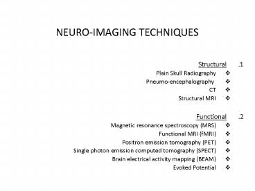
NEURO-IMAGING TECHNIQUES
- Structural
- Plain Skull Radiography
- Pneumo-encephalography
- CT
- Structural MRI
- Functional
- Magnetic resonance spectroscopy (MRS)
- Functional MRI (fMRI)
- Positron emission tomography (PET)
- Single photon emission computed tomography
(SPECT) - Brain electrical activity mapping (BEAM)
- Evoked Potential
2
USES OF NEUROIMAGING
- Indications in Clinical Practice
- Neurological Deficits
- Dementia
- Indications in Clinical Research
- Analysis of Clinically Defined Groups of Patients
- Analysis of Brain Activity during Performance of
Specific Tasks
3
Normal CT Brain
4
POINTS TO REMEMBER
- CT images determined only by degree to which
tissues absorb X-ray - Bone, clotted blood, calcified tissue, contrast
material appear white CSF black - The only component of brain better seen on CT
scan is Calcification, which may be invisible on
MRI
- Plain
- Diagnostic accuracy 82
- Contrast
- IV iodinated contrast medium
- Diagnostic accuracy 92
5
CRITERIA FOR CONTRAST
Contd
- Patients with H/O seizure
- Patients with H/O cerebro-vascular accident
- Suspicion of intracranial SOLs including
granulomas, CNS tumours, metastatic lesions
6
CLNICAL INDICATIONS OF CT BRAIN IN PSYCHIATRY
- Confusion / or dementias of unknown cause
- First episode of psychosis
- First episode of major affective disorder after
50 years of age - Personality changes after 50 years of age
- Psychiatric symptoms following head injury
- Prolonged catatonia
- To rule out complications due to possible head
trauma - Co existence of seizure in psychiatric symptoms
- Movement disorders of unknown etiology
- Focal neurological signs accompanying psychiatric
symptoms
7
CT ADVANTAGES v/s DISADVANTAGES
- ADVANTAGES
- Compared to MRI
- Simpler, cheaper, more accessible
- Tolerated by claustrophobics
- No absolute contraindications
- Fewer pitfalls in interpretation
- Better than MR for bone detail
- DISADVANTAGES
- Ionizing radiation
- IV contrast complications
- Limited range of tissue contrasts
8
MRI
Liquid Helium Cooled 1.5 Tesla Solenoid Magnet
9
THE NORMAL HUMAN BRAIN AS SEEN BY MRI
Data sources The Whole-brain Atlas, K. A.
Johnson and J. A. Becker, Harvard
10
TYPES OF IMAGES
- T1 WEIGHTED IMAGES
- An SE sequence with a short TR (200 1000
milliseconds) and a short TE (20-25 milliseconds) - CSF, cortical bone, air rapidly flowing blood
have negligible signals ? appear dark - Fat bone marrow have high signal intensity ?
appear white - Useful in evaluation of cerebropontine angle
cistern pituitary fossa
- T2 WEIGHTED IMAGES
- An SE sequence with a long TR (2000 2500
milliseconds) and a long TE (gt75 milliseconds) - CSF has bright signal intensity relative to a
dark signal from grey white matter - Useful in demyelination, edema tumour
infiltration - Reveal brain pathology most clearly
11
T1 WEIGHTED IMAGES
T2 WEIGHTED IMAGES
12
MRI IMPORTANT POINTS
- INDICATIONS
- To rule out organic cause of psychiatric illness
- Abrupt change in mental state
- New onset memory loss
- New onset dementia
- ADVANTAGES
- Does not expose the patient to ionizing
radiations - Generates images in three planes
- Demyelinating disease can be assessed reliably
- To study posterior fossa structures
- DISADVANTAGES
- Avoided in patients wearing metallic devices
- Claustrophobia
- Does not pick up bony abnormalities
- Difficult in uncooperative patients
13
TOMOGRAPHIC IMAGES ARE IN A SPECIFIC PLANE
SAGITTAL
AXIAL
CORONAL
RT
RT
14
(No Transcript)
15
Lateral ventricles
16
(No Transcript)
17
(No Transcript)
18
Brain CT
- Note that we take axial slices beginning from the
skull base., parallel to a standard line
(orbito-meatal or canthomeatal line). - The thickness of the slice (the distance between
a slice picture- and the following slice
picture-) is 10mm or as determined. - The skull base is a bony area with much small
details, so we take the slices with less
thickness (5mm) to show al the details. - You have to recognize the following
- 1- Cerebral hemispheres
- 2- Brainstem
- 3- Ventricular system
- 4- Basal ganglia and thalamus
- 5- Basal cisterns (subarachnoid space)
19
Cerebral Hemispheres (Lobes) Brain Stem
- Lobes in the cerebral hemispheres are the
frontal, temporal, parietal, and occipital lobes. - Note that the white matter appears grey, and the
grey matter appears white. - Brainstem is composed of the mid brain, Pons
medulla oblongata.
20
Brain edema
- Appears as hypodense area on CT scan.
- Two main types
- Cytotoxic
- Results from cell death.
- Involves the gray matter.
- Vasogenic
- Results from disruption of the BBB.
- Mainly involves the white matter.
21
Trauma
- Axial injury
- Concussion
- Brain damage at the microscopic level.
- Usually associated with normal imaging
- Contusion
- Focal area of edema that can be associated with
hemorrhage.. - Usually involves the fronto-temperal lobes
22
Trauma
- Shear injury(diffuse axonal injury)
- significant brain damage results from
acceleration/deceleration mechanism. - Associated with poor prognosis.
- MRI is more accurate in evaluating the extent of
injury.
23
Trauma
- Extra-axial injury
- Blood can accumulate in different spaces around
the brain. - Subarachnoid hemorrhage is usually has a benign
self-limiting course. - Its presence is suggestive of significant trauma.
24
Trauma
- Subdural hematoma
- Usually of venous origin.
- Slowly enlarging blood collection between the
dura and the subarachnoid space. - Has the characteristic crescent shape.
- It crosses the suture line.
25
Trauma
- Extradural (Epidural) hematoma
- More than 90 occurs supratentorial and more
than 95 are unilateral. - Usually attain their final size quickly.
- Only 23 of EDH will enlarge, mostly within 36
hours. - Has the characteristic lucent period.
26
Trauma
- Extradural (Epidural) hematoma
- Usually associated with skull fracture(85-95).
- Results from injury to middle meningeal artery or
one of its branches. - About 10 are of venous origin.
- It has the characteristic biconvex shape.
- Limited by the suture lines.
27
Neoplasm
- Divided into two major categories
- Intra-axial
- The tumor is within brain parenchyma.
- Metastasis is the most common etiology in adults.
- Extra-axial
- Arising from the brain coverings or nerve sheaths.
28
Inta-axial Tumors
- Primary brain neoplasm
- Gliomas are the most common types.
- Wide variety of pathological types astrocytoma,
oligodendroglioma, ependymoma. - Metastasis
- Lung, breast and colon are common primary sites.
- Usually multiple, can be hemorrhagic.
29
Extra-axial tumors
- Meningiomas are the most common pathological
type. - Nerve sheath tumors are less common, e.g.
schwannoma, neurofibroma. - Metastasis is less common than intra-axial ones.
- (Example shown below)
30
(No Transcript)
31
Tumor in Wernickes Area
The magnetic field used is 30,000 times that
of the earth's magnetic field. It's effect on
the body, however, is harmless and temporary.
The MRI scanner can detect radiation from certain
molecules, which are present in different
concentrations in different tissues.
32
CNS Infection
- Meningitis is the most common form of CNS
infection. - Its clinical and lab diagnosis.
- Imaging is helpful in excluding secondary
complications. - Diffuse meningeal enhancement is a common
finding. - Normal CT or MRI does not exclude the diagnosis.
33
CNS infections
- Sub-dural effusion
- Common in children especially with H. influenza
meningitis. - Can be treated conservatively.
- Brain abscess
- Usually secondary to hematogenous spread of
microbes. - May not be distinguished from brain tumor.
34
CNS infections
- Viral infections
- MRI is very sensitive for diagnosis of viral
encephalitis. - Herpatic encephalitis has a characteristic
bilateral temporal lobe involvement.
35
36
(No Transcript)
37
Pyogenic Abscess Findings An enhancing ring
lesion within the left posterior frontal
lobe.Differential diagnosis Metastasis. Since
abscess cannot be differentiated clearly from
metastasis by MRI imaging, appropriate history
and clinical findings are needed to aid in
accurate diagnosis. A surgical biopsy may
eventually be required.
38
Vascular Disorders
- Stroke is a major source of mortality and
morbidity. - Most stokes are ischemic, result from vascular
occlusion by a thrombus or embolus. - CT is usually the initial modality to evaluate
these patients.
39
Vascular Disorders
- CT
- Usually becomes positive in 12-24hours after
onset of neurological deficit. - Edema in a vascular distribution.
- Helpful to rule out ICH or hemorrhagic conversion.
40
Vascular Disorders
- MRI
- It becomes positive earlier than CT.
- Diffusion weighted images can become positive in
few minutes from onset. - MRA can be obtained at the same time to evaluate
vascular occlusion.
41
Vascular Disorders
- Vascular abnormalities
- Aneurysms
- usually manifest in the form of subarachnoid
hemorrhage. - AVM
- presents with either ICH or headache.
- Can be diagnosed with enhanced CT or MRI.
- Angiogram is diagnostic and therapeutic.