The Endocrine System - PowerPoint PPT Presentation
1 / 81
Title:
The Endocrine System
Description:
The Endocrine System AP Chapter 45 – PowerPoint PPT presentation
Number of Views:305
Avg rating:3.0/5.0
Title: The Endocrine System
1
The Endocrine System
- AP Chapter 45
2
- The endocrine system, along with the nervous
system, is responsible for coordinating our
responses. - The endocrine system is a slower system and the
nervous system is a faster response.
3
Chemical signals
- Chemicals found in both systems and also as part
of other signaling mechanisms bind to specific
receptor proteins on or in target cells.
4
- Secreted chemical signals include
- Hormones produced by endocrine glands,
- travel through the blood stream to target
organs - ex insulin, estrogen
- Local regulators
- (a) paracrine signals act on neighboring
cells, ex. cytokines, interferon,
prostaglandins - (b) autocrine signals act on secreting cells
itself, ex cytokines
5
- Neurotransmitters - secreted by neurons at
synapses - ex- serotonin, nitric oxide (NO)
- Neurohormones secreted by neurosecretory cells,
travel through the blood stream to target organs
or synapses - ex- epinephrine
- Pheromones released into the environment
between individuals - ex insects marking trails,
- mating, etc.
6
(No Transcript)
7
(No Transcript)
8
Fig. 45-2
Blood vessel
Response
(a) Endocrine signaling
Response
(b) Paracrine signaling
Response
(c) Autocrine signaling
Synapse
Neuron
Response
(d) Synaptic signaling
Neurosecretory cell
Blood vessel
Response
(e) Neuroendocrine signaling
9
Chemical Classes of Hormones
- Three major classes of molecules function as
hormones in vertebrates - Polypeptides (proteins and peptides)
- water soluble
- Amines derived from amino acids
- some are water soluble, some are lipid
soluble - Steroid hormones
- lipid soluble
10
Fig. 45-3
Water-soluble
Lipid-soluble
0.8 nm
Steroid Cortisol
Polypeptide Insulin
Amine Epinephrine
Amine Thyroxine
11
- Lipid-soluble hormones (steroid hormones) pass
easily through cell membranes, while
water-soluble hormones (polypeptides and amines)
do not - The solubility of a hormone correlates with the
location of receptors inside or on the surface of
target cells
12
- Water-soluble hormones (hydrophilic) are secreted
by exocytosis, travel freely in the bloodstream,
and bind to cell-surface receptors - Lipid-soluble hormones (hydrophobic) diffuse
across cell membranes, travel in the bloodstream
bound to transport proteins, and diffuse through
the membrane of target cells
13
Fig. 45-5-1
Fat-soluble hormone
Water- soluble hormone
Transport protein
Signal receptor
TARGET CELL
Signal receptor
NUCLEUS
(a)
(b)
14
Fig. 45-5-2
Fat-soluble hormone
Water- soluble hormone
Transport protein
Signal receptor
TARGET CELL
OR
Signal receptor
Cytoplasmic response
Gene regulation
Cytoplasmic response
Gene regulation
NUCLEUS
(a)
(b)
15
Multiple Effects of Hormones
- Hormones can have multiple effects which depends
on - - the type of receptor they bind to
- - the specific signal transduction
- pathway
- - the specific transcription factor they
activate. - A hormone can also have different effects in
different species
16
Fig. 45-8-1
Same receptors but different intracellular
proteins (not shown)
Epinephrine
Epinephrine
? receptor
? receptor
Glycogen deposits
Vessel dilates.
Glycogen breaks down and glucose is released.
(a) Liver cell
(b) Skeletal muscle blood vessel
17
Fig. 45-8-2
Same receptors but different intracellular
proteins (not shown)
Different receptors
Epinephrine
Epinephrine
Epinephrine
Epinephrine
? receptor
? receptor
? receptor
? receptor
Glycogen deposits
Vessel dilates.
Vessel constricts.
Glycogen breaks down and glucose is released.
(a) Liver cell
(b) Skeletal muscle blood vessel
(c) Intestinal blood vessel
18
Negative feedback and antagonistic hormone pairs
are common features of the endocrine system
- Hormones are assembled into regulatory pathways
- A negative feedback loop inhibits a response by
reducing the initial stimulus - Negative feedback regulates many hormonal
pathways involved in homeostasis
19
Fig. 45-11
Pathway
Example
Stimulus
Low pH in duodenum
S cells of duodenum secrete secretin ( )
Endocrine cell
Negative feedback
Blood vessel
Target cells
Pancreas
Bicarbonate release
Response
20
Insulin and Glucagon Control of Blood Glucose
an example of antagonistic hormone pairs
- The pancreas has clusters of endocrine cells
called islets of Langerhans with alpha cells that
produce glucagon and beta cells that produce
insulin - Insulin reduces blood glucose levels by
- Promoting the cellular uptake of glucose
- Slowing glycogen breakdown in the liver
- Promoting fat storage
21
- Glucagon increases blood glucose levels by
- Stimulating conversion of glycogen to glucose in
the liver - Stimulating breakdown of fat and protein into
glucose - Remember Glucagon Glucose ON!
22
Fig. 45-12-2
Body cells take up more glucose.
Insulin
Beta cells of pancreas release insulin into the
blood.
Liver takes up glucose and stores it as glycogen.
STIMULUS Blood glucose level rises.
Blood glucose level declines.
Homeostasis Blood glucose level (about 90 mg/100
mL)
23
Fig. 45-12-4
Homeostasis Blood glucose level (about 90 mg/100
mL)
STIMULUS Blood glucose level falls.
Blood glucose level rises.
Alpha cells of pancreas release glucagon.
Liver breaks down glycogen and releases glucose.
Glucagon
24
Remember!
- GLUCOSE in the BLOOD
- INSULIN GLUCAGON
25
Diabetes Mellitus
- Diabetes mellitus is perhaps the best-known
endocrine disorder - It is caused by a deficiency of insulin or a
decreased response to insulin in target tissues - It is marked by elevated blood glucose levels
26
- Type I diabetes mellitus (insulin-dependent) is
an autoimmune disorder in which the immune system
destroys pancreatic beta cells - Type II diabetes mellitus (non-insulin-dependent)
involves insulin deficiency or reduced response
of target cells due to change in insulin receptors
27
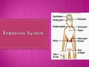
Fig. 45-10
Major endocrine glands
Hypothalamus
Pineal gland
Pituitary gland
Organs containing endocrine cells
Thyroid gland
Thymus
Parathyroid glands
Heart
Liver
Adrenal glands
Stomach
Pancreas
Kidney
Testes
Small intestine
Kidney
Ovaries
28
Coordination of Endocrine and Nervous Systems in
Vertebrates
- The hypothalamus receives information from the
nervous system and initiates responses through
the endocrine system - Attached to the hypothalamus is the pituitary
gland composed of the posterior pituitary and
anterior pituitary
29
Who is the boss?
- The hypothalamus has neurosecretory cells
which secrete releasing and inhibiting hormones
which control the pituitary gland which in turn
controls other glands. - RH, such TRH, indicates a releasing
hormone
30
- The posterior pituitary stores and secretes
hormones that are made in the hypothalamus - The anterior pituitary makes and releases
hormones under regulation of the hypothalamus
31
Fig. 45-14
Cerebrum
Thalamus
Pineal gland
Hypothalamus
Cerebellum
Pituitary gland
Spinal cord
Hypothalamus
Posterior pituitary
Anterior pituitary
32
Hypothalamus
- The hypothalamus secretes two hormones which are
stored in the posterior pituitary. - 1) oxytocin induces uterine contractions
during birth and milk production - 2) ADH which decreases urine volume
33
Fig. 45-15
Hypothalamus
Neurosecretorycells of thehypothalamus
Axon
Posterior pituitary
Anterior pituitary
HORMONE
Oxytocin
ADH
Kidney tubules
TARGET
Mammary glands,uterine muscles
34
The anterior pituitary gland secretes releasing
hormones and inhibiting hormones.
- TSH thyroid stimulating
- FSH and LH stimulates gonads
- ACTH - stimulates adrenal cortex
- Prolactin milk production
- MSH stimulates production of melanocytes (skin
pigments) - GH growth hormone
35
Fig. 45-17
Tropic effects onlyFSHLHTSHACTH
Neurosecretory cellsof the hypothalamus
Nontropic effects onlyProlactinMSH
Nontropic and tropic effectsGH
Hypothalamicreleasing andinhibitinghormones
Portal vessels
Endocrine cells ofthe anterior pituitary
Posterior pituitary
Pituitary hormones
HORMONE
FSH and LH
TSH
ACTH
Prolactin
MSH
GH
TARGET
Testes orovaries
Thyroid
Adrenalcortex
Mammaryglands
Melanocytes
Liver, bones,other tissues
36
Tropic Hormones
- A tropic hormone regulates the function of
endocrine cells or glands - The four strictly tropic hormones are
- Thyroid-stimulating hormone (TSH)
- Follicle-stimulating hormone (FSH)
- Luteinizing hormone (LH)
- Adrenocorticotropic hormone (ACTH)
- (FSH and LH are reproductive hormones.)
37
Thyroid Gland
- T3 and T4 thyroxin, regulates metabolism (needs
dietary iodine to function properly goiter if
not enough iodine) - Calcitonin lowers calcium in blood deposition
in bones and secretion into kidney filtrate - tone down the calcium
38
T3 and T4
- act by binding to thyroid receptors that are
distributed in almost every organ. - Typically, this process regulates gene
transcription and the subsequent production of
various proteins that are involved in
development, growth, and cellular metabolism
39
Graves Disease
- an autoimmune disorder that leads to overactivity
of the thyroid gland (hyperthyroidism) and causes
the thyroid to increase in size. Other symptoms
are anxiety, tiredness, insomnia, irregular heart
rhythms, bulging eyes.
40
Parathyroid Gland
- PTH parathormone raises calcium levels in blood
from bones and reuptake in kidneys
41
Fig. 45-20-2
Activevitamin D
Stimulates Ca2uptake in kidneys
Increases Ca2 uptake in intestines
PTH
Parathyroid gland(behind thyroid)
Stimulates Ca2 release from bones
STIMULUS Falling bloodCa2 level
Blood Ca2 level rises.
Homeostasis Blood Ca2 level(about 10 mg/100 mL)
42
Adrenal medulla
- Epinephrine (adrenaline) raises metabolic rate,
fight or flight - Norepinephrine (noradrenaline) controls blood
pressure
cortex
medulla
43
Adrenal cortex
- Glucocorticoids (cortisol) glucose from noncarb
sources, such as muscles - Mineralocorticoids (aldosterone) induces
kidneys to reabsorb water and salts - Both of these deal with long-term stress
44
Cushings DiseaseCushing's syndrome is a
hormonal disorder caused by prolonged exposure of
the body's tissues to high levels of the hormone
cortisol.
- severe fatigue
- weak muscles
- high blood pressure
- high blood glucose
- increased thirst and urination
- irritability, anxiety, depression
- a fatty hump between the shoulders
- moon face
45
Fig. 45-21c
Adrenal cortex
Adrenalgland
Kidney
(b) Long-term stress response
Effects ofmineralocorticoids
Effects ofglucocorticoids (cortisol)
1. Retention of sodium ions and water by
kidneys
1. Proteins and fats broken down and
converted to glucose, leading to increased
blood glucose
STRESS!
2. Increased blood volume and blood
pressure
2. Possible suppression of immune system
46
The production of these hormones is controlled by
the hypothalamus and anterior pituitary
47
Testes
- Androgens (testosterone) gender, male secondary
sex characteristics
48
Ovaries
- Estrogen maintenance of female reproductive
system and development of secondary female
characteristics - Progesterone prepares uterus for child
49
Pineal Gland
- Melatonin biological clock
50
Hormonal pathways work with the hypothalamus and
anterior pituitary to coordinate responses
- In regulating metabolism by the thyroid
Also, notice the positive AND negative
feedbacks here.
51
Hormonal pathways work with the hypothalamus and
anterior pituitary to coordinate responses
- Ex in the gonads
- GnRH (hypothalamus) affects FSH and LH (anterior
pituitary) which affects estrogens and androgens
(ovaries/testes)
52
Testosterone Synthesis
53
Estrogen and progesterone synthesis
54
Which endocrine gland?
- Too little of my hormone and you will feel tired
and sluggish and probably gain weight.
THYROID
55
- A malfunction in this gland can result in a
giant.
Anterior Pituitary
56
- This gland prepares me for an emergency
situation by increasing my heartrate.
Adrenal Glands
57
- This gland is also used in the digestive
system. It also comes into play when I eat lots
of M and Ms!
PANCREAS
58
- This gland is called the master gland
because it secretes nine hormones many of which
control other endocrine glands by feedback
control.
Pituitary Gland
59
- If this gland is not working properly,
diabetes can result.
Pancreas
60
- If this gland is not working properly, your
nerves and muscles will not function properly
either due to calcium deficiency.
Parathyroid Gland
61
- These glands do not function properly in
chromosomal mutations such as in Turners and
Klinefelters syndrome.
Gonads
62
- This gland makes me wake up in the morning and
ready to go!
Pineal Gland
63
Fig. 45-10
Major endocrine glands
Hypothalamus
Pineal gland
Pituitary gland
Organs containing endocrine cells
Thyroid gland
Thymus
Parathyroid glands
Heart
Liver
Adrenal glands
Stomach
Pancreas
Kidney
Testes
Small intestine
Kidney
Ovaries
64
Name of Gland Hormone Function
A pineal Daily rhythms
B hypothalamus Regulates blood volume and pressure by affecting kidneys
C Growth hormone growth
D thyroid calcitonin
E parathyroid Raises calcium levels
F Hormones for immune system immune
G adrenal adrenaline
H insulin Lowers blood glucose levels
I ovaries Testosterone/
J testes Testosterne/androgens Secondary female sexual characteristics
65
Hormones in the reproductive system
- GnRH from the hypothalamus directs the anterior
pituitary to produce FSH and LH that regulate
gametogenesis and sex hormone production in males
and females - Sex hormones
- - androgens male
- - estrogens - female
66
- So in males, FSH and LH stimulate the production
of sperm and secretion of testosterone
67
Fig. 46-13
Hypothalamus
GnRH
Anterior pituitary
FSH
LH
Secrete testosterone
Nourish developing sperm
Negative feedback
Negative feedback
Leydig cells
Sertoli cells
Inhibin
Spermatogenesis
Testosterone
Testis
68
Fig. 46-11
Seminalvesicle(behind bladder)
(Urinarybladder)
Prostate gland
Bulbourethralgland
Urethra
Erectile tissueof penis
Scrotum
Vas deferens
Epididymis
Testis
(Urinarybladder)
(Urinaryduct)
Seminal vesicle
(Rectum)
(Pubic bone)
Vas deferens
Erectiletissue
Ejaculatory duct
Prostate gland
Urethra
Penis
Bulbourethral gland
Glans
Vas deferens
EpididymisTestisScrotum
Prepuce
69
- In females, these control the reproductive cycle
the uterine cycle (menstrual cycle) and the
ovarian cycle - In the uterus, this results in the build-up of
the inner layer of the uterus called the
endometrium which will be shed (menstruation) if
fertilization does not occur
70
Fig. 46-10b
Oviduct
Ovaries
Follicles
Corpus luteum
Uterine wall
Uterus
Endometrium
Cervix
Vagina
71
- In the ovary, these control the development of
the egg in the follicle, the release of the egg
(ovulation), and the disintegration of the egg
follicle (corpus luteum)
72
Fig. 46-14
(a)
Control by hypothalamus
Inhibited by combination ofestradiol and
progesterone
Hypothalamus
Stimulated by high levelsof estradiol
GnRH
1
Anterior pituitary
Inhibited by low levels of estradiol
FSH
LH
2
Pituitary gonadotropinsin blood
(b)
6
LH
FSH
FSH and LH stimulatefollicle to grow
LH surge triggersovulation
3
Ovarian cycle
(c)
8
7
Corpusluteum
Degeneratingcorpus luteum
Growing follicle
Maturingfollicle
Follicular phase
Ovulation
Luteal phase
Estradiol secretedby growing follicle
inincreasing amounts
Progesterone andestradiol secretedby corpus
luteum
4
Ovarian hormones in blood
Peak causesLH surge
(d)
5
10
Progesterone
Estradiol
9
Progesterone and estra-diol promote
thickeningof endometrium
Estradiol levelvery low
Uterine (menstrual) cycle
(e)
Endometrium
Secretory phase
Menstrual flow phase Proliferative phase
Days
0
5
10
14
20
25
28
15
73
Ovarian cycle
- Follicular phase FSH stimulates follicle growth
- After LH surge, ovulation occurs
- Luteal Phase ruptured follicle becomes a corpus
luteum which secretes progesterone
74
Hypothalamus
GnRH
Ant Pituitary
LH
FSH
Ovarian Cycle
Ovulation
Luteal Phase
Follicular phase
after LH surge
75
Uterine cycle
- Proliferative Phase Estrogens from growing
follicle stimulate the growth of the endometrium - Secretory Phase - After ovulation, progesterone
causes the increased vascularization and
development of secretory glands - Menstrual flow phase rapid drop of hormones
cause endometrium to disintegrate
76
progesterone
estradiol
Promote thickening of endometrium
endometrium
Uterine Cycle
Proliferative Phase
Secretory Phase
Menstrual Cycle
When levels fall, menstrual Cycle begins
77
Fig. 46-14b
(d)
Ovarian hormones in blood
Peak causesLH surge
Progesterone
Estradiol
Ovulation
Estradiol level very low
Progesterone and estra-diol promote
thickeningof endometrium
(e)
Uterine (menstrual) cycle
Endometrium
Menstrual flow phase Proliferative phase
Secretory phase
Days
0
14
15
5
10
20
25
28
78
Corpus Luteum maintains production of
est/progesterone
Follicle produces estrogen
79
What happens if female becomes pregnant?
- Implantation takes place around 7 days after
conception - Embryo secretes hCG human chorionic gonadotropin
maintains est/prog by corpus luteum - In 2nd trimester of pregnancy, placenta takes
over that job
80
How do birth control pills work?
- Synthetic est/prog combination that works by neg
feedback to inhibit GnRN production and thus FSH
and LH and no ovulation
81
(No Transcript)
Recommended
CrystalGraphics Presentations