
Blood? Connective Tissue - PowerPoint PPT Presentation
1 / 32
Title:
Blood? Connective Tissue
Description:
Blood Connective Tissue Body s primary means of transportation Respiratory System provides O2 Digestive System provides nutrients Urinary/Excretory System ... – PowerPoint PPT presentation
Number of Views:91
Avg rating:3.0/5.0
Title: Blood? Connective Tissue
1
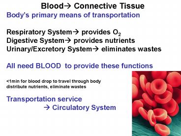
Blood? Connective Tissue Bodys primary means of
transportation Respiratory System? provides
O2 Digestive System? provides nutrients Urinary/Ex
cretory System? eliminates wastes All need BLOOD
to provide these functions lt1min for blood drop
to travel through body distribute nutrients,
eliminate wastes Transportation service
? Circulatory System
2
Blood Structure and Function Blood fluid w/
chemicals dissolved cells Plasma liquid
part Serum
liquid part w/ clotting factors removed (in
clot) Formed Elements cellular components
3
(No Transcript)
4
Formed Elements Red Blood Cells (RBCs)
Erythrocytes (5 million/mm3) White Blood Cells
(WBCs) Leukocytes (7500/mm3)
Granulocytes (granules in cytoplasm)
Neutrophils Eosinophils
Basophils
Nongranulocytes (no granules in cytoplasm)
Lymphocytes
Monocytes Platelets Thrombocytes (300,000/mm3)
5
RBCs, WBCs and Platelets continuously
destroyed Body must continually make new
ones (few million RBCs/sec) Where are
cellular components of blood made? In 2
types of Connective Tissue Myeloid Tissue
(Red Bone Marrow) Forms all types of
blood cells except some lymphocytes
monocytes adults sternum, ribs, hips,
vertebrae, clavicles, cranial bones
Lymphatic Tissue (Lymph Nodes, Thymus,
Spleen) Forms most lymphocytes
monocytes
6
(No Transcript)
7
(No Transcript)
8
(No Transcript)
9
(No Transcript)
10
Anemia Disease caused by inability of blood to
carry sufficient O2 to body cells Results from
?RBCs hemorrage (injury, ulcer, surgery)
OR blood forming cells dont make enough
RBCs (cancer, radiation, x-ray,
infection, disease) ?
Hemoglobin (protein that carries O2)
(Fe needed to make Hgb)
Anemia occurs with ? RBCs or adequate RBCs NOT
carrying enough O2 (b/c low Hgb) Ex.Thalassemia
Major/Minor
11
Pernicious Anemia ?RBCs due to lack of Vit.
B12
Sickle Cell Anemia Inherited Abnormal Hgb s
produced less soluble crystallizes? ? O2
carrying capacity Varying amounts mild, severe,
fatal RBCs sickle shaped (crisis? transfuse)
temporary
12
Polycythemia Bone Marrow produces too
many RBCs Blood becomes too
thick to flow through vessels properly
Therapeutic phlebotomy (blood letting)
13
Hematocrit (Hct) RBC volume in blood
sample Normal Hct 45 (of blood volume RBCs)
low Hct Anemia high Hct Polycythemia
Centrifuge RBCs heavier?bottom Buffy Coat
WBCs Platelets (middle) Plasma/Serum
lighter ?top
14
- Everest climbers Decreased O2 in atm
- Rest camps allow climbers to acclimate
- polycythemic (adjust to low O2 levels)
- Blood Doping Transfusion before event ?
- Intention to ? ? O2 levels to
- enhance muscle performance
- Transfusion Risk Need Vs Risk
15
Leukopenia ? WBC Diseases affecting Immune
system can
? of WBCs
(Buffy Coat for Differential) AIDS? marked
leukopenia
Leukocytosis ? WBC Bacterial infections (some
viruses)
16
Leukemia Malignant disease
Marked WBC ?? Buffy coat thicker
Thrombocytosis ? Platelets ?
Thrombocytopenia Platelets ?
17
macrocytes
Red Blood Cell Morphology
Anisocytosis varied size Poiklocytosis varied
shape Normocytes normal Microcytes
small Macrocytes big
microcytes
crenated RBCs
tears
targets
ovalocytes
18
Hypochromic low Hgb in RBC
Normal RBCs
Hyperchromic high Hgb in RBC
19
RBC Function (120 days) Morphology (no
nucleus/few organelles) Bi-concave disc? large SA
for gas exhange Transport O2 Hgb ?
oxyhemoglobin CO2 Hgb
?carbaminohemoglobin Fe needed for Hgb (protein)
molecule Low Fe? low Hgb ?anemia ?? O2 to cells
? ? break down of nutrients ? ? energy (tired)??
cellular fxs (metabolism) Hemorrage vs slow
gradual bleed
20
? RBCs ? ? O2 levels in blood ? Kidneys release
Erythropoietin ? stimulates Red Bone Marrow ? ?
Erythropoiesis ? more RBCs produces ? ? O2
carrying capacity
21
WBC Function Defend body from infection
Monocytes 4-8 agranulocytes Phagocytes that
become macrophages in tissues ? in chronic
infections
Neutrophils (most numerous 40-70)
granulocytes Phagocytes ? during infection
phagocytes? phagocytosis (AT) engulf microbes
toxic granulation of neuts (infection)
22
Lymphocytes (non granulocytes) 20-45 B lymphs ?
plasma memory cells ? produce Abs to destroy
Ags T-lymphs ? produce lymphotoxins
lymphikines to destroy Ag/s and
activates B lymphs Protect vs infection via
Immune Mechanism NOT phagocytosis Microbe
s invade body? stimulate lymphs to multiply
transform into Plasma Cells Specific microbe
stimulates specific kind of lymph to form 1 type
of plasma cell that makes specific antibody
(protein) vs specific pathogen.
lymphs
plasma cell
23
Eosinophils 1-4 (granulocytes) Protect body vs.
irritants that cause allergies parasitic worms
(can also phagocytize) Inactivates some
inflammatory chemicals
Basophils (granulocytes) Function in allergic rx
(like eos) Granules contain histamine
(vasodilator chemical) discharged at sites of
inflammation Secrete substances (including
heparin anticoagulant)
24
Platelets 150,000-450,000/mm3 3rd type of blood
cell Essential role in
initiating clotting cascade
for coagulation Chain of
Rapid reactions Help control
blood loss from broken vessel
Bleeding time tests Platelet function
25
Hemostasis Prevention of Blood
Loss Contraction Platelets release serotonin
? Smooth muscles in the wall of
blood vessels reduce flow of blood
Vasoconstriction
Platelet plug temporary plug which
adheres to the opening in the vessel Blood
Clots Cascade of factors (proteins) ? clotting.
26
Blood Clotting Cascade (overhead) Injury? Rough
spot ?Platelets become sticky Aggregate at site
of injury release platelet factors Damaged
tissue releases thromboplastin Prothrombin Plt
Factors thromboplastin Ca ?
Thrombin Fibrinogen (soluble) Thrombin ?
Fibrin (insoluble) clot
Plts, and RBCs enmeshed in fibrin
prothrombin, thrombin, fibrinogen proteins Pg
242-243 SF diagram
27
(No Transcript)
28
How do you speed clotting at site of
injury? Gauze? rough, more plts break ? releasing
more clotting factors Importance of Vit
K (accelerates clotting) Stimulates liver cells
to make more prothrombin Result Thrombin forms
faster ? faster clot formation
29
Blood Clotting ? Coagulation Balance occurs
between Procoagulants promote
clotting
Anticoagulants prevent clotting Substances
which dissolve clots are also present Normally
anticoagulants prevail After surgery Patients
on Coumadin (warfarin) to ? ? Clotting Time
Also, Cardiac and stroke risk patients Diet
must watch Vit K intake, counteracts medicine
(geen leafy veggies)
30
Clots in vessels, heart, brain, lung, organs ?
can lead to sudden death, shutting off blood
supply to area Thrombus clot stays in place
where it formed
?Thrombosis Embolis part of clot dislodges into
blood circulation ? Embolism
31
Drugs prevent thrombosis (Ex. coumadin
heparin) Some block stimulating effect of Vit K
on liver ? liver produces less
prothrombin Tests for deep vein thrombosis
embolism ?D-Dimer
32
Blood Plasma and Serum Plasma liquid part of
blood H2O, dissolved substances,
proteins, albumin, transferrin
(carries iron), salts, nutrients,
chemicals, wastes, hormones,
antbodies (proteins/protect) Amount of blood
volume depends on size,
gender, age Most adults? 5.5L of blood (7-9 of
body wt) Plasma volume a little more than ½
volume of whole blood based on HCT Slightly
alkaline 7.4
Recommended
CrystalGraphics Presentations


