
Heart Anatomy - PowerPoint PPT Presentation
1 / 57
Title:
Heart Anatomy
Description:
Heart Anatomy Approximately the size of your fist Location Superior surface of diaphragm Left of the midline Anterior to the vertebral column, posterior to the sternum – PowerPoint PPT presentation
Number of Views:624
Avg rating:3.0/5.0
Title: Heart Anatomy
1
Heart Anatomy
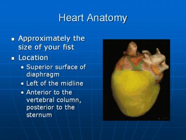
- Approximately the size of your fist
- Location
- Superior surface of diaphragm
- Left of the midline
- Anterior to the vertebral column, posterior to
the sternum
2
Heart Anatomy
Figure 18.1
3
Coverings of the Heart Anatomy
- Pericardium a double-walled sac around the
heart composed of - A superficial fibrous pericardium
- A deep two-layer serous pericardium
- The parietal layer lines the internal surface of
the fibrous pericardium - The visceral layer or epicardium lines the
surface of the heart - They are separated by the fluid-filled
pericardial cavity
4
(No Transcript)
5
Coverings of the Heart Physiology
- The pericardium
- Protects and anchors the heart
- Prevents overfilling of the heart with blood
- Allows for the heart to work in a relatively
friction-free environment
6
Pericardial Layers of the Heart
Figure 18.2
7
Heart Wall
- Epicardium visceral layer of the serous
pericardium - Myocardium cardiac muscle layer forming the
bulk of the heart - Fibrous skeleton of the heart crisscrossing,
interlacing layer of connective tissue - Endocardium endothelial layer of the inner
myocardial surface
8
(No Transcript)
9
Cardiac Muscle Bundles
Figure 18.3
10
External Heart Major Vessels of the Heart
(Anterior View)
- Vessels returning blood to the heart include
- Superior and inferior venae cava
- Right and left pulmonary veins
- Vessels conveying blood away from the heart
- Pulmonary trunk, which splits into right and left
pulmonary arteries - Ascending aorta (three branches)
11
(No Transcript)
12
Figure 18.4d
13
Aorta
Superior vena cava
Left pulmonary artery
Right pulmonary artery
Left atrium
Pulmonary trunk
Left pulmonary veins
Right atrium
Right pulmonary veins
Mitral (bicuspid) valve
Fossa ovalis
Aortic valve
Pectinate muscles
Pulmonary valve
Left ventricle
Tricuspid valve
Papillary muscle
Right ventricle
Chordae tendineae
Interventricular septum
Myocardium
Trabeculae carneae
Visceral pericardium
Inferior vena cava
Endocardium
(e)
Figure 18.4e
14
Atria of the Heart
- Atria are the receiving chambers of the heart
- Each atrium has a protruding auricle
- Pectinate muscles mark atrial walls
- Blood enters right atria from superior and
inferior venae cavae and coronary sinus - Blood enters left atria from pulmonary veins
15
Ventricles of the Heart
- Ventricles are the discharging chambers of the
heart - Papillary muscles and trabeculae carneae muscles
mark ventricular walls - Right ventricle pumps blood into the pulmonary
trunk - Left ventricle pumps blood into the aorta
16
Right and Left Ventricles
Figure 18.6
17
Pathway of Blood Through the Heart and Lungs
- Right atrium ? tricuspid valve ? right ventricle
- Right ventricle ? pulmonary semilunar valve ?
pulmonary arteries ? lungs - Lungs ? pulmonary veins ? left atrium
- Left atrium ? bicuspid valve ? left ventricle
- Left ventricle ? aortic semilunar valve ? aorta
- Aorta ? systemic circulation
18
(No Transcript)
19
http//www.pbs.org/wnet/redgold/journey/phase2_a1.
html
Figure 18.5
20
Coronary Circulation
- Coronary circulation is the functional blood
supply to the heart muscle itself - Collateral routes ensure blood delivery to heart
even if major vessels are occluded
21
Coronary Circulation Arterial Supply
Figure 18.7a
22
Coronary Circulation Venous Supply
Figure 18.7b
23
Heart Valves
- Heart valves ensure unidirectional blood flow
through the heart - Atrioventricular (AV) valves lie between the
atria and the ventricles - AV valves prevent backflow into the atria when
ventricles contract - Chordae tendineae anchor AV valves to papillary
muscles
24
Heart Valves
- Aortic semilunar valve lies between the left
ventricle and the aorta - Pulmonary semilunar valve lies between the right
ventricle and pulmonary trunk - Semilunar valves prevent backflow of blood into
the ventricles
25
Heart Valves
Figure 18.8a, b
26
Heart Valves
Figure 18.8c, d
27
(No Transcript)
28
Atrioventricular Valve Function
Figure 18.9
29
Semilunar Valve Function
Figure 18.10
30
Microscopic Anatomy of Heart Muscle
- Cardiac muscle is striated, short, fat, branched,
and interconnected - The connective tissue endomysium acts as both
tendon and insertion - Intercalated discs anchor cardiac cells together
and allow free passage of ions - Heart muscle behaves as a functional syncytium
31
Microscopic Anatomy of Cardiac Muscle
Figure 18.11
32
Cardiac Muscle Contraction
- Heart muscle
- Is stimulated by nerves and is self-excitable
(automaticity) - Contracts as a unit
- Cardiac muscle contraction is similar to skeletal
muscle contraction
33
Heart Physiology Intrinsic Conduction System
- Autorhythmic cells
- Initiate action potentials
- Have unstable resting potentials called pacemaker
potentials - Use calcium influx (rather than sodium) for
rising phase of the action potential
http//www.phschool.com/science/biology_place/bioc
oach/cardio1/intconduct.html
34
Pacemaker and Action Potentials of the Heart
Figure 18.13
35
Heart Physiology Sequence of Excitation
- Sinoatrial (SA) node generates impulses about 75
times/minute - Atrioventricular (AV) node delays the impulse
approximately 0.1 second - Impulse passes from atria to ventricles via the
atrioventricular bundle (bundle of His)
36
Heart Physiology Sequence of Excitation
- AV bundle splits into two pathways in the
interventricular septum (bundle branches) - Bundle branches carry the impulse toward the apex
of the heart - Purkinje fibers carry the impulse to the heart
apex and ventricular walls
37
(No Transcript)
38
Cardiac Intrinsic Conduction
Figure 18.14a
39
Heart Excitation Related to ECG
SA node generates impulse atrial excitation
begins
Impulse delayed at AV node
Impulse passes to heart apex ventricular excitati
on begins
Ventricular excitation complete
SA node
AV node
Purkinje fibers
Bundle branches
http//www.phschool.com/science/biology_place/bioc
oach/cardio1/intconduct.html
Figure 18.17
40
Heart Excitation Related to ECG
SA node generates impulse atrial excitation
begins
SA node
Figure 18.17
41
Heart Excitation Related to ECG
Impulse delayed at AV node
AV node
Figure 18.17
42
Heart Excitation Related to ECG
Impulse passes to heart apex ventricular excitati
on begins
Bundle branches
Figure 18.17
43
Heart Excitation Related to ECG
Ventricular excitation complete
Purkinje fibers
Figure 18.17
44
Heart Excitation Related to ECG
SA node generates impulse atrial excitation
begins
Impulse delayed at AV node
Impulse passes to heart apex ventricular excitati
on begins
Ventricular excitation complete
SA node
AV node
Purkinje fibers
Bundle branches
Figure 18.17
45
Extrinsic Innervation of the Heart
- Heart is stimulated by the sympathetic
cardioacceleratory center - Heart is inhibited by the parasympathetic
cardioinhibitory center
Figure 18.15
46
Electrocardiography
- Electrical activity is recorded by
electrocardiogram (ECG) - P wave corresponds to depolarization of SA node
- QRS complex corresponds to ventricular
depolarization (contracting) - T wave corresponds to ventricular repolarization
(go back to normal state)
PLAY
InterActive Physiology Intrinsic Conduction
System, pages 36
47
Heart Sounds
Figure 18.19
48
Electrocardiography
Figure 18.16
49
Heart Sounds
- Heart sounds (lub-dup) are associated with
closing of heart valves - First sound occurs as AV valves close and
signifies beginning of systole - Second sound occurs when SL valves close at the
beginning of ventricular diastole
50
Atrial fibrillation
Ventricular fibrillation
Ventricular tachycardia
51
Cardiac Cycle
- Cardiac cycle refers to all events associated
with blood flow through the heart - Systole contraction of heart muscle
- Diastole relaxation of heart muscle
52
Phases of the Cardiac Cycle
- Ventricular filling mid-to-late diastole
- Heart blood pressure is low as blood enters atria
and flows into ventricles - AV valves are open, then atrial systole occurs
53
Phases of the Cardiac Cycle
- Ventricular systole
- Atria relax
- Rising ventricular pressure results in closing of
AV valves - Ventricular ejection phase opens semilunar valves
54
Phases of the Cardiac Cycle
- Isovolumetric relaxation early diastole
- Ventricles relax
PLAY
InterActive Physiology Cardiac Cycle, pages
318
55
Figure 18.20
56
Examples of Congenital Heart Defects
Figure 18.25
57
Age-Related Changes Affecting the Heart
- Sclerosis and thickening of valve flaps
- Decline in cardiac reserve
- Fibrosis of cardiac muscle
- Atherosclerosis
Recommended
CrystalGraphics Presentations


