
Chapt 21 DNA Replication II: Mechanisms; telomerase - PowerPoint PPT Presentation
1 / 27
Title:
Chapt 21 DNA Replication II: Mechanisms; telomerase
Description:
Chapt 21 DNA Replication II: Mechanisms; telomerase Student learning outcomes: Describe how replication initiates proteins binding specific DNA sequences – PowerPoint PPT presentation
Number of Views:252
Avg rating:3.0/5.0
Title: Chapt 21 DNA Replication II: Mechanisms; telomerase
1
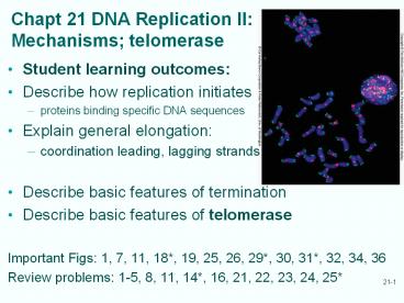
Chapt 21 DNA Replication IIMechanisms
telomerase
- Student learning outcomes
- Describe how replication initiates
- proteins binding specific DNA sequences
- Explain general elongation
- coordination leading, lagging strands
- Describe basic features of termination
- Describe basic features of telomerase
- Important Figs 1, 7, 11, 18, 19, 25, 26, 29,
30, 31, 32, 34, 36 - Review problems 1-5, 8, 11, 14, 16, 21, 22, 23,
24, 25
2
21.1 Initiation and Priming in E. coli
- Initiation of DNA replication requires primers
- Different organisms use different mechanisms
for primers - Primosome - - proteins needed to make primers to
replicate DNA (A. Kornberg) - E. coli primosome
- DNA helicase (DnaB)
- Primase (DnaG)
- Primosome assembly at origin of replication, oriC
is multi-step
3
E. Coli primosomePriming at oriC
primase
- DnaA binds to unique oriC at sites called dnaA
boxes cooperates with RNAP, HU protein to melt
nearby DNA region - DnaB binds to open complex, facilitates
binding of primase to complete primosome - DnaB helicase activity unwinds DNA
- Primosome remains with replisome, repeatedly
primes Okazaki fragment synthesis on lagging
strand
4
Key proteins at the DNA replication fork
Figure 6.13 of Hartl Jones Role of key
proteins in DNA replication draw 5 and 3
ends, leading, lagging strands
5
Priming in Eukaryotes
- Eukaryotic replication is more complex
- Bigger size of eukaryotic genomes, most are
linear - Slower movement of replicating forks
- Each chromosome must have multiple origins
- Model monkey virus SV40 (5200 nt genome)
- Later yeast ARS (centromere) regions
6
Replication of SV40 is bidirectional Isolate
replicating molecules cleave with EcoRI that has
1 site look at molecules in EM (A-j increasing
replication).
Fig. 21.2
7
Origin of Replication in SV40
- SV40 ori adjacent to transcription control region
- Initiation of replication needs viral large T
antigen (major product of early transciption)
binding to - Region within 64-bp ori core
- Two adjacent sites
- T antigen helicase activity opens up replication
bubble within ori core - Priming carried out by primase associated with
host DNA pol a
8
Point mutations define critical regions of SV40
ori AT regions T-Ag binding site
lt -Early genes
Late genes -gt
Fig. 21.4
9
ARS is Yeast Origin of Replication permit
replication of gene in yeastlinker scanning
mutants define critical regions plasmid has
centromere, URA gene grow non-selective and then
check Ura (Fig. 7)
- Autonomously replicating sequences (ARSs)
- 4 important regions
- Region A - 15 bp long with11-bp consensus
sequence highly conserved in ARSs - B1 and B2
- B3 may permits important DNA bend within ARS1
10
21.2 Elongation and processivity
- Once primer in place,DNA synthesis begins
- Coordinated synthesis of lagging and leading
strands keeps pol III holoenzyme on template - Replication is highly processive, very rapid
- pol III holoenzyme in vitro 730 nt/sec (in
vivo 1000 nt/sec) - Pol III core alone is poor polymerase after 10
nt it falls off - Takes time to reassociate with template and
nascent DNA - Missing from core enzyme is processivity factor
- sliding clamp, b-subunit of holoenzyme (see
Table 20.2)
11
Processivity agentb-Subunit is clamp keeps pol
III on DNA
Fig. 12 b-dimer on DNA
- Core plus b-subunit replicates DNA processively
- ( 1,000 nt/sec)
- Dimer formed by b-subunit is ring-shaped
- Ring fits around DNA template
- Interacts with a-subunit of core to tether whole
polymerase and template (Fig. 9) - Holoenzyme stays on template with b-clamp (Fig.
11)
12
Pol III Holoenzyme Table 20.2
Pol III core has 3 subunits Pol III g complex
has 5 subunits DNA-dependent ATPase Pol III
holoenzyme includes b subunit
13
DNA pol III subunits bind each other a, e core
g ATPase, b clamp Purified subunits mixed and
chromatographed to separate complexes from free
proteins SDS-PAGE Western blot tests which
proteins in which complexes Also assayed DNA
polymerase activity
Fig. 21.10
14
Eukaryotic processivity factor
- PCNA forms trimer, a ring that encircles DNA and
holds DNA polymerase on the template
Fig. 14
Fig. 13 b-dimer on DNA in E. coli
15
b Clamp and g Clamp Loader
- b-subunit needs help from g complex to load onto
DNA - This g complex acts catalytically to form
processive adb complex - g not remain associated with complex during
processive replication - Clamp loading is ATP-dependent
- Energy from ATP changes conformation of loader so
d-subunit binds one b-subunits - Binding opens clamp, allows it to encircle DNA
16
Pol III subassembly has 2 cores, one g and no b
- Recall from table 2
- Core pol III has 3 subunits
- is polymerase
- is exonuclease
- t dimerizes core
Fig. 17 g complex has 5 subunits
17
Simultaneous Strand Synthesis by double-headed
pol III
- 2 core polymerases attached through 2 t-subunits
to g complex - One core responsible for continuous synthesis of
leading strand - Other core performs discontinuous synthesis of
the lagging strand - g complex serves as clamp loader to load b clamp
onto primed DNA template - After loading, b clamp loses affinity for g
complex instead associates with core polymerase
Fig. 18
18
Lagging Strand Replication
- g complex and b clamp help core polymerase with
processive synthesis of Okazaki fragment - When fragment completed, b clamp loses affinity
for core - b clamp g complex acts to unload clamp
- Now clamp recycles
Fig. 25
19
21.3 Termination of replication
Fig. 26
- Straightforward for phage like l that produce
long, linear concatemers (rolling circle) - Grows until genome-sized piece cut off, packaged
into phage head - Bacterial replication 2 replication forks
approach each other at terminus region - 22-bp terminator sites bind specific proteins
(terminus utilization substance, TUS) - Replicating forks enter terminus region, pause
- 2 daughter duplexes entangled, must separate
20
Decatenation Disentangling Daughter DNAs in
Bacteria
Fig. 27
- End of replication, circular bacterial
chromosomes are catenanes decatenated in 2
steps - Melt unreplicated double-helical turns linking
two strands - Repair synthesis fills in gaps
- Decatenated by topoisomerase IV
21
Termination in Eukaryoteslinear
chromosomesrole of telomerase
Fig. 29
- Eukaryotes have problem filling gaps left when
RNA primers are removed after DNA replication - DNA cannot be extended 3?5 direction
- No 3-end upstream (unlike circular bacterial
chromosome) - If no resolution, DNA strands get shorter each
replication
22
Telomeres
- Telomeres - special structures at ends of
chromosomes - One strand of telomeres is tandem repeats of
short, G-rich regions (sequence varies among
species) - G-rich telomere strand is made by enzyme
telomerase - Telomerase contains a short RNA that is template
for telomere synthesis - C-rich telomere strand is synthesized by ordinary
RNA-primed DNA synthesis - Process like lagging strand DNA replication
- Ensures chromosome ends are rebuilt, do not
suffer shortening each round of replication
23
- Tetrahymena cells have telomerase activity
- Greider Blackburn
- Cell extracts, synthetic oligo 32P-dNTPs,
other dNTP - Conclusion
- enzyme adds 6-bp units
- Only needs GTP, TTP
- (lanes 3, 6)
- template (TTGGGG)4
Fig. 21.30
24
Telomere Formationtelomerase makes DNA from RNA
templateTERT, telomerase reverse
transcriptase proteins p43 and p123 1
RNA templateTelomerase activity is highin
normal cells S phase, in cancer cells always
Telomere sequences vary Tetrahymena
TTGGGG Vertebrates TTAGGG Yeast
TTGGG
Fig. 31
25
Telomere Structure
Fig. 32
- Eukaryotes protect telomeres from nucleases and
ds break repair enzymes - Ciliates have TEBP (telomere end-binding protein)
to bind and protect 3-single-strand telomeric
overhang - Budding yeast has Cdc13p which recruits Stn1p and
Ten1p that all bind ss telomeric DNA - Mammals and fission yeast have protein similar to
TEBP binding to ss telomeric DNA
26
Mammalian Telomeres
- T loop protects ss telomeric DNA (G-rich 3 end
loops) - Proteins TRF1 and TRF2 help telomeric DNA form
loop in which ss 3-end of telomere invades ds
telomeric DNA - TRF1 may bend DNA into shape for strand invasion
- TRF2 binds at point of strand invasion, may
stabilize displacement loop
Fig. 36
27
Review questions
- 2. List the components of E. coli primosome and
roles in primer synthesis. - 4. Outline strategy for identify yeast ARS
sequence. - 14. How can discontinuous synthesis of lagging
strand keep up with continuous synthesis of
leading strand? - 21. Why do eukaryotes need telomeres, but
prokaryotes do not?
Recommended
CrystalGraphics Presentations


