
Fetal Gas Exchange and Circulation - PowerPoint PPT Presentation
1 / 73
Title:
Fetal Gas Exchange and Circulation
Description:
Patient s medical history is the foundation of ... HPI is a narrative description in detail of ... Interviewer should ask about exposure to family or friends with ... – PowerPoint PPT presentation
Number of Views:340
Avg rating:3.0/5.0
Title: Fetal Gas Exchange and Circulation
1
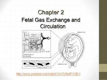
Chapter 2
Fetal Gas Exchange and Circulation
http//www.youtube.com/watch?vOV8wtPYGE-I
2
Introduction
- The fetus in utero shares the mothers
circulation for gas exchange - However the maternal and fetal vascular networks
are separate systems and no blood is shared
between the two - When a zygote (fertilized egg) first travels to
the uterus, it has no nutrient source. The
developing cells here are called Blastocyst,
which must implant into the uterine lining for
nourishment
3
Introduction
- The outer surrounding layer of the blastocyst is
the trophoblast which combines with tissue from
the endometrium to form the chorionic membrane
around the blastocyst
4
(No Transcript)
5
Introduction
- Inside the blastocyst a group of cells arrange
on one side in the shape of a figure eight - The central portion is the embryonic disk which
forms three embryonic germ layers which contain
origins for the below structures - The ECTODERM CNS (brain, spinal cord) PNS
(craniel nerves/spinal nerves, eyes, inner ears,
nose, glandular tissues, skin, teeth - The MESODERM Cardiovascular system, heart/blood
vessels, lymphatics, connective tissues/blood
cells, bone, skeletal muscle, skin,
kidneys/ureters, reproductive tissues, spleen - The ENDODERM Digestive system, respiratory
system, urinary system, liver/pancreas - http//www.youtube.com/watch?vlXN_sDnd1ng
- http//www.youtube.com/watch?vpp2mWgWAnc8
6
(No Transcript)
7
(No Transcript)
8
(No Transcript)
9
Introduction
- The outer or top of the figure eight envelops the
embryonic structure and forms the amniotic sac,
the inner layer forms the yolk sac which then
turns into the embryo, the amniotic sac then
surrounds the embryo. - The embryo attaches to the outer layer through
the umbilical stalk later the umbilical cord - The umbilical cord connects to the finger like
projections in the outer lining of the
chorion/chorionic villi - A capillary network connects the umbilical cord
to the chorionic villi. - http//www.youtube.com/watch?vjLTkCQkbkKg
- Abnormal implantation Ectopic Pregnancy
- http//www.youtube.com/watch?v45HYJpOF6-0
10
Introduction
- The villi intertwine into the blood filled
lacunar cavities of the endometrium of the
maternal uterus - O2, CO2, and nutrients diffuse though the vast
capillary surface area of this indirect
connection between the mother and fetus
11
Maternal-Fetal Gas Exchange
- As fetal development continues, the region of
this interface becomes limited to the
discus-shaped placenta - The umbilical cord connects the placenta to the
fetus with one large vein and two smaller
arteries - As the cord grows the vessels tend to spiral
- Whartons jelly helps protect the vessels and
prevents kinking of the cord
12
(No Transcript)
13
Maternal-Fetal Gas Exchange
- Embryo
- Umbilical stalk
- Umbilical cord
- 2 small arteries
- 1 large vein
- Whartons jelly (for protection)
- Chorion (chorionic villi)
- Endometrium (uterus)
- Becomes placental unit
- http//www.youtube.com/watch?vzvNPw7m74HE
14
Cardiovascular Development
- Heart
- First organ to form
- Begins during third week of gestation
- Completed by week 8
15
Cardiovascular Development (cont.)
- http//www.youtube.com/watch?vaZUDePgRQqI
- Cardiovascular system develops from the mesoderm
layer - By day 22, cardiac contractions are detectable
and bidirectional blood flow begins
16
Cardiovascular Development (cont.)
Fourth week of gestation heart tubes continue to
merge into three structures bulbus cordis,
ventricular bulge and the arterial bulge which
empty into the sinus venosus, which receives
oxygenated, nutrient rich blood from the placenta
Continuation of folding, bending and dilation
continue giving the heart a S shape
17
Cardiovascular Development (cont.)
- Simultaneous external changes occur the septum
primum begins to separate the primitive atrium.
At the same time endocardial cushions develop
which will separate the atriums from the
ventricles. - The left atrium incorporates the pulmonary veins,
the superior vena cava develops . By end of the
4th week the dilating ventricular spaces fold
onto each other creating the ventricular septum
and the base of the bulboventricular loop
18
Cardiovascular Development (cont.)
- Blood flow matures into a unidirectional path as
the myocardium contiues to strengthen by
recruiting myocytes from surrounding mesenchymal
tissue. - Weeks 5-6 internal and external structures mature
quickly - By week 6 the foramen ovale is present (source of
fetal shunting) - Fetal heart rate is about 95 bpm
- http//www.youtube.com/watch?vu1x24IdN7VA
19
(No Transcript)
20
Cardiovascular Development (cont.)
Week 7-8 the ventricular septum is finished
forming A small intraventricular foramen remains
and blood flows between the two ventricles until
the endocardial cushions fuse with the
ventricular septum Tricuspid and Mitral valves
develop
21
(No Transcript)
22
Fetal Circulation
23
(No Transcript)
24
Introduction
- The fetal circulation is markedly different from
the adult circulation - In the fetus, gas exchange does not occur in the
lungs but in the placenta - The placenta must therefore receive deoxygenated
blood from the fetal systemic organs and return
its oxygen rich venous drainage to the fetal
systemic arterial circulation - the fetal cardiovascular system is designed in
such a way that the most highly oxygenated blood
is delivered to the myocardium and brain
25
Introduction
- These circulatory adaptations are achieved in the
fetus by both the preferential streaming of
oxygenated blood and the presence of intracardiac
and extracardiac shunts - fetal circulation can be defined as a
shunt-dependent circulation - In the fetus, deoxygenated blood arrives at the
placenta via the umbilical arteries and is
returned to the fetus in the umbilical vein.
26
Introduction
- Oxygenated blood travels from the placenta to the
fetus through the umbilical vein - The ductus venosus, the first fetal shunt,
appears continuous with the umbilical vein,
shunting 30-50 of the oxygenated blood around
the fetal liver - The amount of shunting through the ductus venous
appears to decrease with gestational age - The shunted oxygen rich blood empties into the
inferior vena cava and mixes with venous blood as
it flows to the right atrium
27
(No Transcript)
28
Fetal Cardiac Shunts
- Foramen ovale
- Between right and left atria bypass the right
ventricle - Ductus arteriosus
- Pulmonary artery to aorta to bypass the right
ventricle - Ductus venosus
- Shunts blood past liver
- the ductus venosus shunts approximately half of
the blood flow of the umbilical vein directly to
the inferior vena cava. Thus, it allows
oxygenated blood from the placenta to bypass the
liver. In conjunction with the other fetal
shunts, the foramen ovale and ductus arteriosus,
it plays a critical role in preferentially
shunting oxygenated blood to the fetal brain. It
is a part of fetal circulation - http//www.youtube.com/watch?vcgccQVcFLi4
29
Ductus venosus
- The ductus venosus is open at the time of the
birth and is the reason why umbilical vein
catheterization works. Ductus venosus naturally
closes during the first week of life in most
full-term neonates however, it may take much
longer to close in pre-term neonates. Functional
closure occurs within minutes of birth.
Structural closure in term babies occurs within 3
to 7 days. - After it closes, the remnant is known as
ligamentum venosum. - If the ductus venosus fails to occlude after
birth, the individual is said to have an
intrahepatic portosystemic shunt (PSS). The
ductus venosus shows a delayed closure in preterm
infants, Possibly, increased levels of dilating
prostaglandins leads to a delayed occlusion of
the vessel
30
(No Transcript)
31
Patent Foramen Ovale (PFO)
32
http//www.youtube.com/watch?vyDSTONfL4h8
33
Fetal Cardiac Shunts
- Shunted oxygen-rich blood empties into the
inferior vena cava and mixes with venous blood as
it flows to the right atrium - In the right atrium most of the blood received
from the inferior vena cava passes through the
foramen ovale to the left atrium - The remainder of the blood in the right atrium
mixes with desaturated blood from the superior
vena cava and empties into the right ventricle
blood here has slightly higher oxygen partial
pressures. This blood is pumped through the
pulmonary arteries into the developing lungs - The PVR is high in utero due to compression of
the vessels from low lung volumes, and low lung
oxygen concentrations
34
Fetal Cardiac Shunts
- Since the lungs in utero are void of air,
chemical mediators keep vessels constricted in
the pulmonary vascular bed - 13-25 of the fetal blood flow reaches the lungs
- Blood from the pulmonary veins empties into the
left atrium and the flows into the left ventricle
and then out through the atrium to the head,
right arm and coronary circulation - The high PVR keeps most of the pulmonary artery
blood flow from the right ventricle to bypass the
lungs, flowing through the Ductus arteriosus into
the aorta - Deoxygenated blood from the upper torso returns
to the right atrium via the superior vena cava - Blood in the descending and abdominal aorta flows
through the two umbilical arteries and back to
the placenta for oxygenation
35
Transition to Extrauterine Life
- Increase pulmonary blood flow
- Vasodilation
- Initiation of gas exchange
- Increasing PaO2
- Stretching pulmonary units
- Inhabitation of vasoconstrictors
36
(No Transcript)
37
Transition to Extrauterine Life
- Clamping of the umbilical cord vessels removes
the low pressure system of the placenta from the
fetus - During the first breath several factors improve
pulmonary blood flow and reduce PVR - Inflating the lungs initiates gas exchange and
dilates the pulmonary arterioles - Rising PaO2 stimulates release of endogenous
pulmonary vasodilating factors - Stretching of the pulmonary units stretches open
the vascular units and stimulates the release of
anti vasocontricting agents
38
Transition to Extrauterine Life
- Once PVR decreases, pressures in the right side
of the heart decrease and pressures in the left
side increase - The foramen ovale closes once the pressure in the
left exceeds the right this facilitates the
increase of blood flow to the lungs - Pressure in the aorta increases and becomes
greater than the pressure in the pulmonary artery - The shunting in the ductus arteriosus decreases
- The PDA typically closes quickly from increases
in PaO2 and prostaglandin levels - Prostaglandins are mediators and have a variety
of strong physiological effects, such as
regulating the contraction and relaxation of
smooth muscle tissue
39
Transition to Extrauterine Life
- Ductus arteriosus closes typically completely
within 24 hours after birth. If they do not close
it is termed a PDA - PDA affects girls more often than boys. The
condition is more common in premature infants and
those with neonatal respiratory distress syndrome - Infants with genetic disorders, such as Down
syndrome, and whose mothers had rubella during
pregnancy are at higher risk for PDA. - PDA is common in babies with congenital heart
problems, such as hypoplastic left heart
syndrome, transposition of the great vessels, and
pulmonary stenosis.
40
PDA
- A small PDA may not cause any symptoms. However,
some infants may have symptoms such as - Fast breathing
- Poor feeding habits
- Rapid pulse
- Shortness of breath
- Sweating while feeding
- Tiring very easily
- Poor growth
- Babies with PDA often have a heart murmur that
can be heard with a stethoscope. However, in
premature infants, a heart murmur may not be
heard. The health care provider may suspect the
condition if the infant has breathing or feeding
problems soon after birth.
41
PDA
- Changes may be seen on chest x-rays. The
diagnosis is confirmed with an echocardiogram. - Sometimes, a small PDA may not be diagnosed until
later in childhood. - To assess a babies oxygenation after birth the
probe is placed preductal on the right hand or
wrist - We can then compare SpO2 readings pre and post
ductally to assess the severity of a PDA
42
PDA
- If the rest of the baby's heart and blood flow is
normal or close to normal, the goal is to close
the PDA. If the baby has certain other heart
problems or defects, keeping the ductus
arteriosus open may be lifesaving. Medicine may
be used to stop it from closing - Sometimes, a PDA may close on its own. In
premature babies it often closes within the first
2 years of life. In full-term infants, a PDA
rarely closes on its own after the first few
weeks. - When treatment is needed, medications such as
indomethacin or a special form of ibuprofen
are often the first choice. Medicines can work
very well for some newborns, with few side
effects. The earlier treatment is given, the more
likely it is to succeed.
43
PDA
- A transcatheter device closure is a procedure
that uses a thin, hollow tube placed into a blood
vessel. The doctor passes a small metal coil or
other blocking device through the catheter to the
site of the PDA. This blocks blood flow through
the vessel. These coils can help the baby avoid
surgery. - Surgery may be needed if the catheter procedure
does not work or it cannot be used. Surgery
involves making a small cut between the ribs to
repair the PDA. Surgery has risks, however. Weigh
the possible benefits and risks with your health
care provider before choosing surgery.
44
(No Transcript)
45
PFO
- Normally the foramen ovale closes at birth when
increased blood pressure on the left side of the
heart forces the opening to close. - If the atrial septum does not close properly, it
is called a patent foramen ovale. This type of
defect generally works like a flap valve, only
opening during certain conditions when there is
more pressure inside the chest. This increased
pressure occurs when people strain while having a
bowel movement, cough, or sneeze.
46
(No Transcript)
47
PFO
- If the pressure is great enough, blood may travel
from the right atrium to the left atrium. If
there is a clot or particles in the blood
traveling in the right side of the heart, it can
cross the PFO, enter the left atrium, and travel
out of the heart and to the brain (causing a
stroke) or into a coronary artery (causing a
heart attack). - People with PFO do not need any treatment if
there are no associated problems, such as a
stroke. Patients who have had a stroke or
transient ischemic attack (TIA) may be placed on
some type of blood thinner medication, such as
aspirin, plavix (clopidogrel), or coumadin
(warfarin) to prevent recurrent stroke. - Surgical repair may be indicated
48
Development of Baroreceptors and Chemoreceptors
- Baroreceptors
- Baroreceptors are stretch receptors in the wall
of some blood vessels. They are involved in the
control of arterial pressure through the
discharge of impulses to the cardiovascular
centre when there is distension due to a change
in the blood pressure. - Baroreceptors are found in the carotid sinus
(dilation in the left and right internal carotid
arteries), the aortic arch, and the elastic
arteries of the neck and chest and some veins. - http//www.youtube.com/watch?v5-bruUXxGKA
49
(No Transcript)
50
Baroreceptors
- Any decline in the blood pressure stretches the
vascular wall which stimulates the baroreceptors.
- These receptors send impulses to the
cardiovascular center which in turn decreases
parasympathetic stimulation of the heart via the
vagus nerves and increases the sympathetic
stimulation of the heart. - The cardiovascular center stimulates the
secretion of adrenaline and noradrenaline from
the medulla of the adrenal gland. The effect on
the heart and blood vessels is to accelerate
heart rate and contractility and promote
vasoconstriction, resulting in an increase in
blood pressure
51
Baroreceptors
- If there is an increase in blood pressure, the
baroreceptors send impulses to the cardiovascular
center - In response the cardiovascular center increases
the parasympathetic stimulation of the heart, and
decreases its sympathetic stimulation. - The heart rate and contractility will decrease
leading to low cardiac output and the peripheral
resistance will decline due to vasodilation. Low
cardiac output and low peripheral resistance
cause a decrease in blood pressure
52
Baroreceptors
- The baroreceptors in the carotid sinus are
responsible for the regulation of the blood
pressure in the brain, while those in the aortic
arch are responsible for regulation of systemic
blood pressure.
53
Chemoreceptors
- Chemoreceptors are found close to the carotid and
aortic baroreceptors in small structures called
carotid bodies and aortic bodies. - They are sensitive to any change in the chemical
composition of the blood, such as a decrease in
oxygen level and pH of the blood or an increase
in the carbon dioxide level. These receptors send
impulses to the cardiovascular center which in
turn increases the sympathetic stimulation to the
blood vessels causing an increase in blood
pressure. - Chemoreceptors also stimulate the respiratory
centers in the brain to increase the rate of
respiration.
54
- http//www.youtube.com/watch?vDvYWFKAQNS8
55
(No Transcript)
56
Adult vs Fetal Circulation
- Adult circulation sequence
- Non-oxygenated blood enters the right atrium via
the inferior and superior vena cava. - Increase level of blood in the right atrium
causes the tricuspid valve to open and drain the
blood to the right ventricle. - Pressure of blood in the right ventricle causes
the pulmonic valve to open and non-oxygenated
blood is directed to the pulmonary artery then to
the lungs.
57
Adult vs Fetal Circulation
- Adult circulation sequence
- Exchange of gases occurs in the lungs. Highly
oxygenated blood is returned to the heart via the
pulmonary vein to the left atrium. - From the left atrium the pressure of the
oxygenated blood causes the mitral valve to open
and drain the oxygenated blood to the left
ventricle. - Left ventricle then pumps the oxygenated blood
that opens the aortic valve. Blood is then
directed to the ascending and descending aorta to
be distributed in the systemic circulation
58
Adult vs Fetal Circulation
- Fetal Circulation Sequence
- Exchange of gases occurs in the placenta.
Oxygenated blood is carried by the umbilical vein
towards the fetal heart. - The ductus venosus directs part of the blood flow
from the umbilical vein away from the fetal liver
(filtration of the blood by the liver is
unnecessary during the fetal life) and directly
to the inferior vena cava. - Blood from the ductus venosus enters to the
inferior vena cava. Increase levels of oxygenated
blood flows into the right atrium.
59
Adult vs Fetal Circulation
- Fetal Circulation Sequence
- In adults, the increase pressure of the right
atrium causes the tricuspid valve to open thus,
draining the blood into the right ventricle.
However, in fetal circulation most of the blood
in the right atrium is directed by the foramen
ovale (opening between the two atria) to the left
atrium. - The blood then flows to the left atrium to the
left ventricle going to the aorta. Majority of
the blood in the ascending aorta goes to the
brain, heart, head and upper body.The portion of
the blood that drained into the right ventricle
passes to the pulmonary artery.
60
Adult vs Fetal Circulation
- Fetal Circulation Sequence
- As blood enters the pulmonary artery (carries
blood to the lungs), an opening called ductus
arteriosus connects the pulmonary artery and the
descending aorta. Hence, most of the blood will
bypass the non-functioning fetal lungs and will
be distributed to the different parts of the
body. A small portion of the oxygenated blood
that enters the lungs remains there for fetal
lung maturity. - The umbilical arteries then carry the
non-oxygenated blood away from the heart to the
placenta for oxygenation.
61
Anatomic and physiologic differences between the
infant and adult
- The respiratory mechanism of the pediatric
patient varies from the adult in both anatomy and
physiology. As children grow, the airway enlarges
and moves more - caudally as the c-spine elongates. The pediatric
airway overall has poorly developed cartilaginous
integrity allowing for more laxity throughout the
airway. - Another important distinction is the narrowest
point in the airway in adults is at the cords
versus below the cords for children.
62
Anatomic and physiologic differences between the
infant and adult
Anatomy Pediatric Adult
Tongue Large Normal
Epiglottis shape Floppy, omega shaped Firm, flatter
Epiglottis Level Level of C3-4 Level of C5-6
Trachea Smaller, shorter Wider, longer
Larynx Shape Funnel Shaped Column
Larynx Position Angles posteriorly away Straight up and down
Narrowest Point At level of Vocal cords
TLC Ventilator set VT 250 ml 4-6 ml/kg 6 Liters 5-10 ml/kg
63
(No Transcript)
64
Anatomic and physiologic differences between the
infant and adult
- There are also many physiologic differences in
respiratory mechanisms between children and
adults. - Children have a more complaint trachea, larynx,
and bronchi due to poor cartilaginous integrity. - This in turn allows for dynamic airway
compression, i.e. a greater negative inspiratory
force sucks in the floppy airway and decreases
airway diameter. - This in turn increases the work of breathing by
increasing the negative inspiratory pressure
generated.
65
Anatomic and physiologic differences between the
infant and adult
- A vicious cycle is created which may eventually
lead to respiratory failure - Subglottic stenosis ? ? negative inspiratory
force ? airway collapse ? ? subglottic stenosis ?
? negative inspiratory force ? ? work of
breathing ?? respiratory - failure. Pediatric patients also have more
compliant chest walls also increasing the work of
breathing i.e. the outward pull of the chest is
greater..
66
Anatomic and physiologic differences between the
infant and adult
- Infants are dependent on functional diaphragms
for adequate ventilation. The accessory muscles
contribute less to the overall work of breathing
in infants as compared to older children and
adults. - Therefore, a non-functional diaphragm often leads
to respiratory failure. Diaphragmatic fatigue is
one amongst several potential causes of
respiratory failure and apnea in young patients
with RSV bronchilitis. - Finally, the respiratory muscles themselves have
a significant oxygen and metabolite requirement
in children. In pediatric patients the work of
breathing can account for up to 40 of the
cardiac output, particularly in stressed
conditions
67
Thermoregulation
- Hyperthermia is usually secondary to
- overheating due to an external source however it
can be secondary to other factors including
sepsis, hypermetabolism, neonatal abstinence
syndrome, and maternal hyperthermia at delivery. - Clinically hyperthermia may present with
- irritability, poor feeding, flushing,
hypotension, tachypnea or apnea, lethargy and
abnormal posturing, in addition to an elevated
peripheral or core temperature. If untreated then
seizures, coma, neurological damage and
ultimately death may occur
68
Thermoregulation
- Hypothermia All neonates are at risk of
hypothermia within the first twelve hours of
life, particularly the extremely premature and
growth retarded infants. - Other risk factors include abnormal skin
integrity including gastroschisis, exmphalos and
neural tube defects and neonates with
neurological impairment global or to the
hypothalamus in particular. - Hypoglycemic infants or those already
significantly metabolically stressed are also at
risk
69
Thermoregulation
- The mainstay of care is to maintain the newborn
in a neutral thermal environment which ensures
minimal metabolic activity and oxygen consumption
are required to conserve body temperature - Incubators are now specifically designed to
minimize losses by radiation, convection,
conduction and evaporation whilst allowing clear
visibility and access to the patient
70
(No Transcript)
71
Thermoregulation
- Ambient temperature and humidity are easily
controlled. A skin temperature probe is placed
away from regions where brown fat metabolism
occurs and should be reflective if under a
radiant warmer. - All newborns should have a hat to prevent
excessive heat loss from the head. Plastic
wrapping and increased vigilance regarding
maintaining temperature control should be
instigated for any transfers.
72
(No Transcript)
73
(No Transcript)
Recommended
CrystalGraphics Presentations


