
Asepsis is Everything!! - PowerPoint PPT Presentation
1 / 32
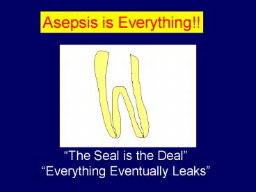
Title: Asepsis is Everything!!
1
Asepsis is Everything!!
The Seal is the Deal Everything Eventually
Leaks
2
Eric M. Rivera, DDS, MS
3
Where to Sear Root Canal Filling Material
VS
Flush With Orifice Level
Below Orifice Level
4
Where to Place Restorative Material
Amalgam as Final Restoratuion - Sufficient
Remaining Tooth Structure
VS
Flush With Orifice Level
Below Orifice Level
Amalgam Plug Not Needed(?)
5
Where to Place Restorative Material
Amalgam as Final Restoratuion - Insufficient
Remaining Tooth Structure
VS
Flush With Orifice Level
Below Orifice Level
Amalgam Plug Needed(?)
6
IntraCoronal Amalgam Use
- With respect to depth of amalgam in the canal
space, it is speculated that it is not necessary
to use amalgam as a coronal-radicular core
material if adequate volume of chamber exists. If
minimal chamber volume exists, may gain
additional retention and seal. - Nayyar A, Walton RE, and Leonard LA. An amalgam
coronal-radicular dowel and core technique for
endodontically treated posterior teeth. J
Prosthet Dent, 1980. 43(5) p. 511-5. - Ulusoy N, Nayyar A, Morris CF, Fairhurst CW.
Fracture durability of restored functional cusps
on maxillary nonvital premolar teeth. J Prosthet
Dent, 1991. 66(3) p. 330-5.
7
Coronal Restoration
- Just as important and many times more important
than Root Canal Filling due to coronal
microleakage - Ray, H.A. and M. Trope, Periapical status of
endodontically treated teeth in relation to the
technical quality of the root filling and the
coronal restoration. Int Endod J, 1995. 28(1) p.
12-8. - The purpose of this study was to evaluate the
relationship of the quality of the coronal
restoration and of the root canal obturation on
the radiographic periapical status of
endodontically treated teeth. - Full-mouth radiographs from randomly selected new
patient folders at Temple University Dental
School were examined. The first 1010
endodontically treated teeth restored with a
permanent restoration were evaluated
independently by two examiners. Post and core
type restorations were excluded. According to a
predetermined radiographic standard set of
criteria, the technical quality of the root
filling of each tooth was scored as either good
(GE) or poor (PE), and the quality of the coronal
restoration similarly good (GR) or poor (PR). The
apical one-third of the root and surrounding
structures were then evaluated radiographically
and the periradicular status categorized as (a)
absence of periradicular inflammation (API) or
(b) presence of periradicular inflammation (PPI).
- The rate of API for all endodontically treated
teeth was 61.07. GR resulted in significantly
more API cases than GE, 80 versus 75.7. PR
resulted in significantly more PPI cases than PE,
30.2 versus 48.6. The combination of GR and GE
had the highest API rate of 91.4, significantly
higher than PR and PE with a API rate of 18.1.
8
Eric M. Rivera, DDS, MS
9
Eric M. Rivera, DDS, MS
10
(No Transcript)
11
PermaFlo Purple
Whats the big deal about coronal seal?
12
Flowable Composite
- May provide added protection against bacterial
contamination, especially if - Temporary restoration leaks or is lost
- Restorative procedures are not performed under
rubber dam isolation - Not recommended as build-up material due to
strength and dimensional stability concerns - Fills the difficult to access intracoronal space
(due to magnification and illumination under
Dental Operating Microscope)
13
Intraorifice Barrier/Sealing
- Intraorifice barriers should be considered
immediately after - Root Canal filling as a secondary seal to prevent
infection/reinfection by microleakage.
14
Name Yr Type Study Amt IO Barrier Results
Roghanizad Jones 1996 Leakage Dye 3.0 mm Amal w Varnish gt Cavit Term gt Control
Pisano et al 1998 Leakage Microbes 3.5 mm Cavit gt IRM Super EBA gt Control (all leaked in lt 49 days)
Wolcott et al 1999 Leakage Microbes 3.0 mm GI (VitrebondGC AmericaKetac bond) gt No Barrier
Belli et al 2001 Leakage Fluid Filtration ? Resins (ClearfilSEBondOneStepCB Metabond) gt IRM gtGP No Sealer
Galvan et al 2002 Leakage Fluid Filtration 3.0 mm Amalgambond gt CB Metabond gt (IRM Eliteflo Palfique) gt Control
Howdle et al 2002 Leakage Dye Transparency ? Bonded Tytin (VitrebondSuperbondD Liner IIPanavia 21) gt Unbonded Tytin
Shindo et al 2004 Leakage Dye 4.0 mm Advantageous sealing ability of Adhesive and Flowable Materials
Shimada et al 2004 Histology Monkey ? No necrosis in any groups. No bacterial penetration along cavity walls in Flowable Composite or Glass Ionomer Cement. Amalgam without Adhesive Liner showed slight bacterial penetration along wall
Yamauchi et al 2005 Abstract Histology Dog left open 2.0 mm Significant periapical inflammation in 90 of samples when plugs not placed. Reduced to 47 w Composite or 37 w IRM Plug.
15
Intraorifice Barrier/Sealing
- Intraorifice barriers should be placed
immediately after - Root Canal filling as a secondary seal to prevent
infection/reinfection by microleakage.
16
(No Transcript)
17
Intraorifice Barrier/Sealing
- Roghanizad N and Jones JJ, Evaluation of coronal
microleakage after endodontic treatment. J Endod,
1996. 22(9) p. 471-3. - A new method is suggested for placing a coronal
seal in the orifice of the root canal right after
root canal therapy. - Root canal therapy was done on 94 extracted human
maxillary centrals. Three mm of the coronal
gutta-percha was replaced by either Cavit, TERM,
or amalgam with cavity varnish. After
thermocycling and 2 wk of immersion in dye, the
amount of dye penetration was measured. - The results showed that amalgam with two coats of
cavity varnish sealed significantly better than
Cavit and TERM. However, Cavit and TERM were
still significantly better than a positive
control group.
18
Intraorifice Barrier/Sealing
- Pisano DM, DiFiore PM, McClanahan SB,
Lautenschlager EP, Duncan JL. Intraorifice
sealing of gutta-percha obturated root canals to
prevent coronal microleakage. J Endod, 1998.
24(10) p. 659-62. - A study was conducted to evaluate Cavit,
Intermediate Restorative Material, and Super-EBA
as intraorifice filling materials to prevent
coronal microleakage. - Root canal instrumentation and obturation was
done on 74 extracted single-rooted teeth. Three
and one-half millimeters of the gutta-percha was
removed from the coronal aspect of the root canal
and replaced with one of the three filling
materials. The teeth were suspended in
scintillation vials containing trypticase soy
broth, and human saliva was added to the pulp
chambers. Microbial penetration was detected as
an increase in turbidity of the broth
corresponding to bacterial growth. - At the end of 90 days, the results showed that
15 of the Cavit-filled orifices leaked, whereas
35 of the Intermediate Restorative Material and
Super-EBA-filled orifices leaked. The
gutta-percha obturated root canals that received
an intraorifice filling material leaked
significantly less than the obturated, unsealed
control group--all of which leaked in lt 49 days.
19
Intraorifice Barrier/Sealing
- Wolcott JF, Hicks ML, Himel VT. Evaluation of
pigmented intraorifice barriers in endodontically
treated teeth. J Endod, 1999. 25(9) p. 589-92. - The purpose of this study was to evaluate the
effectiveness of three pigmented glass ionomer
cements used as intraorifice barriers to prevent
coronal microleakage. - One hundred ten extracted mandibular human
premolars were divided into four experimental
groups of 25 teeth each and two control groups of
5 teeth each. The experimental teeth were
instrumented and obturated using
thermoplasticized gutta-percha and AH26 sealer.
Group 1 teeth received no further treatment.
Teeth in groups 2 through 4 had 1 of 3 pigmented
glass ionomers (Vitrebond, GC America, and
Ketac-Bond) placed as an intraorifice barrier.
Positive control teeth were instrumented but not
obturated. The negative control teeth were
instrumented, obturated, and externally sealed
with epoxy resin. The coronal 3 mm of each root
was sealed into the lumen of an 18-mm segment of
latex surgical tubing. After the apparatus was
sterilized, 2.0 ml of a 24 h growth of Proteus
vulgaris in trypticase soy broth (TSB) was placed
in the coronal reservoir of the tooth. The
inoculated apparatus was placed into a
presterilized test tube containing 1.5 ml of TSB
and incubated for 90 days at 37 degrees C. The
TSB in the lower reservoir was observed daily for
turbidity, which would indicate leakage along the
full length of the obturated root canal. To
determine if differences in microbial leakage
occurred among the four experimental groups,
Pearson's chi 2 and Fisher's exact tests were
performed. The confidence level was set at 95.
The positive and negative controls validated the
microbial testing method. - The teeth without an intraorifice barrier leaked
significantly more than teeth with Vitrebond
intraorifice barriers (p lt 0.05). The difference
in leakage among the experimental glass ionomer
barriers was not significant (p gt 0.05).
20
Intraorifice Barrier/Sealing
- Belli S, Zhang Y, Pereira PN, Pashley DH.
Adhesive sealing of the pulp chamber. J Endod,
2001. 27(8) p. 521-6. - The purpose of this in vitro study was to
evaluate quantitatively the ability of four
different filling materials to seal the orifices
of root canals as a secondary seal after root
canal therapy. - Forty extracted human molar teeth were used. The
top of pulp chambers and distal halves of the
roots were removed using an Isomet saw. The canal
orifices were temporarily sealed with a
gutta-percha master cone without sealer. The pulp
chambers were then treated with a self-etching
primer adhesive system (Clearfil SE Bond), a wet
bonding system (One-Step), a 4-methacryloyloxyethy
l trimellitate anhydride adhesive system (CB
Metabond), or a reinforced zinc oxide-eugenol
(IRM). The specimens were randomly divided into
four groups of 10 each. A fluid filtration method
was used for quantitative evaluation of leakage.
Measurements of fluid movement were made at 2-min
intervals for 8 min. The quality of the seal of
each specimen was measured by fluid filtration
immediately and after 1 day, 1 wk, and 1 month. - Even after 1 month the resins showed an excellent
seal. Zinc oxide-eugenol had significantly more
leakage when compared with the resin systems (p lt
0.05). Adhesive resins should be considered as a
secondary seal to prevent intraorifice
microleakage.
21
Intraorifice Barrier/Sealing
- Galvan RR, West LA, Liewehr FR, Pashley DH.
Coronal microleakage of five materials used to
create an intracoronal seal in endodontically
treated teeth. J Endod, 2002. 28(2) p. 59-56. - The purpose of this study was to quantitatively
compare the sealing effectiveness of five
restorative materials that were used to create an
intracoronal double seal. - Fifty-two extracted mandibular molars were
randomly divided into five groups of 10 teeth,
and one positive and one negative control tooth.
The crowns were removed and the pulpal floor and
canal orifices were sealed with 3 mm of one of
the following materials Amalgabond, CB
Metabond, One-Step Dentin Adhesive with AEliteflo
composite, One-Step with Palfique composite, or
intermediate restorative material (IRM). Each
tooth was affixed to a fluid filtration device
and the seal was evaluated at 0, 1, 7, 30, and 90
days. - The results showed a significant (p 0.0001)
difference in leakage between the materials. At 7
days, IRM, AEliteflo, and Palfique leaked
significantly more than Amalgabond or CB
Metabond. Amalgabond consistently produced the
best seal of all the materials throughout the
duration of the study.
22
Intraorifice Barrier/Sealing
- Howdle, M.D., K. Fox, and C.C. Youngson, An in
vitro study of coronal microleakage around bonded
amalgam coronal-radicular cores in endodontically
treated molar teeth. Quintessence Int, 2002.
33(1) p. 22-9. - OBJECTIVE The aim of this study was to compare
the coronal microleakage of conventional and
bonded amalgam coronal-radicular (Nayyar)
restorations on endodontically treated molar
teeth, because coronal seal is a major factor in
the long-term success of endodontic treatment. - METHOD AND MATERIALS Forty extracted human molar
teeth were root-filled and prepared for
coronal-radicular amalgam restorations. Four
groups of 10 teeth were restored with Tytin
amalgam and Vitrebond, Superbond D Liner II,
Panavia 21, or no adhesive agent. The teeth were
placed in India ink for 1 week, and then
demineralized and rendered transparent. The ink
penetration was assessed with a coded scoring
system. - RESULTS The bonded amalgam groups produced
significantly less leakage than did the nonbonded
group. No statistically significant differences
in leakage were detected among the bonded amalgam
groups. CONCLUSION To prevent the reinfection of
the endodontically treated molar, it may be
preferable to restore the tooth immediately after
obturation by employing a bonded amalgam
coronal-radicular technique.
23
Intraorifice Barrier/Sealing
- Shindo K, Kakuma Y, Ishikawa H, Kobayashi C, Suda
H. The influence of orifice sealing with various
filling materials on coronal leakage. Dent Mater
J, 2004. 23(3) p. 419-23. - The aim of this study was to evaluate the sealing
ability of materials filled in the orifice after
root canal treatment. - A total of 100 root canal-treated teeth were
divided into six experimental groups 1, Protect
Liner F (PL) 2, Panavia F (PF) 3, DC core-Light
cured (DCL) 4, DC core-Chemically cured (DCC)
5, Super-EBA (SE) 6, Ketac (KC). The materials
were filled--to a depth of 4 mm--in the coronal
part of the root canals, and evaluated for
microleakage. - The number of teeth that failed to stop dye
penetration in the filled materials differed
statistically between PL and DCL or SE or KC, PF
and SE or KC, DCC and KC, DCL and KC. The mean
distance of dye penetration differed
significantly between PL and SE or DCC, PF and SE
or DCC. Hence, these results indicated the
advantageous sealing ability of adhesive and
flowable materials.
24
Intraorifice Barrier/Sealing
- Shimada Y, Seki Y, Sasafuchi Y, Arakawa M, Burrow
MF, Otsuki M, Tagami J. Biocompatibility of a
flowable composite bonded with a self-etching
adhesive compared with a glass lonomer cement and
a high copper amalgam. Oper Dent, 2004. 29(1) p.
23-8. - This study evaluated the pulpal response and
in-vivo microleakage of a flowable composite
bonded with a self-etching adhesive and compared
the results with a glass ionomer cement and
amalgam. - Cervical cavities were prepared in monkey teeth.
The teeth were randomly divided into three
groups. A self-etching primer system (Imperva
FluoroBond, Shofu) was applied to the teeth in
one of the experimental groups, and the cavities
were filled with a flowable composite
(SI-BF-2001-LF, Shofu). In the other groups, a
glass ionomer cement (Fuji II, GC) or amalgam
(Dispersalloy, Johnson Johnson) filled the
cavity. The teeth were then extracted after 3, 30
and 90 days, fixed in 10 buffered formalin
solution and prepared according to routine
histological techniques. Five micrometer sections
were stained with hematoxylin and eosin or Brown
and Brenn gram stain for bacterial observation. - No serious inflammatory reaction of the pulp,
such as necrosis or abscess formation, was
observed in any of the experimental groups.
Slight inflammatory cell infiltration was the
main initial reaction, while deposition of
reparative dentin was the major long-term
reaction in all groups. No bacterial penetration
along the cavity walls was detected in the
flowable composite or glass ionomer cement except
for one case at 30 days in the glass ionomer
cement. The flowable composite bonded with
self-etching adhesive showed an acceptable
biological com- patibility to monkey pulp. The in
vivo sealing ability of the flowable composite in
combination with the self-etching adhesive was
considered comparable to glass ionomer cement.
Amalgam restorations without adhesive liners
showed slight bacterial penetration along the
cavity wall.
25
Intraorifice Barrier/Sealing
- Yamauchi S, Shipper G, Buttke T, Yamauchi M,
Trope M. Effect of Orifice Plugs on the
Periapical Inflammation in Dogs. J Endod, 2005.
Abstract. - Gutta-percha and sealer do not resist coronal
leakage thus placing the burden on the filling
above it. The purpose of this study was to
evaluate the effect of orifice plugs using
dentin-bonding composite resin (C) (Clearfil SE
Bond and Clearfil Photo CoreKuraray Medical Inc)
or IRM in resisting coronal leakage as assessed
by periapical inflammation in vivo. - 60 premolar roots in 3 beagle dogs were
instrumented to at least size 40 and were filled
with gutta-percha (GP) and AH26 Sealer (S) and
the coronal 2mm was removed with a heated
plugger. In group 1 and 2 C and IRM respectively
were used as plugs in the prepared 2mm space. In
group 3 no plugs were placed and served as
control. The access cavities were kept open for 8
months after which the dogs were killed. The
periapical regions of the roots were prepared for
histologic examination. - Significant periapical inflammation was observed
in 90 of the samples where plugs were not placed
(GPS), but in those with plugs, the occurrence
was decreased to 47 (GPSC) and 37 (GPSIRM),
respectively. - The poor seal of gutta-percha and sealer was
confirmed in this study. The placement of an
orifice plug with composite resin or IRM
significantly improved resistance to coronal
leakage but are still not sufficient to provide
adequate resistance to bacterial penetration. - Supported by Kuraray Medical Inc
26
Experimental Procedure
Instrumentation/Obturation
Placement of Orifice Plug
Removal of G/S
Plug (IRM or Composite)
2 mm
8 months
Histology
27
Evaluation of periapical inflammation
No inflammation
Mild inflammation
Severe inflammation
28
(No Transcript)
29
Flowable Composite
30
Flowable Composite
Flowable Composite Not Placed In Canals Where
Post or Plug Needed
31
Flowable Composite
Flowable Composite Not Placed In Canals Where
Post Needed
Post Space Preferably Created with Heated Plugger
(do not allow to cool) May also use Rotary
Instruments, Carefully!! Endodontist will provide
Post Space if Requested
32
We Strive To Please theReferring Dentist!!
- Communication
- Biological Principles
- Communication
- Asepsis
- Communication
- Literature Support
- Communication
33
Eric M. Rivera, DDS, MS
34
Returned to Restorative Dentist
- Please Read Chart and/or Referral Letter
- Root Canal Filling Material Used
- Restoration Placed
- Cotton Pellet Placed
- Please Review Postoperative Radiograph
- Level of Root Canal Fill
- Space between Root Canal Fill and Restoration
35
Returned to Restorative Dentist
36
Returned to Restorative Dentist
If it were possible to place a material to the
anatomic apex that prevented leakage and had
dimensional stability, we would use this material.
37
Returned to Restorative Dentist
Significant Loss of Tooth Structure
38
Returned to Restorative Dentist
Significant Loss of Tooth Structure
39
Returned to Restorative Dentist
Amalgam placed when Access is through Intact
Crown/Onlay Restoration
40
Eric M. Rivera, DDS, MS
41
Thank You!
42
Questions??
I appreciate your feedback!!
43
How To Contact Us
University of North Carolina School of
Dentistry Department of Endodontics and Endodontic
Dental Faculty Practice
1098 Old Dental Building, CB 7450 Chapel Hill,
NC 27599-7450 919-966-2707 (Office) 919-966-6344
(Fax) 919-966-2115 (Dental Faculty
Practice) first_last_at_dentistry.unc.edu
Recommended
CrystalGraphics Presentations


