PowerPoint PPT Presentation
1 / 26
Title:
1
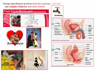
Human reproduction involves intricate anatomy
see text and complex behavior previous
lecture
2
Fertilization of egg initiates development what
initiates ovulation?
humans and most laboratory species are
spontaneous ovulators Spontaneous ovulations
are primarily the result of an LH surge
from anterior pituitary induced by ovarian
steroid hormones regular cycling
next slide
3
http//www.campuslife.utoronto.ca/services/SEC/fre
pro.html Day 1 of menstrual flow low estrogen
causes the hypothalamus to send a message
to the pituitary gland to secrete FSH. The
ovaries react to the FSH by beginning the
maturation of an egg inside the follicles of
the ovaries. The follicle produces estrogen.
Estrogen causes the cells in the endometrium of
the uterus to multiply and the lining
gradually becomes thicker. The estrogen level
continues to rise for around 10 days, until
it is high enough to stimulate the pituitary
gland to release LH (leutenizing hormone).
This sudden rise in LH (called the LH Surge)
acts on the ovaries and triggers the release
of the egg from the follicle.
Egg release (ovulation) occurs about halfway
between the start of one menstrual flow and the
next, and means the egg is available for
fertilization if sperm are present in the
reproductive tract. After an egg is released,
the follicle becomes a corpus luteum (yellow
body). The corpus luteum produces both estrogen
and progesterone. with the addition of
progesterone from the corpus luteum, the
lining changes to form distinct layers with tiny
blood vessels Progesterone also causes LH and
FSH to drop gradually. When the level of LH is
low enough, the corpus luteum stops producing
progesterone and estrogen. This causes the uterus
to shed its lining as the mesntrual flow - which
is Day 1 of the next cycle. The low level of
estrogen also signals the hypothalamus and the
pituitary gand to start secreting more FSH
again, thereby beginning the next reproductive
cycle.
Oral contraceptive pills consist of estrogen and
progesterone (progestin), or progesterone only.
Estrogen prevents the release of an ovum.
Progesterone makes the the cervical mucus very
thick which makes it more difficult for
sperm to reach the uterus. Progesterone also
makes the endometrium of the uterus unfavourable
for implantation.
4
the leading hypothesis is that it has selected
for the emergence of long term pair bonds,
where the man had to stick around, form a
relationship with the woman, have sex with
her on a regular basis, simply because he
didnt know when she would be ovulating. And I
think its not coincidence that these three
things have co-evolved together.
Concealed ovulation, long-term pair bonds,
and heavy male parental investment.
5
Spermatogenesis is continuous and prolific in the
adult human males. Each ejaculation of a human
male contains 100 to 650 million sperm cells,
males can ejaculate daily w/o loss of
fertility sperm is NOT limiting.
6
In humans, oogenesis differs from spermatogenesis
in three major ways 1. In meiotic divisions of
oogenesis, almost all the cytoplasm is
monopolized by a single daughter cell, the
secondary oocyte, which forms the ova the
other products of meiosis, smaller cells called
polar bodies, degenerate. w/ spermatogenesis
all four products of meiosis develop into mature
sperm. 2. Spermatogonia (stem cells) continue
mitosis throughout the males life, this is
not the case for oogenesis At birth, an
ovary already contains all the primary oocytes it
will ever have. 3. Oogenesis is cyclic,
spermatogenesis is not.
7
http//www.ucalgary.ca/UofC/eduweb/virtualembryo
/db_tutorial.html
In humans The fertilization process takes about
24 hours. A sperm can survive for up to 48 hours.
It takes about 10 hours to navigate the female
productive track, moving up the vaginal
canal, through the cervix, and into the fallopian
tube where fertilization begins. Though 300
million sperm may enter the the vagina, only
1, 3 million, enter the uterus.
8
After penetrating the zona, incoming sperm
enter the perivitelline space surrounding
the egg and land on the egg plasma membrane,
where the sperm head initiates sperm-egg
fusion.
http//arbl.cvmbs.colostate.edu/hbooks/pathphys/re
prod/fert/fert.html Prior to fertilization, the
egg is in a quiescent state, arrested in
metaphase of the second meiotic division. Upon
binding of a sperm, the egg rapidly undergoes a
number of metabolic and physical changes
that are collectively called egg Activation.
Prominent effects include completion of the
second meiotic division and the so-called
cortical reaction.
The Cortical Reaction a signal transduction
cascade causes the endoplasmic reticulum to
release Ca2 , causing the cortical granules
to fuse w/ the plasma membrane and release
enzymes viltelline layer becomes the
fertilization envelope - slow block to
polyspermy.
Sperm cells do not contribute any materials
required for activation. Unfertilized eggs can
be artificially activated by the injection of
Ca2 or by a variety of mildly injurious
treatments, such as temperature shock. It is
possible to artificially activate an egg that has
had its own nucleus removed mRNA coding for
new proteins are stockpiled in an inactive form
in the cytoplasm of the unfertilized egg.
9
The zygote's first cell division begins a series
of divisions, with each division occurring
approximately every twenty hours. In
mammals, haploid sperm oocyte nuclei dont fuse
until after 1st division Each blastomere within
the zona pellucida becomes smaller and smaller
with each division. molecules in cytoplasm
partitioned to establish polarity
When cell division has generated about sixteen
cells, the zygote becomes a morula (mulberry
shaped). It leaves the fallopian tube and
enters the uterine cavity three to four days
after fertilization. http//www.ucalgary.ca/UofC/e
duweb/virtualembryo/db_tutorial.html
10
4 days post-ovulation the morula enters the
uterine cavity.
Cell division continues, and a cavity known as a
blastocel forms in the center of the morula.
Cells flatten and compact on the inside of the
cavity while the zona pellucida remains the
same size. With the appearance of the cavity in
the center, the entire structure is now
called a blastocyst.
The presence of the blastocyst indicates that two
cell types are forming the embryoblast
(inner cell mass on the inside of the
blastocele), and the trophoblast (the cells
on the outside of the blastocele). http//www.ucal
gary.ca/UofC/eduweb/virtualembryo/db_tutorial.html
11
5 - 6 days post-ovulation The trophoblast cells
secretes an enzyme which erodes the epithelial
uterine lining and creates an
implantation site for the blastocyst.
In a cyclical process of hormonal stimulation,
the ovary is induced to continue producing
progesterone by human chorionic gonadotropin
(hCG) released by the trophoblast cells of
the implanting blastocyst. detected in pregnancy
tests later hCG may protect the fetus from
rejection by the maternal immune system by
increasing the expression of Indoleamine
2,3-dioxygenase (IDO), which promotes
degradation of tryptophan
Endometrial glands in the uterus enlarge in
response to the blastocyst and the
implantation site becomes swollen with new
capillaries. Circulation begins, a process
needed for the continuation of pregnancy. http//w
ww.ucalgary.ca/UofC/eduweb/virtualembryo/db_tutori
al.html
12
7 - 12 days post-ovulation Trophoblast cells
engulf and destroy cells of the uterine lining
creating blood pools, both stimulating new
capillaries to grow and foretelling the
growth of the placenta. Ectopic pregnancies can
occur at this time
The inner cell mass divides, rapidly forming a
two-layered disc. The top layer of cells
epiblast will become the embryo and amniotic
cavity, while the lower cells hypoblast
will become the yolk sac. Formation of the
placenta. http//www.ucalgary.ca/UofC/eduweb/virtu
alembryo/db_tutorial.html
13
16 days post-ovulation gastrulation continues
changing the two-layered disc into a
three-layered disc. The ectoderm grows rapidly
over the next few days
The three layers of epiblast will eventually
give rise to from fate mapping Endoderm that
will form the lining of lungs, tongue, tonsils,
urethra and associated glands, bladder and
digestive tract. Mesoderm that will form the
muscles, bones, lymphatic tissue, spleen,
blood cells, heart, lungs, and reproductive and
excretory systems. Ectoderm that will form the
skin, nails, hair, lens of eye, lining of
the internal and external ear, nose, sinuses,
mouth, anus, tooth enamel, pituitary
gland, mammary glands, and all parts of the
nervous system. http//www.ucalgary.ca/UofC/eduweb
/virtualembryo/db_tutorial.html
14
Images at UNSW are from the Kyoto Collection,
reproduced with the permission of Prof. Kohei
Shiota
Fetus by Leonardo da Vinci
15
(No Transcript)
16
Sonic Hedgehog Shapes The Brain
The size
and shape of brain structures can be controlled
by a
signaling molecule known as Sonic Hedgehog,
University
of Chicago researchers show
in a paper the March 16,
2001, issue of Science. During development, the
brain becomes organized into highly specialized
groups of neurons, called brain nuclei,
each expressing its own set of genes this
process can be coordinated by the secretion of a
single molecule, Sonic Hedgehog, that
operates as a 'positional signal'.
"A positional signal is a neat mechanism for
creating patterns of different types of cells,"
said Cliff Ragsdale, principal investigator in
the study. "Target cells respond differently to
a signaling molecule according to their
distance from the source of the signal."
http//www.chembio.uoguelph.ca/educmat/chm736/diff
ertx.htm Sonic hedgehog, Shh is the human homolog
of Drosophila hedgehog, Hh. Other Hh related
genes include Indian hedgehog, Ihh, which acts in
cartilage, and Desert hedgehog, Dhh, which
acts in testicular tissue. Hh homologs have been
reported in zebrafish (Mrs. Tiggywinkle) and
nematodes (warthog).
17
Recovery from injury is something like
re-development
The morphogen Sonic hedgehog is an indirect
angiogenic agent upregulating two families
of angiogenic growth factors. Pola et al. NATURE
MEDICINE 7 (6) 706-711 JUN 2001AbstractSonic
hedgehog (Shh) is known to regulate
epithelial/mesenchymal interactions during
embryonic development. the hedgehog-signaling
pathway is present in adult cardiovascular
tissues and can be activated in vivo. Shh
was able to induce robust angiogenesis, Shh
also augmented blood-flow recovery and limb
salvage following operatively induced
hind-limb ischemia blood deprivation in aged
mice. Shh might have potential therapeutic use
for ischemic disorders.
The role of hedgehog signalling in
tumorigenesis.Wicking C, McGlinn E. CANCER
LETTERS 173 (1) 1-7 NOV 8 2001Abstract
members of this pathway are crucial to the
processes of tumorigenesis. mutations in the
gene encoding the hedgehog receptor molecule
patched are responsible for both familial
and sporadic forms of basal cell carcinoma (BCC),
as well as a number of other tumour types.
It is now known that a number of key members of
the hedgehog cascade are involved in
tumorigenesis, and dysregulation of this pathway
appears to be a key element in the aetiology
of a range of tumours.
18
Hox gene topology key to limb development. At
some point in evolution the Hox genes,
responsible for organizing structures along the
trunk axis, were co-opted to regulate limb
development. Intriguingly, Hox genes for digits
are found at one extremity of the HoxD cluster,
suggesting that limb skeletal organization is
somehow controlled by the Hox genes' genomic
topology. In this issue, the mechanism by which
this collinearity produces digit morphology is
unravelled.
The cover (courtesy J. Zakany) shows digital
morphology revealed in a clay impression of a
mouse's 'foot'.
Serial deletions and duplications suggest a
mechanism for the collinearity of Hoxd genes in
limbsMARIE KMITA et al.Nature 420, 145150
(2002)
First come, first served. Hox genes are
arranged and expressed in the same order as the
body parts they help to produce. New work ROLF
ZELLER JACQUELINE DESCHAMPS Nature 420,
138139 (2002)
19
The NIH developed this primer to help
readers understand A. What are stem cells
and why are they important? they are
unspecialized cells that renew themselves
through cell division. can be induced to
become cells with special functions such as
heart muscle or the insulin-producing cells
of the pancreas. B. How are embryonic stem cells
grown in the laboratory? transferring the
inner cell mass of 4-5 day blastocyst
into a plastic laboratory culture dish
coated with mouse embryonic skin cells
called a feeder layer If scientists can
direct the differentiation of embryonic
stem cells into specific cell types, they
may be able to use the resulting,
differentiated cells to treat certain diseases
Parkinsons Disease Type
I Diabetes
20
Immunology at the maternal-fetal interface
lessons for T cell tolerance and
suppression.Mellor AL, Munn DHAnnual Review Of
Immunology 18 367-391 2000
fetal alloantigens encoded by polymorphic
genes inherited from the father ought to
provoke maternal immune responses leading to
fetal rejection soon after blastocyst
implantation in the uterine wall. No other
tissue, when surgically transplanted between
genetically different individuals, enjoys the
impunity from lethal host (maternal)
immune responses that characterize the
maternal-fetal relationship. This unique
relationship resembles a parasitic condition
in which the fetus is nurtured and given
immunological protection
Originally, Medawar proposed three broad
hypotheses to explain the paradox of
maternal immunological tolerance to her fetus
(a) physical separation of mother and fetus
(b) antigenic immaturity of the fetus and (c)
immunologic inertness of the mother.
reject
reject
yes - but how?
The Holy Grail of reproductive immunology has
been to elucidate the fundamental processes
that explain fetal survival in all mammalian
species.
21
Prevention of T cell-driven complement activation
and inflammation by tryptophan catabolism
during pregnancy.Mellor et al. NATURE IMMUNOLOGY
2 (1) 64-68 JAN 2001Indoleamine 2,3 dioxygenase
(IDO) produced by fetal trophoblast
protects developing fetuses from maternal immune
responses in mice. IDO inhibitor treatment
triggered extensive inflammation at the
maternal-fetal interface in susceptible mating
combinations These data show that IDO activity
protects the fetus by suppressing T cell-driven
inflammatory responses to fetal alloantigens.
Tryptophan degradation by human placental
indoleamine 2,3-dioxygenase regulates
lymphocyte proliferation.Kudo et al. JOURNAL OF
PHYSIOLOGY-LONDON 535 (1) 207-215 AUG 15
2001 CD4 T helper lymphocyte division was
specifically suppressed by indoleamine 2,3
-dioxygenase-mediated tryptophan depletion. Our
results show that mechanisms are present in the
human placenta which are able to regulate
cellular proliferation of the maternal immune
system. This mechanism is dependent both
on placental indoleamine, 2,3-dioxygenase-mediated
tryptophan degradation and on tryptophan
sensing systems within lymphocytes.
22
Genetic Conflicts In Human-pregnancy.Haig D.
Quarterly Review Of Biology 68495-532 DEC 1993.
AbstractFetal genes will be selected to
increase the transfer of nutrients to their
fetus, and maternal genes will be selected to
limit transfers in excess of some maternal
optimum.
During implantation, fetally derived cells
(trophoblast) invade the maternal
endometrium and remodel the endometrial spiral
arteries into low-resistance vessels that
are unable to constrict. the fetus gains
direct access to its mother's arterial blood.
Placental hormones, including human placental
lactogen (hPL) increase maternal
resistance to insulin. This action, however,
is countered by increased maternal production of
insulin. Gestational diabetes develops if
the mother is unable to mount an adequate
response to fetal manipulation.
Similarly, fetal genes are predicted to enhance
the flow of maternal blood through the
placenta by increasing maternal blood pressure.
Preeclampsia a complex disease w/ hypertension
can be interpreted as an attempt by a
fetus to increase its supply of nutrients by
increasing the resistance of its mother's
peripheral circulation.
23
Genomic imprinting might have something to
do with genetic conflicts immune tolerance.
Basics of gametic imprinting.Ruvinsky A. JOURNAL
OF ANIMAL SCIENCE 77 228-237, Suppl. 2 1999
Imprinted genes in mammals are expressed from
only one chromosome either the gene
inherited from the mother or that from the father
is silenced.
Such genes include Igf2 insulin-like growth
factor, maternal suppressed and other genes
involved in growth control here, imprinting
might work to balance maternal and paternal
demands on the size of an embryo.
The differential labelling of cytosine guanine
base pairs with methyl groups distinguishes
the two copies of an imprinted gene. This
differential methylation usually originates in
the parental germ cells methylation of a
gene ensures that it cannot be transcribed. Inheri
table mechanisms that control gene expression
without affecting gene sequence are said to be
epigenetic.
24
Frequent loss of imprinting of IGF2 and MEST
in lung
adenocarcinoma.Kohda et al. 2001. Molecular
Carcinogenesis 31184-191.
Abstract Accumulating evidence suggests that
deregulation of imprinted genes,
including loss of imprinting (LOI), plays a
role in oncogenesis.
we investigated allelic expression of six
imprinted genes in human lung
adenocarcinomas as well as in matched normal lung
tissue. LOI of the insulin-like growth factor 2
gene (IGF2) and mesoderm-specific transcript
(MEST, also known as paternally expressed gene 1)
was noted in 47 (7 of 15) and 85 (11 of
13) of informative cases, respectively.
Monoallelic expression was maintained in all the
matched normal tissues examined. These findings
indicated that independent deregulation took
place in imprinted genes and suggested
that aberrant imprinting of IGF2 and MEST
was involved in the development of lung
adenocarcinoma.
25
Nausea and vomiting in early pregnancy Its role
in placental development.Huxley RR Obstetrics
and Gynecology 95 (5) 779-782 MAY 2000
Nausea and emesis in early pregnancy is a common
phenomenon affecting between 50 and 70 of
pregnant women, but little is known about
the etiology and possible function
Morning sickness a positive effect on pregnancy
outcome and is associated with a
decreased risk of miscarriage, preterm birth,
low birth weight (LBW), and perinatal death.
Meaning of morning sickness still
unsettled.Pirisi A. LANCET 357 (9264) 1272-1272
APR 21 2001
26
Ascription of resemblance of newborns by
parents and nonrelatives.McLain et al.
2000.Evolution and Human Behavior 2111-23.
It has been hypothesized that human females
ascribe the resemblance of their infants to the
father or his relatives to promote assurance
of paternity renders fathers more likely
to invest in and less likely to harm mothers and
their children.
mothers were significantly more likely to
ascribe resemblance to the domestic father than
to themselves. This bias was exaggerated in the
presence of domestic fathers.
Yet, judges matched photographs of these mothers
to their newborns significantly more
frequently than they matched domestic fathers to
newborns. The bias in how mothers remark
resemblance does not reflect actual resemblance
and may . assure domestic fathers of their
paternity.
The low rate with which newborns are matched to
fathers may be biologically significant.
Concealment of paternity may be favored when
suspicion of cuckoldry leads fathers to
abandon or harm newborns. Genomic imprinting is
one mechanism by which resemblance of newborns
could be biased toward mothers. men
selected to hide paternity ?
Reactions to children's faces - Resemblance
affects males more than females.Platek et al.
EVOLUTION AND HUMAN BEHAVIOR 23 (3) 159-166 MAY
2002

