
Prezentacja programu PowerPoint - PowerPoint PPT Presentation
1 / 199
Title:
Prezentacja programu PowerPoint
Description:
Title: Prezentacja programu PowerPoint Author: X Last modified by: Maciej Zi tek Created Date: 3/16/2002 3:09:45 PM Document presentation format – PowerPoint PPT presentation
Number of Views:404
Avg rating:3.0/5.0
Title: Prezentacja programu PowerPoint
1
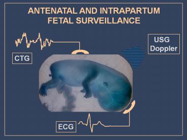
ANTENATAL AND INTRAPARTUM FETAL SURVEILLANCE
USG Doppler
CTG
ECG
2
MAJOR CAUSES OF PERINATAL LOSS Fetal
abnormality Preterm delivery Chronic utero
placental insufficiency Acute Hypoxia
Unexplained intrauterine death Others
3
MODALITIES OF ANTENATAL FETAL
SURVEILLANCE Detection of fetal abnormalities
Assessment of fetal growth Monitoring of
fetal well-being Others
4
APPROACH TO ANTENATAL MONITORING OF FETAL
GROWTH AND WELL-BEING Confirmation of
gestational age Clinical methods of monitoring
Investigations
5
DYNAMIC PARAMETERS To monitor fetal dynamic
response to chronic utero-placental insufficiency
AFI renal perfusion Umbilical arterial flow
(A/B ratio) fetal peripheral and
placental circulation Cerebral arterial flow
(MCA) head sparing effect
6
FETAL MONITORING
Fetal distress is defined in terms of the
manifestation of the fetal hypoxia (by changes
in the fetal heart rate FHR or fetal blood
pH) SIGNIFICANCES OF FETAL DISTRESS Hypoxic
damage to the foetus is difficult to
quantify, but the effect can be devastating. -
neurological abnormalities - cerebral palsy and
mental retardations - fetal death results
from severe intrapartum asphyxia
7
BASIC DEFINITIONS
HYPOXAEMIA - decrease in the oxygen content
of the arterial blood alone HYPOXIA - decrease
in the oxygen content that affects the
peripheral tissues ASPHYXIA - general oxygen
deficiency that affects the high priority
organs as well
8
PATHOPHYSIOLOGY OF FETAL HYPOXIA
In the absence of stress, the fetus is neither
acidotic nor hypoxic. An adequate delivery of
oxygen to the tissues occurs despite the low
fetal arterial partial pressure of oxygen
(pO2). The transfer of oxygen across the
placenta to the fetus is enhanced by following
mechanisms - fetal cardiac output and
systematic blood flow rates are higher than
those of the adult. - the affinity of fetal
blood for the oxygen and - the fetal oxygen
caring capacity, both of which are greater
than those of an adult
9
PATHOPHYSIOLOGY OF CHRONIC FETAL
COMPROMISE utero-placental insufficiency
reduced pO2 to fetal CNS redistribution
of fetal cardiac output perfusion to CNS
heart perfusion to peripheral viscera renal
perfusion visceral circulation OLIGOHYDRAMNIOS
bowel distension umbilical arterial
resistance meconium peritonitis necrotising
enterocolitis etc
10
Sequelae of Chronic Oligohydramnios 1. fetal
demise 2. pulmonary hypoplasia 3. facial
deformities 4. skeletal deformities
11
A decreased amniotic fluid volume is
frequently one of the first clues to an
underlying fetal abnormality. The
sonographer/sonologist should, therefore, have a
basic understanding of the mechanisms responsible
for normal amniotic fluid production. Once the
derivation of amniotic fluid is understood, the
potential mechanisms that can result in
oligohydramnios can be better appreciated.
12
Etiology of Oligohydramnios 1. intrauterine
growth restriction 2. post-term pregnancies 3.
preterm rupture of the membranes 4. fetal
anomalies and/or aneuploidy 5. iatrogenic
13
30 week gestation. A single deepest pocket of
amniotic fluid (7 cm), indicating a normal
amniotic fluid volume.
Visually normal amniotic fluid volume at 18
weeks' gestation.
1.8 cm pocket of amniotic fluid
indicating oligohydramnios. The color box
confirms that umbilical cord is not present in
the pockets of amniotic
Subjective assessment of amniotic fluid volume.
20 week fetus with a unilateral multicystic
kidney and congenital absence of the other
kidney, resulting in anhydramnios
14
Oligohydramnios due to bilateral renal
agenesis. Transabdominal ultrasound at 20 weeks'
gestation.
15
An amniotic fluid index of 4.2 cm, indicating
oligohydramnios there is an absence of amniotic
fluid in the upper quadrants. The color box to
the right of the image indicates the presence of
umbilical cord.
16
19 week fetus with Turner's syndrome, cystic
hygroma (arrows) and oligohydramnios.
15 week fetus with posterior urethral valves.
The bladder (b) is massively distended.
Enlarged "key-hole" bladder associated with
posterior urethral valves.
17
Amniotic fluid has a number of important roles
in embryo/fetal development 1.
Permitting fetal movement and the development of
the musculoskeletal system. 2. Swallowing of
amniotic fluid enhances the growth and
development of the gastrointestinal tract. 3. The
ingestion of amniotic fluid provides some
fetal nutrition and essential nutrients.
18
Amniotic fluid has a number of important
roles in embryo/fetal development 4.
Amniotic fluid volume maintains amniotic
fluid pressure thereby reducing the loss of lung
liquid an essential component to pulmonary
development. 5. Protects the fetus from external
trauma. 6. Protects the umbilical cord from
compression. 7. It's constant temperature helps
to maintain the embryo's body temperature. 8.
It's bacteristatic properties reduces
the potential for infection.
19
STRESSED FETUS
When perfusions is decreased because of impaired
uterine or umbilical blood flow , the transfer of
oxygen to the fetus is diminished, and the
results is an accumulations of carbon dioxide in
the fetus. 1. Increased carbon dioxide causes
an increase in the partial pressure of carbon
dioxide (pCO2) and a concomitant fall in pH,
analogous to adult respiratory acidosis. 2.
Continued hypoxia deprives the fetus of
sufficient oxygen to perform the aerobic
reactions, resulting in buildup of organic acids
with the accumulation of pyruvic and lactic acids
resulting in metabolic acidosis
20
STRESSED FETUS
3. Transient decreases in the fetal or
uterine perfusions usually cause a short-lived
respiratory acidosis, whereas more prolonged or
profound decreases results in a combined
respiratory and metabolic acidosis 4. fetal
oxygen deprivation usually results in the FHR or
fetal bradycardia
21
FETAL DEFENCE MECHANISMS
- Increased tissue oxygen extraction
- Reduced non-essential activity
- Increased sympathetic activity
- Redistribution of blood flow
- Anaerobic metabolism with the metabolism
- of blood sugar glucolysis, and
- glycogen - glycogenolisis
22
More effective uptake of oxygen Reduced
activity Decrease in growth rate Maintained
energy balance
sO2
Hipoxaemia
Hypoxia
Asphyxia
days and weeks hours minutes
t
FETAL RESPONSE TO HYPOXAEMIA
23
Surge of stress hormones Redistribution of blood
flow Peripheral tissue anaerobic
metabolism Maintained energy balance
sO2
Hipoxaemia
Hypoxia
Asphyxia
days and weeks hours minutes
t
FETAL RESPONSE TO HYPOXIA
24
Alarm reaction Anaerobic metabolism in the
central organs The heart fails to function
sO2
Hipoxaemia
Hypoxia
Asphyxia
days and weeks hours minutes
t
FETAL RESPONSE TO ASPHYXIA
25
ANTENATAL AND INTRAPARTUM FETAL SURVEILLANCE
- Cardiotocography
- Contraction stress test
- Nonstress test
- Biophysical profile
- Fetal movement counts
- Umbilical artery Doppler velocimetry
- Fetal electrocardiography
- Fetal pulse oxymetry
- Fetal blood sampling
26
Fetal Neurodevelopment and Sequence of Fetal
Deterioration
CSTcontraction stress test NSTnonstress test
BPPbiophysical profile.
27
BIOPHYSICAL PROFILE
biophysical profile was first introduced in the
late 1970s. It requires more expensive equipment
and more highly trained personnel than the other
testing modalities. The study is based on the
concept that hypoxic fetuses lose certain
behavioral parameters in the reverse order in
which they were acquired in the course of fetal
development It evaluates indicators of chronic
fetal hypoxia and placental function, such as
amniotic fluid volume, in addition to more acute
indicators, such as fetal breathing, movements
and tone.
28
PATHOPHYSIOLOGY OF BIOPHYSICAL VARIABLES
The gradual hypoxia concept Fetal CNS
centres embriogenesis FT cortex FM cortex-nucl
ei FBM ventral surface of 4th ventricle FHR post
erior hypothalamus medulla hypoxia
29
BIOPHYSICAL PROFILE
30
(No Transcript)
31
INTERNAL FETAL HEART MONITORING IS DONE TO -
help determine whether the stress of labor is
threatening the health of the fetus. -
measure the strength and duration of labor
contractions.
32
CONTINUOUS ELECTRONIC FETAL MONITORING
33
(No Transcript)
34
Indications for continuous electronic fetal
monitoring
CC cord compression FA fetal anaemia FS
fetal sepsis Other other mechanisms RFR
reduced fetal nutritional reserves RUPO
reduced uterine perfusion or oxygen delivery (no
vascular disease) UPVD uteroplacental
vascular disease
35
FetalIntrauterine growth restrictionCongenital
anomaliesFetal cardiac arrhythmiasIsoimmunizatio
nHydrops fetalisFetal infections such as
parvovirus, coxsackievirus B, syphilis, toxo
CONDITIONS THAT PLACE FETUSES AT RISK FOR ADVERSE
OUTCOMES
Pregnancy-relatedPoorly controlled gestational
diabetesMultiple gestationsPregnancy-induced
hypertensionCholestasis of pregnancyPremature
rupture of the membranes (preterm)Unexplained
elevated maternalserum alpha-fetoproteinPolyhydr
amniosOligohydramniosPlacental
abruptionAbnormal placentationPostdatesUnexplai
ned stillbirth in a prior pregnancy
MaternalChronic hypertensionCollagen-vascular
diseasesSickle cell anemiaCurrent substance
abuseImpaired renal functionAsthmaPneumoniaSig
nificant cardiac diseaseSeizure
disordersDiabetesAcute febrile
illnessesSignificant anemia (hematocrit lt26)
36
ANTENATAL RISK FACTORS
37
INTRAPARTUM RISK FACTORS
38
INTRAPARTUM RISK FACTORS
39
ELECTRONIC FETAL MONITORING
40
(No Transcript)
41
(No Transcript)
42
(No Transcript)
43
(No Transcript)
44
CATEGORISATION OF FETAL HEART RATE TRACES
45
CATEGORISATION OF FETAL HEART RATE FEATURES
46
ELEMENTS OF FHR PATTERN
The baseline FHR is the steady rate fetal that
occurs during and between contractions in the
absence of accelerations and decelerations . The
normal baseline FHR is 120-150 beats per minute.
At 16 weeks , the average baseline is 160 BPM.
The baseline FHR decreases approximately 24
BPM from 16weeks to term .
47
BEAT TO BEAT VARIABILITY
- represents the continuous interaction of the
sympathetic - and parasympathetic nervous system in adjusting
the - FHR to fetal metabolic or hemodynamic conditions.
- decreased variability may signify loss of fine
autonomic - control of FHR
- - good variability usual predict a good fetal
outcome
48
ABNORMAL FHR
DECREASED VARIABILITY Asphyxia Drugs Prematurity
Tachycardia Sleep state of fetus Cardiac and
CNS abnormalities Arrythmias
FETAL TACHYCARDIA Asphyxia Maternal
fever Fetal infection Prematurity Drugs Fetal
stimulations Arrythmias Maternal
anxiety Thyreotoxicosis Idiopatic
FETAL BRADYCARDIA Asphyxia Drugs Reflex
(pressure on fetal head) Hypothermia Arrythmias
Idiopatic
49
BASELINE RATE LESS THAN 110/MIN
50
ACCELERATION
51
DECELERATIONS
- Periodic changes in the FHR assume importance in
- defining the mechanism and intensity of asphyxia
insults. - There are 3 patterns of periodic decelerations
based on - the configuration of the waveform and the timing
of the - deceleration in relation to the uterine
contraction. - EARLY DECELERATIONS
- LATE DECELERATIONS
- VARIABLE DECELERATION
52
EARLY DECELERATIONS
- are not caused by systemic hypoxia
- do not appear to be associated with poor fetal
outcome - occur with fetal head compressions
- begin with the onset of uterine contractions
- reach their lowest point at the peak of the
contraction - Return to baseline as the contraction ends
53
EARLY DECELERATIONS
54
LATE DECELERATIONS
- Occur in situations
- - ablation of placentae
- maternal acute hypotension
- hyper stimulation during oxitocin infusion
- acute decrease in the intervillous space flow
- Are found with increased frequency
- preeclampsia
- hypertension
- diabetes mellitus
- intrauterine growth retardation
- other disorders associated with chronic
placental - insufficiency .
55
LATE DECELERATIONS
- usually are associated with acute/chronic
fetoplacental insufficiency - occur after the peak of the uterine contraction
- are precipitated by hypoxemia (which slows the
FHR as results of CNS asphyxia) or direct
myocardial depression and associated with mixed
respiratory and metabolic acidosis
56
LATE DECELERATIONS
57
VARIABLE DECELERATIONS
- are inconsistent in configurations
- have no uniform temporal relationship to the
onset - of the contraction
- usually are the results of compressions of the
- umbilical cord between fetal parts and
surrounding - maternal tissues
- often are associated with oligohydramnions (ex.
PROM) - may be associated with profound combined
acidosis
58
VARIABLE DECELERATIONS
59
VARIABLE DECELERATIONS
60
REDUCED VARIABILITY
61
REDUCED VARIABILITY
62
REDUCED VARIABILITY
63
In the presence of abnormal FHR patterns and
uterine hypercontractility (not secondary to
oxytocin infusion) tocolysis should be
considered. A suggested regimen is
subcutaneous terbutaline 0.25 mg. In cases of
suspected or confirmed acute fetal compromise,
delivery should be accomplished as soon as
possible, accounting for the severity of the FHR
abnormality and relevant maternal factors. The
accepted standard has been that, ideally, this
should be accomplished within 30 minutes.
64
FETAL BLOOD SAMPLING
65
FETAL BLOOD SAMPLING
66
FETAL BLOOD SAMPLING
67
CLASSIFICATION OF FETAL BLOOD SAMPLE RESULTS
68
FETAL MOVEMENT COUNTS
Perception of fetal movement is an inexpensive,
noninvasive method of assessing fetal
well-being. Generally, the patient is asked to
relax on her left side 30 minutes after eating
and to concentrate on fetal movement. The
patient should record the time that she starts
the test and note each time the baby kicks or
moves. A healthy fetus should move approximately
three to five times within one hour in this
setting.
69
FETAL MOVEMENT COUNTS
An alternative method is the Cardiff
Count-to-Ten chart, whereby the patient records
fetal movements during the course of usual daily
activity. A period of 12 hours without at least
10 perceived movements is considered a warning
signal. If the test result is not reassuring,
the patient should be evaluated and should
undergo further testing, such as evaluation with
a nonstress test.
70
FETAL MOVEMENT COUNTS
Studies have shown that fetal movement counts
are an effective screening measure, with reported
reductions in fetal mortality from 8.7 deaths per
1,000 live births to 2.1 deaths per 1,000 live
births. Although the ideal method for performing
the test, including how often it should be
repeated, has not been defined, it is clear that
complaints of decreased fetal movement are
significant and warrant further evaluation.
71
NONSTRESS TEST
Nonstress test is an indirect measurement of
uteroplacental function. The patient is usually
seated in a reclining chair, slightly tilted to
the left to avoid supine hypotension. A Doppler
ultrasound transducer and a tokodynamometer are
used to monitor the fetal heart rate and uterine
activity simultaneously. Fetal movements can also
be recorded during the test. A reassuring, or
reactive, nonstress test exhibits at least
two accelerations in the fetal heart rate in a
20-minute period that are at least 15 beats per
minute above the baseline and last at least 15
seconds. A nonreactive test does not meet these
criteria.
72
NONSTRESS TEST
Fetal heart rate reactivity is a reflection of
the balance between the fetus's sympathetic and
parasympathetic tone. It is an acquired neurologic
reflex and is therefore dependent on gestational
age about 65 of healthy fetuses will have a
reactive nonstress test at 28 weeks of gestation,
85 t at 32 weeks of gestation and 95 at 34
weeks of gestation. Fetal heart rate
accelerations are coupled to fetal movement as
the fetus matures consequently, they will be
seen more frequently when the fetus is awake or
in an active sleep state. Since fetuses can have
normal sleep cycles lasting up to 40 minutes, a
nonstress test might require over an hour to
complete if it is initially nonreactive. It is
important to differentiate whether a nonreactive
tracing truly represents a compromised fetus or
merely reflects a temporary behavioral state.
73
CONTRACTION STRESS TEST
74
Doppler ultrasound in high risk
pregnancies(Protocol for a Cochrane Systematic
Review)
Department of Obstetrics and Gynaecology of the
Center for Integral Attention to Womens Health
(CAISM) State University of Campinas (UNICAMP),
Brazil
75
The first Doppler ultrasound report using
continuous wave assessment of umbilical artery
flow was published in 1977 (Fitzgerald 1977).
With the use of colour Doppler, in 1987, it was
possible to study the middle cerebral artery in
fetuses and compare to umbilical artery
pulsatility index (PI) ratio to demonstrate
centralization of the fetal circulation
(Wladimiroff 1987). Waveforms in the ductus
venosus, was recognized as a key examination to
predict right heart failure in the hypoxic fetus
and an important indicator of imminent fetal
demise (Kiserud 1991). The relationship between
abnormal uterine artery Doppler velocimetry
and pre-eclampsia, intra-uterine growth
retardation and adverse pregnancy outcome is well
established (Aquilina 1996).
76
When the fetus is hypoxic, the cerebral arteries
tend to become dilated in order to preserve the
blood flow to the brain. In the middle cerebral
artery, the systolic to diastolic (A/B) ratio
will decrease (due to an increase in diastolic
flow) in the presence of chronic hypoxic
insult to the fetus. This increase in blood flow
can be evidenced by Doppler ultrasound of the
middle cerebral artery. This effect has been
called "brain sparing effect" and is demonstrated
by a lower value of the pulsatility index.
77
UMBILICAL ARTERY DOPPLER VELOCIMETRY
... is based on the observation that flow
velocity waveforms in the umbilical artery of
normally growing fetuses differ from those of
growth-restricted fetuses. Specifically, the
umbilical flow velocity waveform of normally
growing fetuses is characterized by high-velocity
diastolic flow, whereas with intrauterine
growth restriction, there is diminution of
umbilical artery diastolic flow. In some cases of
extreme intrauterine growth restriction, flow is
absent or even reversed.
78
Normal Doppler waveform of umbilical artery
79
Normal Doppler waveform of umbilical artery
80
Abnormal Doppler waveform of umbilical
artery showing absent end diastolic flow (AEDF)
81
Abnormal Doppler waveform of umbilical
artery showing reversed end diastolic flow (REDF)
82
Normal Doppler waveform of uterine artery
83
Abnormal Doppler waveform of uterine artery
Notch
84
Normal Power-Doppler waveform of middle cerebral
artery
85
Normal Doppler waveform of middle cerebral artery
86
Abnormal Doppler waveform
of middle cerebral artery
high diastolic velocities
Normal
87
Normal ductus venosus waveform at 25 weeks of
gestation
88
Normal flow velocity waveforms of the ductus
venosus
89
Normal Ductus venosus sonogram. Positive A wave.
Abnormal Ductus venosus sonogram. Reverse A wave
S systole D diastole A atrial contraction
90
Abnormal waveform with reversal of flow during
atrial contraction
91
T/QRS rise
Biphasic ST type 1
Biphasic ST type 2
Biphasic ST type 3
92
FETAL ELECTROCARDIOGRAPHY
93
Inadequate quality CTG poor contact from external
tranducer? FSE not working or detached?
Uterine hypercontactility Is the mather receiving
oxytocin? Has the mother recently
received vaginal prostaglandins?
Maternal tachycardia/pyrexia maternal
infection? Tocolytic infusion? Dehydrated?
Other maternal factors what is the maternal
position ? Is the mother hypotensive? Has she
just had a vaginal examination? Has she had a
vasovagal episode? Has she just had an epidural?
Stop oxytocin infusion consider tocolysis
IF Tgt38 consider screening and treatement If
pulse gt140/min. reduce Tocolytic infusion
Check blood pressure give crystalloid if
appropriate
Check maternal pulse, position of
transducer, consider applying FSE.
Suspicious CTG Pathological CTG
Encourage mother to adopt left lateral
position Check blood pressure give crystalloid
if appropriate
gt7,25 FBS should be repeated if the FHR
abnormality persists 7,21-7,24 repeat FBS within
30 min. or consider delivery if rapid fall
since last sample lt7,20 Delivery
indicated (all scalp pH estimations should be
interpreted taking into the previous pH
measurment, the rate of progress in labour,
clinical features of the mother and baby)
Fetal blood sampling indicated
Encourage mother to adopt left lateral
position Check blood pressure give crystalloid
if appropriate
Fetal blood sampling inappropriate
Encourage mother to adopt left lateral
position Check blood pressure give crystalloid
if appropriate
Expedite delivery
Urgency of delivery should take into account the
severity of the FHR abnormality and
relevant internal factors
94
Are any of the following risk factors
present? Maternal problems previous cesarean
section Pre-eclampsia Post-term pregnancy
(gt42weeks) Prolonged membrane rupture
(gt24hs) Induced labour Diabetes Antepartum
haemorrhage Other maternal medical disease Fetal
problems Fetal growth restriction Prematurity Oli
gohydramnios Abnormal Doppler artery
velocimetry Multiple pregnancy Meconium-stained
liquor Breech presentation
Intremittent ausculation for full minute after a
contraction
Abnormal FHR on ausculation Baseline lt110bpm or gt
160bpm Any decelerations
No
Cardiotocograph classification NORMAL A CTG where
all four features fall into the reassuring
category SUSPICIOUS A CTG where features fall
into one of the non-reassuring categories and
the remainder of the features are
reassuring PATHOLOGICAL A CTG where features fall
into two or more of the abnormal
categories Fetal heart rate feature
classification Baseline(bpm) variability(bpm) De
celeration Acceleration Reassuring 110-160 ?5 non
e present Non-reassuring 100-109 lt5 for?40 early
161-180 lt90minutes variable single
prolonged up to 3 minutes Abnormal lt100 lt5
for?90 Atypical variable gt180 minutes late sinus
oidal single prolonged pattern for
?10min. greater than 3 min.
Yes
CONTINUOUS ELECTRONIC FETAL MONITORING
Offer and recommend continuous EFM
The absence of accelerations with an otherwise
normal CTG is of uncertain significance
Intrapartum risk factors oxytocin
augmentation epidural analgesia vaginal bleeding
in labour maternal pyrexia fresh meconium-stained
liquor
Yes
Yes
95
PHYSIOLOGY OF LABOUR
There are still those who think that the
delivery of a woman is easy. François
Mauriceau, 1694
96
Maternal
Fetal
UTERUS Distension Contractions Force
Coordination
BIRTH CANAL Bony pelvis
Soft tissue Size
Distensibility Symmetry
Tension Sacrum Friction
Spines Vaginal-vulval
Coccyx angle
Canal length
Size Presentation Anomalies Head
volume Attitude Plastic modelling moulding Caput
DELIVERY
97
Conditions for successful delivery
98
PELVIMETRY SHOULD BE DONE AS A MATTER OF
ROUTINE IN ALL PRIMIPARAE
Following pelvic measurements, should be taken
The intercristal The interspinous The
intertrochanteric The external conjugate
99
28 cm 25 cm 31 cm
The intercristal The interspinous The
intertrochanteric
EXTERNAL DIMENSIONS OF THE BONY PELVIS
100
THE BONY PELVIS
101
ILIUM
Iliac crest provides attachments to the iliac
fascia, abdominal muscles ,and fascia
lata. Anterior superior and inferior spine
superior spine provides the point of fixation of
the inguinal ligament Posterior superior and
inferior spine superior spine is the point of
attachment for the sacrotuberous ligament and
the posterior sacral iliac ligament. Arcuate line
marks the pelvic brim and lies between the
first two segments of the sacrum Iliopectineal
eminence (linea terminalis) the line of
junction of the ilium and the pubis. Iliac fossa
the smooth anterior concavity of the
ilium, covered by the iliacus muscle.
102
ISCHIUM
Ischial spine delineates the greater and lesser
sciatic notch above and below it. It is the
point of fixation for the sacrospinous ligament.
The ischial spine represents an important
landmark in the performance of pudental nerve
block and sacrospinous ligament vaginal
suspension vaginal palpation during
labor allows detection of progressive fetal
descent. Ischial ramus joins that of
the pubis to encircle the obturator foramen
provides the attachment for the inferior fascia
of the urogenital diaphragm and the perineal
musculofascial attachments.
103
PUBIS
Body formed by the midline fusion of the
superior and inferior pubic rami. Symphysis
pubis a fibrocartilaginous symphyseal
joint where the bodies of the pubis meet in the
midline allows for some resilience and
flexibility, which is critical during
parturition. Superior and inferior pubis rami
join the ischial rami to encircle the obturator
foramen. Provide the origin for the muscles of
the thigh and leg. Provide the attachment for
the inferior layer of the urogenital diaphragm. P
ubic tubercle a lateral projection from the
superior pubic ramus, to which the inguinal
ligament, rectus abdominis, and pyramidalis
attach.
104
1- TRUE CONJUGATE DIAMETER ( 10,5 - 11,5 cm ) 2-
OBSTETRIC CONJUGATE ( 10 - 11 cm ) 3- DIAGONAL
CONJUGATE DIAMETER ( 12,5 cm )
1 2 3
PELVIC DIAMETERS
105
- For obstetrical purposes, the pelvis is described
as having 3 imaginary planes - Plane of the inlet Four diameters have
been described. - Anteroposterior diameter This is the distance
between the sacral promontory and the symphysis
pubis it is designated the obstetrical
conjugate. This conjugate normally measures
approximately 10 cm or more, but it may be
shortened considerably in an abnormal pelvis. - Transverse diameter This is the greatest
distance between the linea terminalis on either
side of the pelvis. This imaginary line usually
intersects the obstetrical conjugate at a point
approximately 4 cm in front of the promontory. - Two oblique diameters Each of these diameters
extends from one of the sacroiliac joints to the
iliopectineal eminence on the opposite side of
the pelvis. These diameters normally average less
than 13 cm each.
106
Plane of the mid pelvis This is the plane of
the smallest dimensions. This plane is extremely
important following engagement of the head in
obstructed labor. The interspinous diameter
(approximately gt10 cm) usually is the smallest
diameter of the pelvis.
107
- Plane of the pelvic outlet This consists of 2
triangular areas created from the connection of
an imaginary line between the 2 ischial
tuberosities. The apex of the posterior triangle
is at the tip of the sacrum, and the apex of the
anterior triangle is under the pubic arch. The
following 3 diameters of the outlet are of
importance - Anteroposterior diameter This normally is
9.5-11.5 cm and extends from the lower margin of
the symphysis pubis to the tip of the sacrum. - Transverse diameter This commonly is 11 cm and
is the distance between the inner edges of the
ischial tuberosities. - Posterior sagittal diameter This usually exceeds
7 cm and extends from the tip of the sacrum to a
right-angle intersection with the line between
the ischial tuberosities.
108
Inlet Midcavity Outlet
11x13cm 12x12cm 11.5-x11cm
109
600
SAGITAL SECTION OF THE BONY PELVIS
110
The 3 planes of pelvis
111
Male pelvis Female pelvis
112
NORMAL
FLAT
CHILDS
NARROW
TYPES OF PELVIS
113
PLATYPELLOID
GYNECOID
ANDROID
ANTRTHROPOID
CALDWELL-MOLOY PELVIC TYPES
114
Pelvis of Mrs H extremely distorted by Mollities
Ossium, a disease of adult life, which leads to
softening of the bones of the body. Brim
triangular - nearly closed on the left
side. Successful Caesarean section performed,
May 1849.
115
Transversely Contracted Pelvis. "Robert" pelvis.
Note the faulty development of the sacral alae.
The patient from whom this pelvis was removed
was admitted to St. Mary's Hospital in June
1867 and delivered by Caesarean Section. Mother
died Child- lived
116
Small Round Pelvis. The pelvis of Mrs B, aged 27
years who died of rupture of the uterus as a
result of dystocia, 1st October, 1853.
117
Kyphotic or Funnel shaped Pelvis. The
transverse diameters diminish from above
downwards, being most contracted at the outlet.
The conjugate of the outlet is also affected
owing to the tilting forwards of thelower end of
the sacrum.
118
Model of female pelvis of Mrs M. distorted by a
large exostosis springing from and intimately
connected with the sarum. This large bony
tumour occupied the cavity of this bone and also
that of the coccyx. Caesarean section was
performed Sept 1829, Woman's age at time of
birth 26. Duration of Labour about 30
hours Mother - Died Child - Dead before operation.
119
Kyphotic or Funnel shaped Pelvis
120
The pelvis of Elizabeth Thompson is in the
museum of St Mary's Hospital, Manchester, and was
presented by me. There is a block of oak carved
on the model which, with others, I have given to
the aforesaid hospital. I have endeavoured to
bring a mutilated infant through it, but I have
never succeeded. However, it is one thing to
operate on an inanimate machine, a block of wood,
let it be ever so accurately formed, and another
topo erate on the pelvis of a living woman. I
deny the possibility of bringing a mutilated
full-grown child through such a pelvis, whatever
appliances are used." Thomas Radford
121
The passage of a baby through a normal
pelvis during birth.
The attempted passage of a baby through an
abnormal pelvis during birth. Note how natural
birth would have been impossible.
122
DIAMETERS OF THE FOETAL SKULL
MENTO-OCCIPITAL (13.5cm) SUBOCCIPITO-BREGMAT
IC (9.5 cm) FRONTO-OCCIPITAL (12.0 cm)
123
LAMBOID S.
POSTERIOR FONTANEL
SAGITAL S.
ANTERIOR FONTANEL
CORONAL S.
124
STAGES IN THE MECHANISM OF LABOURIN THE VERTEX
PRESENTATION
- Descent with engagement of the head and
- increased flexion
- Internal rotation
- Extension, resulting in the birth of the head
- Restitution, or the untwisting of the neck
- External rotation of the head, accompanied with
internal rotation of the shoulders - Delivery of the shoulders
- Expulsion of the rest of the body of the foetus
125
DESCENT
- The movement of descent is brought about
- by two factors
- ? General contents pressure of the uterus
- before rupture of the membranes
- ? Foetal axis pressure which comes into
- effect after the rupture of the membranes
126
DESCENT
- In normal cases the head engages in what
- is known as a synclitic manner
- the sagittal suture of the head lies in one
- or other of the oblique diameters of the
- pelvic brim, so that the parietal bones on
- either side are at the same level.
127
DESCENT
- where abnormalities of mechanism occur, the
- sagittal suture may be pushed towards
- the symphysis pubis (posterior asynclitism
- Litzmanns obliquity)
- or
- the sacral promontory (anterior asynclitism
- Naegelis obliquity)
128
The fetal head descending through the pelvis in
labour
129
The concept of the fetal head descending through
the pelvis in labour is checked by vaginal
examination when the level of the presenting part
is assessed against the level of the
ischial spines (in centimetres) vertically
130
ENGAGEMENT OF FETAL HEAD A- maximum
diameter of head is above inlet of pelvis
and head is not engaged B- engagement has
taken place (maximum diameter of head is
below inlet of pelvis) C- head is not
engaged D- when mother sits up on her
elbows,the head sinks in, an indication
that the head will engage when labour starts
131
INTERNAL ROTATION
- ?The shape of the pelvis the forward incline of
- the walls of the pelvic cavity helps to
rotate - forwards the most dependent part of the
- presenting pole.
- ?The tendency to forward rotation is helped by
- the contour of the musculo-fascial forming
the - pelvic floor.
132
INTERNAL ROTATION
- ?The impetus given by the spine of the
- ischium is another dominant causative
- factor in this phenomenon.
- ?The effective contractions of the uterus
- are essential to promote internal rotation.
133
EXTENTION
- is the resultant of forces the effect of
- ? the uterine contractions from above
- and
- ? the elastic resistance of the pelvic floor
- from below
134
EXTENTION
- ?The occiput hitches against the symphysis
- pubis, the face sweeps over the perineum,
- and the successive parts of the foetal head
- to be born are the sinciput.
135
RESTITUTION
- ?As soon as the head is free outside the vulval
- outlet it rotates through 1/8 of a circle,
and - thus the neck is untwisted and the chin
rotates - towards the right (left occipito-ant.
position) or - the left (right occipito-ant. position)
136
EXTERNAL ROTATION
- ? After the untwisting of the neck has occurred,
- the next movement is one of internal
rotation - of the shoulders. This brings the anterior
- shoulder underneath the symphysis pubis,
- and with this movement occurs external
- rotation of the head
- the bisacromial diameter is brought
- into the antero-posterior diameter of
- the pelvic outlet.
137
? Once the shoulders have rotated into the
antero-posterior diameter of the outlet,
descent continues with the uterine
contractions, until the anterior shoulder
hitches underneath the symphysis pubis and
the posterior shoulder sweeps over the
perineum by a process of latero- flexion of
the spine and is delivered first.
EXTERNAL ROTATION
138
VAGINAL EXAMINATIONIN LABOUR
- 1. Condition of the vulva, the vagina the
- extent to which they are dilatable and the
- pressure of any lubricating mucus.
- 2. The condition of the cervix whether the
- cervical canal is dilated and the extend to
- which the external os is dilated or
dilatable. - 3. The condition of the bladder and rectum
139
VAGINAL EXAMINATIONIN LABOUR
4. Whether the membranes are entire or ruptured.
If present, the nature of the bag and whether
the membranes are tough. 5. The presenting
part - whether it is the head or any other
part of foetus, and the particular details
concerning the presenting part. 6. The presence
of a caput and the degree of moulding in
cephalic presentations.
140
VAGINAL EXAMINATIONIN LABOUR
- 7. The exact position of the presenting part
- with reference to the maternal pelvis is
- whether the head is at the brim, or through
- the inlet in the cavity, or at the outlet.
- 8. Whether in cases of cephalic presentation
- the occiput has rotated, and if so, to what
- extent.
141
VAGINAL EXAMINATIONIN LABOUR
- 9. Whether the sacral promontory can be
- palpated or not.
- 10. The presence of abnormalities, such a
- prolapsed cord, or placenta praevia.
142
- WHEN SHOULD THE MEMBRANES BE RUPTURED
ARTFICIALLY?
When the cervix is fully dilated and the bag of
waters remains entire owing to tough
membranes. In some cases of antepartum
haemorrhage, rupturing the membranes controls
bleeding.
143
WHEN SHOULD THE MEMBRANES BE RUPTURED
ARTFICIALLY?
- As a method of induction of labour
- As a preliminary to operative delivery
144
STAGES OF LABOUR
I - the stage of dilatation
II - the stage of expulsion III
- the stage of placental delivery
and uterine contraction and
retraction
145
THE FIRST STAGE
- True uterine contractions, or labour pains
- A muco-sanguinous discharge or the show
- The dilatation of the cervical canal, so that
- both the internal and the external os become
- completely dilated
- The fixation of the head at the brim of the
- pelvis and its progressive descent
- Rupture of membranes
146
Latent (a) and active (b) phase of labour in a
multiparous and a primiparous woman, as shown on
partogram
147
(No Transcript)
148
THE SECOND STAGE
- The occurrence of the characteristic uterine
contractions - The coming into action of the accessory muscles
of labour - The progressive descent of the presenting part
- The dilatation of the vagina and vulva with
stretching of the pelvic floor - The expulsion of the foetus
149
When the cervix has dilated to 10 cm, the mother
has an uncontrollable urge to push
150
(No Transcript)
151
STEPS OF NORMAL DELIVERY
152
THE EPISIOTOMY ISSUE
153
THE CAUSES OF PERINEAL LACERATIONS ARE
- Relative disproportion in size between the
presenting part and the vaginal outlet. - Too rapid expulsion of the presenting part, so
that enough time is not allowed for gradual
stretching of the perineum - Faulty mechanism, whereby a larger diameter of
the presenting part emerges through the outlet
154
THE THIRD STAGE
- The separation of the placenta after the
formation of a retroplacental haematoma - The expulsion of the placenta
- The control of the haemorrhage
- The permanent contraction and retraction of the
uterus
155
(No Transcript)
156
THE CAESAREAN SECTION
Horizontal incision Vertical incision
157
C-section - Indications
Transverse position Placenta abruptio
Breech presentatio Placenta previa
158
C-section Emergency placental indications
Placenta previa Placenta abruptio
Ruptured uterus
159
PROLAPSED UMBILICAL CORD
160
C-section
161
C-section
162
C-section
The surgeon reaches into the abdominal
incision and lifts the babys head as an
assistant pushes down on the upper uterus.
163
C-section
The surgeon will clamp and cut the umbilical
164
C-section
1 day after 1 year after
165
Forceps delivery Simpson forceps
166
FORCEPS Naegele type
1 cm 9,5 cm
4 cm
9 cm
40,5cm
15cm
Franz Karl Naegele 1777-1851
167
- Indications for operative vaginal
deliveries are identical for - forceps and vacuum extractors. The
following indications - apply when no contraindications exist
- Prolonged second stage (1) This includes
nulliparous woman with failure to descend for 2
hours without, and 3 hours with, conduction
anesthesia. (2) multiparous woman with failure to
descend for 1 hour without, and 2 hours with,
conduction anesthesia. - Suspicion of immediate or potential fetal
compromise is an indication. - Shortening of the second stage for maternal
benefits Maternal indications include, but are
not limited to, exhaustion, bleeding, cardiac or
pulmonary disease, and history of spontaneous
pneumothorax. - In expert hands, fetal malpositions, including
the after-coming head in breech vaginal delivery,
can be indications for forceps delivery.
168
- Prerequisites for forceps delivery
include the following - The head must be engaged.
- The cervix must be fully dilated and retracted.
- The position of the head must be known.
- The type of pelvis should be known.
- The membranes must be ruptured.
- No disproportion should be suspected between
the size of - the head and the size of the pelvic inlet and
mid pelvis. - The patient must have adequate anesthesia.
- Adequate facilities and supportive elements
should be - available.
- The operator should be fully competent in the
use of the - instruments and the recognition and management
of - potential complications.
- An operator should be present who knows when to
stop, to - not force the issue, and to not aggressively
use both forceps - and vacuum in combination because this has
been shown to - increase morbidity for both the mother and
fetus.
169
Forceps delivery technique
170
Forceps delivery The left handle held in the left
hand (Simpson forceps)
171
Forceps delivery Introduction of the left blade
into the left side of the pelvis
172
Forceps delivery Left blade in place
introduction of the right blade by the
right hand
173
Forceps delivery A median or mediolateral
episiotomy may be performed at this point. A
left mediolateral episiotomy is shown here.
174
Forceps delivery The forceps have been locked.
The inset shows a left occipitoanterior fetal
position
175
Forceps delivery Horizontal traction with the
operator seated
176
Forceps delivery Upward traction
177
Forceps delivery Disarticulation of the branches
of the forceps beginning modified Ritgen
maneuver
178
- Contraindications to forceps-assisted
- vaginal deliveries
- Any contraindication to vaginal delivery .
- Refusal of the patient to consent to the
procedure - Cervix not fully dilated or retracted
- Inability to determine the presentation and
fetal - head position or pelvic adequacy
- Suspected cephalopelvic disproportion
- Unsuccessful trial of vacuum extraction
- Absence of adequate anesthesia
- Inadequate facilities and support staff
- Inexperienced operator
179
- Maternal complications associated with
- forceps-assisted vaginal deliveries
- Early (ie, acute) complications include (1)
lacerations to the cervix, vagina, perineum, or
bladder (2) extension of episiotomies (3)
increase in blood loss (4) hematomas - and (5) intrapartum rupture of the unscarred
uterus. - Late complications mainly are related to injury
to the pelvic support tissues and organs and
include (1) urinary stress incontinence, (2)
fecal incontinence, (3), anal sphincter injuries,
and (4) pelvic organ prolapse.
180
Fetal complications associated with
forceps-assisted vaginal deliveries
- Transient facial forceps marks, bruising,
- lacerations, and cephalohematomas are possible.
- Skull fractures, intracranial hemorrhage with
falx, - or tentorial lacerations also have been
reported. - Cerebral palsy, mental retardation, and
behavioral - problems tend to be more related to hypoxic
episodes - or other intrapartum, environmental, or
congenital - factors.
181
FORCEPS AND VACUUM EXTRACTION
- CONDITIONS
- Cervix - fully dilated
- Membranes - ruptured
- Position and station of fetal head
- known and engaged
- Fetus - alive
- Maternal pelvis - evaluated and found
- appropriate
182
FORCEPS AND VACUUM EXTRACTION
- INDICATIONS
- Delay in the second stage of labour
- Prophylaxis (to shorten the second stage of
labour) - Fetal distress
- Maternal distress
183
FORCEPS INJURIES
- BABY
- Intracranial haemorrhage
- Shoulder dystocia
- Fractures of the skull
- Paresis of the facial nerve
- MOTHER
- Tears of the cervix and vagina
- Uterine rupture
- Damage of the bladder and rectum
- Infection
184
VACUUM INJURIES
- BABY
- Abrasion to the scalp
- Cephalhaematoma
- Retinal haemorrhage
- MOTHER
- Local injuries are significantly less
- common then with forceps
185
- The fetal malpresentations include
- Breech presentation.
- Face presentation.
- Brow presentation.
- Occipito posterior position.(Back labour)
- Transverse lie.
- Shoulder presentation.
186
VARIETIES OF BREECH PRESENTATION
- - Breech with extended legs
- - Complete breech
- - Footling
187
BREECH PRESENTATION- DIAGNOSIS
- PRESENTING PART- the head is round, hard and
regular. The breech is small, softer and
irregular. - EXTERNAL EXAMINATION- the head in the fundus is
ballotable (first Leopold maneuver). - FETAL HEART is detected higher in the abdomen
(above the umbilicus).
188
BREECH PRESENTATION- PROGNOSIS
- Perinatal mortality is 5 times that of the
cephalic presentation ! - REASONS
- Hypoxia
- Difficulty with delivery of the aftercoming head
- Birth trauma
189
BREECH PRESENTATION- MANAGEMENT
- What is the correct treatment ?
- Caesarean section
- External version
- Elective breech delivery
?
190
EXTERNAL (CEPHALIC) VERSION
- PROFITS
- Reduction of caesarean sections
- RISK
- Placental separation
- Premature rupture of the membranes
- Premature labour
- Entanglement of the cord
- Fetomaternal red cell transfer
- Fetal trauma
191
EXTERNAL (CEPHALIC) VERSION
- CONDITIONS
- Relax uterus and abdominal wall
- Breech mobile above brim
- Complete breech
- Multiparity
- The fetal heart should be monitored
- CONTRAINDICATIONS
- Caesarean section is to be carried out (the
breech is easier then the head to deliver via a
uterine incision) - Antepartum bleeding
- Multiple pregnancy
- Ruptured membranes
- Oligo- and ahydramnion
- Fetal death
192
THE SUGGESTED CRITERIA FOR A VAGINAL BREECH
DELIVERY
- Normal labour curve
- Estimated fetal weight between 2500-3800g
- Complete breech presentation
- A reassuring fetal heart tracking
- An adequate maternal pelvis by clinical
pelvimetry - Normally flexed fetal head
193
TRANSVERSE LIE
- AETIOLOGY
- Uterine malformations (bicornuate, subseptate
uterus) - Polyhydramnion
- Praevial attachment of the placenta
- Multiparity
- Tumours of the uterus
- Multiple pregnancy
194
TRANSVERSE LIE
- DIAGNOSIS
- Ultrasound !
- Contour of the abdomen - lateral expansion
- No fetal pole in the fundus or lower uterus
- Vaginal examination- very carefully !
- Possibility of placenta praevia and possibility
of rupturing of membranes accidentally.
195
TRANSVERSE LIE - MANAGEMENT OF LABOUR AND DELIVERY
- Caesarean section - the procedure of choice
- Internal version followed by breech extraction -
associated with a high fetal mortality rate and
the risk of uterine rupture - There is no normal mechanism of labour. Only
small babies may occasionally be delivered by
spontaneous evolution
196
DEFLEXION ATTITUDES OF THE FETUS
- BREGMA PRESENTATION
- (military attitude , Eyes front attitude)
- DIAGNOSIS
- anterior frontanelle (bregma) will be felt
first, the smaller posterior frontanelle will be
difficult to reach or out of touch - Diameter of the fetal skull - Occipitofrontal
(11-12 cm) - Leading point - vertex (bregma)
- The prognosis for vaginal delivery is poor.
Nowadays, caesarean section would be preferred to
manual flexion followed by forceps delivery
197
DEFLEXION ATTITUDES OF THE FETUS
- BROW PRESENTATION
- DIAGNOSIS
- Palpation of the fetal trunk - the chest is
pushed forward and may be on the opposite side to
the breech. The fetal heart is often heard easily
over the prominent chest - Vaginal examination - central forehead will be
felt, with the frontal suture running across it. - Diameter of the fetal skull - mentovertical (14
cm) - Leading point - brow (sinciput)
- Vaginal delivery is very difficult. Nowadays
Caesarean section is the preferred manoeuvre
(procedure of choice)
198
DEFLEXION ATTITUDES OF THE FETUS
- FACE PRESENTATION
- is the most extreme form of deflexion
- DIAGNOSIS
- External examination - the deep groove between
fetal - beck and occiput
- The fetal heart is heart easily over the
prominent chest - Vaginal examination - partly soft, partly firm,
irregular contour will be noted (compare with
breech, anencephalus) - Diameter of the fetal skull - submentobregmatic
- (9,5 cm - the same as the most favourable when
the - head is fully flexed suboccipito-bregmatic)
- Leading point - face (root of the nose)
199
THANK YOU
Recommended
CrystalGraphics Presentations


