Musculari PowerPoint PPT Presentations
All Time
Recommended
Galvani, L. De Viribus Electricitatis in Motu Musculari. 1792
| PowerPoint PPT presentation | free to view
Histology of the Digestive System Basic Histological Layers Mucosa a. Epithelium b. Lamina Propria c. Muscularis Mucosae Submucosa a. Submucosal plexus
| PowerPoint PPT presentation | free to download
Anatomy & Physiology II Tony Serino, Ph.D. ... deglutition Mucosa: Str. Squamous Muscularis: Skeletal Esophagus Function: Deglutition Two sphincters: ...
| PowerPoint PPT presentation | free to download
Gastric and duodenal ulcer disease Ulcer disease ulcer is a defect of gastric or duodenal mucosa which interfere over lamina muscularis mucosae, submucosa or ...
| PowerPoint PPT presentation | free to download
PEPTIC ULCER Ulcers are defined as a breach in the mucosa of the alimentary tract, which extends through the muscularis mucosa into the submucosa or deeper.
| PowerPoint PPT presentation | free to download
PEPTIC ULCER DISEASE (PUD) By Dr. Abdelaty Shawky Assistant professor of pathology * Ulcer is defined as a breach in the mucosa , which extends through the muscularis ...
| PowerPoint PPT presentation | free to download
Digestive System MCQs Inner circular and outer longitudinal layers are characteristic of the ____ layer of the digestive tract. lamina propria muscularis mucosa ...
| PowerPoint PPT presentation | free to view
Parietal cells. Chief cells. Enteroendocrine cells. Submucosa. Muscularis. What goes down must come... Controlled by PNS (X), secretin, distension ...
| PowerPoint PPT presentation | free to view
78 y/o male with a PMHx of HTN initially presented with ... usually in an adenomatous polyp or gland. As they grow. usually invade. the muscularis mucosa ...
| PowerPoint PPT presentation | free to view
structure and tissue. muscularis of uterus. Name this structure. umbilical cord. fluid. amniotic fluid. structure. placenta. region of uterus. region of uterus. cervix ...
| PowerPoint PPT presentation | free to view
Muscularis Externa. Mucosa. PERISTALSIS. Substance P and Acetycholine. VIP and Nitric Oxide. Relaxation of Longitudinal Muscles Contraction of Circular Muscles ...
| PowerPoint PPT presentation | free to view
A REKESZ S A NYEL CS SEB SZETE DIAPHRAGMA - ANATOMIA elv lasztja a has- s mell reget centrum tendineum (v. cava inf.) pars muscularis, pars costalis, pars ...
| PowerPoint PPT presentation | free to download
Department of Biology and Marine Biology. Basic histological plan of ... lobule. lobule. lobule. Pancreas -- acinus. interlobular duct. connective tissue. acini ...
| PowerPoint PPT presentation | free to view
Note: depending on the region of esophagus sectioned there may be skeletal ... Jaw Bone. Peridontal ligament. Cementum. Czura 2005. Tooth (decalcified) 4X Objective ...
| PowerPoint PPT presentation | free to view
DIGESTIVE SYSTEM 3 major components: Oral cavity. Alimentary canal. Associated Glands: Salivary glands. Liver. Pancreas. * * * * Wall of the Alimentary Canal Four ...
| PowerPoint PPT presentation | free to view
Histology for Pathology Gastrointestinal System and Exocrine Pancreas Theresa Kristopaitis, MD Associate Professor Director of Mechanisms of Human Disease
| PowerPoint PPT presentation | free to download
Both are present in the small intestine as specializations to ... Interspersed among enterocytes. Appear empty because mucus is washed out in preparation ...
| PowerPoint PPT presentation | free to view
-p . , - - Ovarium ...
| PowerPoint PPT presentation | free to view
lamina propria - glands present at lower end = Cardiac Glands (shallow esophageals) ... Appendix - 3' finger-like diverticulum off the cecum; epithelium with many DNES ...
| PowerPoint PPT presentation | free to view
characterized by peripheral eosinophilia, eosinophilic invasion of the ... of intestinal eosinophil infiltration (Gastroenterology 1996;110:768-774) ...
| PowerPoint PPT presentation | free to view
Urinary Tract Part Deux Boggusrl@email.uc.edu
| PowerPoint PPT presentation | free to download
... THE LIVER: the hepatic lobule. OUR FRIEND, THE LIVER. Branch of the bile duct. Branch of the hepatic artery. Branch of the hepatic portal vein. Portal Triad ...
| PowerPoint PPT presentation | free to view
The small intestine is split into 3 parts the duodenum, jejunum and the ileum. ... Lacteal-Pertaining to, or containing, chyle; as, the lacteal vessels ...
| PowerPoint PPT presentation | free to view
Crown Neck (Found Below Gum Line) Root 6 1 5 What Tooth? What Tooth? What Tooth? Incisor Bicuspid Cuspid (Canine) 1 2 3 4 5 6 7 Enamel Dentin Cementum and Periodontal ...
| PowerPoint PPT presentation | free to view
Chapter 13 Digestive tract---Digestive system: Digestive tract Digestive gland 1. Components of digestive tract ---oral cavity ---pharynx ---esophagus ...
| PowerPoint PPT presentation | free to view
Purpose: This collection of digital images was created to assist students in ... cuboidal cells of DCT have lighter stained nuclei and distinct luminal border ...
| PowerPoint PPT presentation | free to view
Respiratory Epithelium (nasal cavity) 1. Respiratory epithelium (ciliated pseudostratified) ... 3. hyaline cartilage c-ring. Lung. 1. alveoli (not tested) ...
| PowerPoint PPT presentation | free to view
Chapter 24 The Digestive System BIO 211 Lab Instructor Dr. Gollwitzer * Small Intestine 3 Regions Duodenum (10 in.) First part; connects to pylorus Mixing bowl ...
| PowerPoint PPT presentation | free to download
Title: PowerPoint Presentation Author: The Left Coast Group, Inc. Last modified by: Jacki Created Date: 7/9/2002 7:45:27 PM Document presentation format
| PowerPoint PPT presentation | free to view
Common structural plan tubular & saccular organs. 4 ... Exception: Duodenal Glands of Brunner. 4 cell types present at all levels. Simple Columnar Cells ...
| PowerPoint PPT presentation | free to view
The Esophagus Gross anatomy ... C)appendix. D) jejunum. Another name for serosa is A)adventitia. B) visceral peritoneum. C)serous gland. D) mucosa.
| PowerPoint PPT presentation | free to view
... Pemeriksaan najis tahunan Garis Panduan Saringan Kanser Kolon (Menyediakan kaedah pencegahan kanser ...
| PowerPoint PPT presentation | free to download
Typical electrolytes (Na, K, Cl, phosphate, bicarbonate) ... Deciduous vs. permanent. Incisors. Canines (cuspids) Premolars (bicuspids) Molars. Going down ...
| PowerPoint PPT presentation | free to view
TUBO DIGESTIVO Marisa Vignote Unidad ... como son las epidemiologicas ya que la prevalencia de ambas esta elevada en las zonas de alto riesgo, ... a veces sangrado En ...
| PowerPoint PPT presentation | free to download
Title: LESIONES QU STICAS PANCRE TICAS: APORTACI N DE LA ECOENDOSCOPIA EN EL DIAGN STICO Y TRATAMIENTO Author: argue001 Last modified by: Javi
| PowerPoint PPT presentation | free to download
Lab 41 Digestive System Anatomy For Lab Practical 2 Be able to identify the following tissues microscopically: esophagus, stomach, small intestine (identify section ...
| PowerPoint PPT presentation | free to view
(1) Preparation before lab studying these images will help you in the lab ... acinus. Liver 40X. lobule. triad region. central vein * Liver 100X. lobule. triad region ...
| PowerPoint PPT presentation | free to view
Propulsion movement through alimentary canal (swallowing, peristalsis) ... Diagrammatic view of lobular organization. Histology of the G.I. Tract. A Mucous membrane ...
| PowerPoint PPT presentation | free to view
Every day, around 1700 Americans die of the disease ... It is known as the 'Cotswold System' or 'Modified Ann Arbor Staging System' ...
| PowerPoint PPT presentation | free to download
The Digestive System
| PowerPoint PPT presentation | free to download
Digestion I. Lab 42 Alimentary Canal. THE BEAST. Functions of digestive system. Ingestion of food ... Tube within a tube. Inside the abdominal cavity (coelom) ...
| PowerPoint PPT presentation | free to view
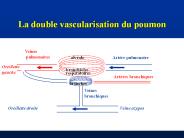
La double vascularisation du poumon Veines pulmonaires Art re pulmonaire alv ole Oreillette gauche bronchioles respiratoires Art res bronchiques bronches
| PowerPoint PPT presentation | free to download
Calyx. Renal Pelvis. Renal Hilus. Nephron. Glomerulus. Bowman's capsule. Proximal convoluted tubule ... Calyx. Renal pelvis. Ureter. Bladder. Urethra. Out of ...
| PowerPoint PPT presentation | free to view
Achalasia. Normal Oesophagus. Cancer of the Oesophagus. Type and Location of Tumours of Oesophagus ... Total gastrectomy and Roux-en-Y reconstruction ...
| PowerPoint PPT presentation | free to view
... giant cell carcinoma, choriocarcinoma, carcinomas arising in endometriosis, ... pTNM STAGING Ramifications (i.e., optional subdivisions of existing TNM ...
| PowerPoint PPT presentation | free to view
Tis Carcinoma in situ: intraepithelial or invasion of lamina propria ... Based on clinical data PLUS surgery and pathology report information ...
| PowerPoint PPT presentation | free to view
Disorders of the Digestive System. Inflammatory bowel disease. Inflammation of ... The Digestive System in Later Life. Middle age gallstones and ulcers ...
| PowerPoint PPT presentation | free to view
Title: automatisme de l'intestin et controle nerveux intrinseque et extrinseque Author: toutain 2005 Last modified by: Pierre-Louis TOUTAIN Created Date
| PowerPoint PPT presentation | free to view
Transpyloric : 9th costal cartilages. Transtubercular / Interspinous. Abdominal Regions ... General Disposition of Viscera. Digestive system, peritoneum ...
| PowerPoint PPT presentation | free to view
Histology Slides for the Urinary System Slides are presented in order of magnification As you view the following s make sure you can accomplish these goals:
| PowerPoint PPT presentation | free to download
Total gastrectomy and Roux-en-Y reconstruction. Thorascopic eosphagectomy ... Chyle leaks. The septic patient. DVT and PE. Medical comorbidity. Post Operative Leaks ...
| PowerPoint PPT presentation | free to view
Tutorial on Computational Optical Imaging
| PowerPoint PPT presentation | free to view
THE DIGESTIVE SYSTEM Chapter 19
| PowerPoint PPT presentation | free to view
Accessory digestive organs teeth, tongue, gallbladder, salivary glands, liver, and pancreas. Digestive System: Overview. Figure 23.1. Histology of the ...
| PowerPoint PPT presentation | free to view


