Photomicrograph PowerPoint PPT Presentations
All Time
Recommended
Photomicrograph of illite, showing fibrous structure. Photomicrograph of Attapulgite ... SEM Photomicrograph of Montmorillonite. SEM of Illite. PREVIOUS. SEM ...
| PowerPoint PPT presentation | free to view
Photomicrographs of pancreatic islets ... Photomicrograph of islet grafts from Tg (-) littermates ... Photomicrographs of islet grafts from H-AS GAD NOD mice ...
| PowerPoint PPT presentation | free to view
TEM PHOTOMICROGRAPHS OF AEROSOL DERIVED NANOPARTICLES. SILICA PARTICLES ... TEM PHOTOMICROGRAPH OF AEROSOL DERIVED MULLITE. 100 nm. MULLITE PARTICLES. 9/4/09 ...
| PowerPoint PPT presentation | free to view
"Copy link here : good.readbooks.link/pwr/1496316762 Master histology with idealized and actual photomicrography!This thirteenth edition of Atlas of Histology with Functional Correlations (formerly diFiore’s ) provides a rich understanding of the basic histology concepts that medical and allied health students need to know. Realistic, full-color illustrations as well as actual photomicrographs of histologic structures are complemented by concise discussions of their most important functional c"
| PowerPoint PPT presentation | free to download
Photomicrograph of blood cells in a hypotonic solution; the ... Photomicrograph of blood cells in an isotonic solution. Cells in Hypotonic Solution ...
| PowerPoint PPT presentation | free to view
On a photomicrograph, draw straight lines all the same in length and count the ... can then be calculated by using the magnification of the photomicrograph. ...
| PowerPoint PPT presentation | free to view
(3) this photomicrograph represents the equilibrium microstructure at 180 C (355 F) ... Probably not exact as 20% from photomicrograph was estimation ...
| PowerPoint PPT presentation | free to view
A photomicrograph of a polycrystalline specimen exhibiting these characteristics ... (b) Photomicrograph of the surface of a polished and etched polycrystalline ...
| PowerPoint PPT presentation | free to download
Photomicrograph of a corner of an EEV CCD. Structure of a CCD 6. OD. OS ... Photomicrograph of the on-chip amplifier of a Tektronix CCD and its circuit diagram. ...
| PowerPoint PPT presentation | free to view
On the basis of the photomicrograph for the lead-tin alloy shown in Figure 10.15 ... Can use a photomicrograph of an alloy to estimate its composition simply by ...
| PowerPoint PPT presentation | free to view
... (e.g., cryptospiridium, giardia), or has chemical water ... Cryptosporidium, Giardia, HPC, Legionella, Viruses ... Photomicrographs of Giardia cysts: ...
| PowerPoint PPT presentation | free to view
Salientia (Frogs and Toads) Caudata (Salamanders and Newts) Gymnophiona (Caecilians) ... Frog Inner Ear. Photomicrographs. Rana pipiens tympanic membrane ...
| PowerPoint PPT presentation | free to view
Charge Coupled Devices (CCDs) were invented in October 19, ... Photomicrograph of a corner of an EEV CCD. Edge of. Silicon. 160mm. Image Area. Serial Register ...
| PowerPoint PPT presentation | free to download
Photomicrograph of Mount Saint Helens Dome Rock ... .gov/Volcanoes/MSH/Eruption04/Petrology/dome_rock_comparison_1986_to_2004.h tml ...
| PowerPoint PPT presentation | free to view
Figure 2 is a nissl-stained photomicrograph showing the subnuclei of the pig PVN. Figure 3 is a photomicrograph of a Nissl-stained section of the PVN. ...
| PowerPoint PPT presentation | free to view
High-power photomicrograph can show the conidiophores with the characteristic ... Eosinophilic inflamatory infiltrate and fibrosis with no tissue or vascular ...
| PowerPoint PPT presentation | free to view
hormones released into the bloodstream travel throughout the body ... Photomicrograph of Thyroid Gland. 18-42. Formation of Thyroid Hormone ...
| PowerPoint PPT presentation | free to view
Photomicrograph provided by Gordon J. King. Placental ... newborn will get these antibodies from the first milk - Colostrum. Allows storage in allantoic ...
| PowerPoint PPT presentation | free to view
combines 3 important processes: , because blood vessels are open; ... This photomicrograph shows several thin-walled capillary blood vessels growing ...
| PowerPoint PPT presentation | free to view
DNA (yellow) was stained with Hoechst 33342. Note how rapid is the collapse of ... This photomicrograph clearly shows that the green-fluorescent lectin staining ...
| PowerPoint PPT presentation | free to download
???? : ??? ?? Dr.Shih-Chieh Chen. ???? : ??? Chun-Chih Liu. ??? ... Photomicrograph Taken by. Department of anatomy, Kaohsiung Medical University. Nasal septum ...
| PowerPoint PPT presentation | free to view
host-defense response to invading substance; ... This low power photomicrograph shows numerous discrete, uniformly sized, round ...
| PowerPoint PPT presentation | free to view
Photomicrograph of sodium chloride crystallizing from a stock ... Photomicrograph of .15 M NaCl solution to which 10 mg/mL Bovine Serum Albumin has been added. ...
| PowerPoint PPT presentation | free to download
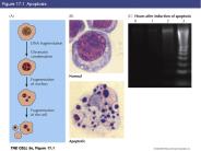
Figure 17.1 Apoptosis Figure 17.2 Phagocytosis of apoptotic cells Key Experiment 17.1: Photomicrographs of a normal worm (A) and a ced-3 mutant (B) Figure 17.3 ...
| PowerPoint PPT presentation | free to download
Explain how structure affects the general mechanical and optical ... Explain the glass transition temperature, factors affecting it, ... Photomicrograph ...
| PowerPoint PPT presentation | free to view
The dark color of the metastases is due to associated hemorrhage. Right panel: Photomicrograph of one of the brain metastases shows a poorly differentiated, ...
| PowerPoint PPT presentation | free to download
HEREDITY THE CONTINUITY OF TRAITS FROM GENERATION TO GENERATION ... KARYOTYPE PHOTOMICROGRAPH OF AN INDIVIDUALS SOMATIC CELLS CHROMOSOMES (DURING METAPHASE) ...
| PowerPoint PPT presentation | free to view
The bacterial cell wall is a unique structure which surrounds the cell membrane. ... This photomicrograph demonstrates the positive AFB staining characteristics of ...
| PowerPoint PPT presentation | free to view
Hermit the NEWT. Photomicrographs of living and. dividing new lung cell. ... Two centrosome forming radial arrays of microtubules (newt lung cell) right. ...
| PowerPoint PPT presentation | free to view
provides context for recent and anthropogenic climate effects ... photomicrograph of 'thin section' Three Dominant Surface Rocks: Basalt ...
| PowerPoint PPT presentation | free to view
"Copy Link : gooread.fileunlimited.club/pwjul24/1451187424 Histology: A Text and Atlas: With Correlated Cell and Molecular Biology 7th Edition Now in its seventh edition, Histology: A Text and Atlas is ideal for medical, dental, health professions, and undergraduate biology and cell biology students. This best-selling combination text and atlas includes a detailed textbook, which emphasizes clinical and functional correlates of histology fully supplemented by vividly informative illustrations and photomicrographs. Separate, superbly illustrated atlas sections follow almost every chapter and feature large-size, full-color digital photomicrographs with labels and accompanied descriptions that highlight structural and functional details of cells, tissues, and organs.Updated throughout to reflect the latest advances in the field, this &two in one& text and atlas features an outstanding art program with all illustrations completely r"
| PowerPoint PPT presentation | free to download
Transmission of genes from parents to offspring results in similarities among ... Karyotype - A display or photomicrograph of an individual's somatic-cell ...
| PowerPoint PPT presentation | free to view
probability of incurring harm or loss. harm from the environment could include. injury ... Photomicrograph of Oscillatoria rubescens. Lake Washington regional plan ...
| PowerPoint PPT presentation | free to view
You can enter the database through the traditional abstracts ... Photograph; Photomicrograph. Photograph; Satellite Image. Photograph; Study Site Photograph ...
| PowerPoint PPT presentation | free to view
Heredity: Inheritance: transmission of traits from one ... Genes: coded information in hereditary units, given to offspring by ... Photomicrograph of ...
| PowerPoint PPT presentation | free to view
Virtual education at Iowa has been supported by: ... No change in student performance on photomicrograph or glass exams ...
| PowerPoint PPT presentation | free to view
The characterization of the chromosomal complement of an individual or a species, ... A photomicrograph of chromosomes arranged according to a standard classification. ...
| PowerPoint PPT presentation | free to view
Department for Evaluations, Standards and Training (DEST) Thursday ... www.hpa-standardmethods.org.uk. The original Dane photomicrograph Prof. Richard Tedder ...
| PowerPoint PPT presentation | free to view
Propagation delay function of load capacitance and resistance of ... Photomicrograph of early ECL Gate (1967) Digital Integrated Circuits Prentice Hall 1995 ...
| PowerPoint PPT presentation | free to view
DNA wrapped around protein cores (histones) and coiled further into a rod-shaped ... A karyotype is a photomicrograph of the chromosomes in a dividing cell ...
| PowerPoint PPT presentation | free to view
lasts for a few hours, and is followed. by depression and anxiety. EFFECT of ... Dark-field photomicrograph, sagittal plane, of 5-HT immunoreactive axons in the ...
| PowerPoint PPT presentation | free to view
Beamline X26A experimental table. Photomicrograph of roaster oxide grain from Yellowknife showing differences in ... The nematodes detoxify lead through ...
| PowerPoint PPT presentation | free to view
associated with liver enzymes in the plasma. loss of liver ... The original Dane photomicrograph. 42nm Dane particles. 22nm HBsAg spheres and filaments ...
| PowerPoint PPT presentation | free to view
1) The Rock cycle Inter-relationships of Earth's parts ... Picture in page 98: Polarized-light photomicrograph of a thin section of gabbro ...
| PowerPoint PPT presentation | free to view
If the neuron fires, then something is learned. ... released by motor neurons at the muscle cells. Photomicrograph of the neuromuscular junction ...
| PowerPoint PPT presentation | free to download
Each of us began as a single cell, so one important question is: ... Photomicrograph of the chromosomes in a dividing cell found in a human ...
| PowerPoint PPT presentation | free to view
Carry genetic material which is copied and passed on to generations of cells ... A photomicrograph (fancy name for a picture taken by a microscope) of ...
| PowerPoint PPT presentation | free to view
Jerry M. Woodall. National Medal of Technology Laureate, ... EDX mode photomicrograph of 95-5 sample showing. In and Sn in the grain boundaries ...
| PowerPoint PPT presentation | free to view
During one of the major rodeo events of the year, and with thousands of fans in ... photomicrograph of. Bacillus anthracis bacteria. Image source: CDC ...
| PowerPoint PPT presentation | free to download
FIGURE 8-1 Initial events following fracture of a long bone diaphysis. ... B. A photomicrograph of a fractured rat femur three days after injury showing ...
| PowerPoint PPT presentation | free to view
Chair, Virginia Marsh resigned and Anne Raby took over the responsibility of the ... Photomicrograph showing peripheral blood neutrophilia with left-shift, including ...
| PowerPoint PPT presentation | free to view
Humans have 46 (diploid number/2n) in all cells except sex cells ... Autosomes all other chromosomes. Karyotype photomicrograph of chromosomes ...
| PowerPoint PPT presentation | free to view
The microscopes you are most familiar with are called light microscopes or ... Know the difference between a Photomicrograph and a Microphotograph! 4.2- Cell size. ...
| PowerPoint PPT presentation | free to view
DNA is a long, thin molecule that stores genetic information. ... Karyotype. Photomicrograph of the chromosomes in a normal dividing cell found in a human ...
| PowerPoint PPT presentation | free to view
18 years of experience building and managing 20 National and International R&D ... Photomicrograph of MMC produced from alloy 13 and 7 micron diameter alumina ...
| PowerPoint PPT presentation | free to view
NTU GIEE Lab 405. 1. Design of a 10-Bit 55MS/s Analog to Digital ... Die Photomicrograph. NTU GIEE Lab 405. 6. Pre-Layout Simulation. Fs=55Mhz and Fin=5.425Khz ...
| PowerPoint PPT presentation | free to view


