Retin A 05 PowerPoint PPT Presentations
All Time
Recommended
An eye healthcare facility defines the retina as the innermost light-sensitive membrane covering the back wall of the eyeball. When light strikes the retina, photoreceptors transform the light into electrical signals. The electrical signals go from your retina to your brain via the optic nerve.
| PowerPoint PPT presentation | free to download
Eye is made up of Iris, Pupil, Cornea and Retina. The retina is an extremely thin tissue that lines the inside of the back of the eye. It is the light-sensitive portion of the eye. Light from the objects we are looking at, enters the eye. Cornea and the eye lens focus the light image onto the retina. Human eye works like a camera, light striking the retina causes a complex biochemical change within certain layers of the retina and this, in turn, stimulates an electrical response within other layers of the retina.
| PowerPoint PPT presentation | free to download
Data with 9 visual field around the fovea are obtained. ... Blood vessels in the fovea have low contrast in all the polarization properties. ...
| PowerPoint PPT presentation | free to view
Retina. Dr.Nupur Dr. Shruti LAQs. RETINOBLASTOMA. Definition, inheritance, Pathology and clinical stages treatment. Differential diagnosis. 2)Retinal detachment.
| PowerPoint PPT presentation | free to view
Retinal detachment is the retina is the light sensitive film at the back of the eye and retinal detachment is a condition where the retina peels away from the inner wall of the eye.
| PowerPoint PPT presentation | free to download
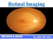
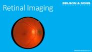
Retinal imaging is useful for photographing inside of the eye, particularly the retina. This is one of the core clinical service from opticians in london which provides a permanent record of the appearance of the eye.
| PowerPoint PPT presentation | free to download
Retinal imaging or retinal photography is a digital picture of back of your eye. It provides permanent record of the appearance of the eye which is helpful to identify the abnormalities of eyes. We at Belson & Sons provides you all the eye care facilities in London and Essex.
| PowerPoint PPT presentation | free to download
By: Nassar Nassar
| PowerPoint PPT presentation | free to download
Title: No Slide Title Author: Judie Walter Last modified by: Office 98 Created Date: 10/21/1999 4:40:40 PM Document presentation format: On-screen Show
| PowerPoint PPT presentation | free to download
fovea. photoreceptors sample the retinal image. aliasing. January 21, 2004. PSY280 - Hamstra ... fields are small, distant field (far from fovea) are large ...
| PowerPoint PPT presentation | free to view
Retinal network. Parallel Pathways- retina to cortex. Contrast sensitivity. Primate visual system cartoon. Human retina section. Retinal network. Bipolar cell types ...
| PowerPoint PPT presentation | free to view
Neovascularisation scatter laser CRVO Ischemic Non-ischemic CRVO Ischemic Non-ischemic Non-ischemic CRVO (Course and Follow-up) Residual signs: Disc ...
| PowerPoint PPT presentation | free to download
The growing prevalence of target diseases, the aging population, the increasing ability to restore vision, rising disposable income in developing countries, rising healthcare costs, and the prolonged use of retinal implants that eliminates the need for additional surgery are all expected to fuel the growth of the retinal implant market in the coming years. However, throughout the projected period, the market expansion is anticipated to be constrained by the absence of medical reimbursements, biocompatibility, and long-term stability of the material utilized to create retinal implants.
| PowerPoint PPT presentation | free to download
Normal and Pathological Images captured from IntuCAM 45
| PowerPoint PPT presentation | free to download
Get Free Report Sample here:- https://bit.ly/2O6DGrg Retinal Disorder Treatment Market report focuses on the global Retinal Disorder Treatment status, future forecast, growth opportunity, key market and key players.
| PowerPoint PPT presentation | free to download
Acromegaly: IgF-1 (the HbA1c of growth hormone) Unusual Cases of Diabetes ... Untreated Acromegaly, significant cardiovascular risk, joint problems, bowel cancer ...
| PowerPoint PPT presentation | free to view
Contact us for appointment or enquiries about lens Implant and Retinal surgery with Dr Vasant Raman in Plymouth.
| PowerPoint PPT presentation | free to download
Title: PowerPoint Presentation Last modified by: USER Created Date: 1/1/1601 12:00:00 AM Document presentation format: On-screen Show Other titles
| PowerPoint PPT presentation | free to view
According to Straits Research, the global retinal imaging devices market is projected to attain a value of USD 3,605 million by the end of 2031 at a CAGR of 5.6%.https://straitsresearch.com/report/retinal-imaging-devices-market/request-sample
| PowerPoint PPT presentation | free to download
According to Straits Research, the global retinal imaging devices market is projected to attain a value of USD 3,605 million by the end of 2031 at a CAGR of 5.6%.https://straitsresearch.com/report/retinal-imaging-devices-market/request-sample
| PowerPoint PPT presentation | free to download
The retina is a layer of tissue within the back of your eye that senses light and sends images to your brain. within the center of this nervous tissue is that the macula. It provides the sharp, visual sense needed for reading, driving and seeing fine detail.
| PowerPoint PPT presentation | free to download
Bharat Book presents the report on “Retinal Vein Occlusion - Pipeline Review” (https://www.bharatbook.com/healthcare-market-research-reports-216410/retinal-vein-occlusion3.html). This report provides customers analysis including current suppliers, procurement prices & quantity being purchased annually.
| PowerPoint PPT presentation | free to download
The retina is a layer of tissue within the back of your eye that senses light and sends images to your brain. within the center of this nervous tissue is that the macula. It provides the sharp, visual sense needed for reading, driving and seeing fine detail.
| PowerPoint PPT presentation | free to download
Retinal vein occlusion market is expected to gain market growth in the forecast period of 2022-2029. Data Bridge Market Research analyses the market to account to grow at a CAGR of 10.70% in the above mentioned forecast period and is likely to reach USD 27,297.9 million by 2029. https://www.databridgemarketresearch.com/request-a-sample/?dbmr=global-retinal-vein-occlusion-market
| PowerPoint PPT presentation | free to download
Retinal Vein Occlusions Morphology CRVO BRVO Hemispheric VO Hemicentral VO Papillophlebitis Macular BRVO CENTRAL RETINAL VEIN OCCLUSION The actual mechanisms ...
| PowerPoint PPT presentation | free to download
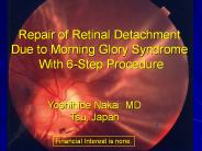
Repair of Retinal Detachment Due to Morning Glory Syndrome With 6-Step Procedure Yoshihide Nakai. MD Tsu, Japan Financial Interest is none. A 51-year-old female.
| PowerPoint PPT presentation | free to download
... components of our prosthesis are assembled, and the stimulating electrode arrays ... Si wafers serve as carriers for these freestanding films during processing. ...
| PowerPoint PPT presentation | free to view
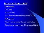
RETINAL VEIN OCCLUSION Epidemiology 51% 65y 10-15%
| PowerPoint PPT presentation | free to download
... Nerve Head that would aid an Ophthalmic clinician in the diagnosis of a patient. ... versus Joint Segmentation for Region Based Image Fusion' J. J. Lewis et ...
| PowerPoint PPT presentation | free to view
The intravenous fluorescein angiogram pattern of an ischemic central retinal ... with the extent of capillary nonperfusion on the fluorescein angiogram. ...
| PowerPoint PPT presentation | free to view
"14 minutes ago - COPY LINK TO DOWNLOAD : flip.ebookmarket.pro/pwjul24/0323930433 | [READ DOWNLOAD] Atlas of Retinal OCT: Optical Coherence Tomography | Atlas of Retinal OCT: Optical Coherence Tomography "
| PowerPoint PPT presentation | free to download
Retinal venous and arterial occlusions are among the more common serious ophthalmic conditions presenting acutely. Should be always considered in the DDx of a pt with ...
| PowerPoint PPT presentation | free to download
Retinal diseases can lead to vision loss and impairment, and retinal drugs aim to address or manage these conditions to prevent further deterioration of vision. AMD is a common retinal disease that affects the macula, the central part of the retina responsible for detailed vision. It is a leading cause of vision loss in older adults. This condition occurs in people with diabetes and can cause damage to blood vessels in the retina, leading to vision problems and, in severe cases, blindness. RVO is a blockage of the veins that drain blood from the retina, causing vision problems due to reduced blood flow. Click here for more information: https://www.htfmarketintelligence.com/report/global-retinal-drugs-market
| PowerPoint PPT presentation | free to download
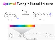
Spectral Tuning in Retinal Proteins hn all-trans 11-cis
| PowerPoint PPT presentation | free to download
Patients with diabetes today have a lot of advantages thanks to the many modern medicines which help them to minimize their condition and live their life in a healthy and happy manner.For more information visit: http://www.opticareoptician.co.uk/eye-care/diabetic-retinal-screening/
| PowerPoint PPT presentation | free to download
In order to register the user who wants to use the programme, the project Detection of Retinal Pigmentosa in Paediatric Age Patients combines deep learning with MySQL.
| PowerPoint PPT presentation | free to download
Shree Ganesh Netralaya is best Retina Eye Specialist Hospital in Indore offering best Retinal treatment for Retinal Detachment, Retinal holes and tears, Macular Hole
| PowerPoint PPT presentation | free to download
Similarity measure between images based on entropy: ... Freedman-Diaconis: 2 * IQR * n-1/3 ... Using freedman-diaconis bin size: At the incorrect registration: ...
| PowerPoint PPT presentation | free to view
1. Eye Health – Caring for your Retina. 2. Diabetes and the prevention of Retinal Problems. 3. Retina Problems can be Associated with Age. 4. Latest Advances in Retina Treatments for Vision Loss.
| PowerPoint PPT presentation | free to download
Structure-Function Relationship of Retinal Proteins
| PowerPoint PPT presentation | free to download
Neural Integrity Retinal Assessment (NIRA ) Problem Photography of the retina for the follow up of diabetic retinopathy is a standard test for advanced changes that ...
| PowerPoint PPT presentation | free to download
Discover the most prevalent retinal disorders affecting adults that can lead to irreversible damage. Best Eye Doctor Arcadia provides expert explanations, diagnosis, and management of common retinal conditions and their symptoms.
| PowerPoint PPT presentation | free to download
What is Virtual Retinal Display Basics of VRD How Work VRD VRD System Overview Key Features Advantages Disadvantages Application Future Scope Conclusion
| PowerPoint PPT presentation | free to download
Retinal receptors (actually receptive fields) require constantly changing stimulus ... Some crystals (e.g. tourmaline) freely transmit light along one crystal axis and ...
| PowerPoint PPT presentation | free to view
Browse - Market Tables, Figures and an in-depth TOC on "Virtual Retinal Display Market". http://industryarc.com/Report/15035/virtual-retinal-display-market.html
| PowerPoint PPT presentation | free to download
1. Take care of your eyes and they will last forever 2. When our general health affects our eyes 3. Ophthalmology Examinations are Important
| PowerPoint PPT presentation | free to download
"China Vitreo Retinal Surgery Devices Market Outlook to 2022", provides key market data on the China Vitreo Retinal Surgery Devices market. The report provides value, in millions of US dollars, volume (in units) and average prices (USD) within market segements - Vitreo Retinal Surgery Packs, Vitreo Retinal Prefilled Silicone Oil Syringes and Vitreo Retinal Per Fluro Carbon Liquids (PFCL).
| PowerPoint PPT presentation | free to download
Retina is a layer of tissue at the back of the eye, which helps to see images focused on it by the cornea and lens. Retinal Detachment is an eye disorder, wherein the retina gets separated from the underlying layer of blood vessels, which supplies oxygen and other nutrients to it.
| PowerPoint PPT presentation | free to download
Entoptic Phenomena of Retinal Origin Page 2.36 Retinal Adaptation The Troxler Effect The Troxler Effect The Troxler Effect Retinal Adaptation Entoptic Perception of ...
| PowerPoint PPT presentation | free to view
DACH (dachshund) Has coiled-coil domain, probably dimerises; shown to interact with eya. ... DACH: dachshund = mouse/human Dach1, Dach2. A conserved team at ...
| PowerPoint PPT presentation | free to view
Protect your rental health with a dedicated optometrist in Escondido. The experts will discuss causes, and symptoms, and provide cutting-edge solutions promptly.
| PowerPoint PPT presentation | free to download
RETINAL BLOOD VESSEL EXTRACTION (SEGMENTATION) Available Image Databases DRIVE and STARE databases are available for the public.
| PowerPoint PPT presentation | free to download
The global retinal imaging devices market is growing at a CAGR of 5.93% and is expected to reach $9322.89 million, during the forecast period of 2023-2032.
| PowerPoint PPT presentation | free to download
Discover effective strategies for managing, preventing & treating Retinal Detachment. Learn how you can utilize the expertise of eye doctors Palm Desert team for timely treatment and optimal outcomes to see clearly for a long time.
| PowerPoint PPT presentation | free to download
Retinal Surgery Device Market will be US$ 2.87 Billion by 2027. Global Forecast, Impact of COVID-19, Industry Trends, by Product, Application, Growth, Opportunity Company Analysis.
| PowerPoint PPT presentation | free to download


