Retinal Imaging PowerPoint PPT Presentations
All Time
Recommended
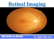
Retinal imaging is useful for photographing inside of the eye, particularly the retina. This is one of the core clinical service from opticians in london which provides a permanent record of the appearance of the eye.
| PowerPoint PPT presentation | free to download
Retinal imaging or retinal photography is a digital picture of back of your eye. It provides permanent record of the appearance of the eye which is helpful to identify the abnormalities of eyes. We at Belson & Sons provides you all the eye care facilities in London and Essex.
| PowerPoint PPT presentation | free to download
Normal and Pathological Images captured from IntuCAM 45
| PowerPoint PPT presentation | free to download
According to Straits Research, the global retinal imaging devices market is projected to attain a value of USD 3,605 million by the end of 2031 at a CAGR of 5.6%.https://straitsresearch.com/report/retinal-imaging-devices-market/request-sample
| PowerPoint PPT presentation | free to download
According to Straits Research, the global retinal imaging devices market is projected to attain a value of USD 3,605 million by the end of 2031 at a CAGR of 5.6%.https://straitsresearch.com/report/retinal-imaging-devices-market/request-sample
| PowerPoint PPT presentation | free to download
... Nerve Head that would aid an Ophthalmic clinician in the diagnosis of a patient. ... versus Joint Segmentation for Region Based Image Fusion' J. J. Lewis et ...
| PowerPoint PPT presentation | free to view
Similarity measure between images based on entropy: ... Freedman-Diaconis: 2 * IQR * n-1/3 ... Using freedman-diaconis bin size: At the incorrect registration: ...
| PowerPoint PPT presentation | free to view
Safety aspects of Fluorescein Angiography ... vascular disorders) Stereo Imaging Indo Cyanine Green Angiography Optical Coherence Tomography Electro Diagnostic ...
| PowerPoint PPT presentation | free to download
Compare proximal stimulus with commands to muscles... Did I tell myself to move? ambiguity if I'm being moved passively. ambiguity if we're both moving ...
| PowerPoint PPT presentation | free to view
The global retinal imaging devices market is growing at a CAGR of 5.93% and is expected to reach $9322.89 million, during the forecast period of 2023-2032.
| PowerPoint PPT presentation | free to download
The global retinal imaging devices market is growing at a CAGR of 5.93% and is expected to reach $9322.89 million, during the forecast period of 2023-2032.
| PowerPoint PPT presentation | free to download
Isometropia - equal refractive errors OD and OS. Anisometropia ... vague and non-specific: asthenopia ('eye-strain'), headaches, photophobia, reading problems. ...
| PowerPoint PPT presentation | free to view
... Magnification ... Spectacle magnification produces a new incident chief ray angle at the eye (for ... Spectacle magnification in myopia decreases the incident chief ...
| PowerPoint PPT presentation | free to view
Active Contours Technique in Retinal Image. Identification of the ... Threshold: Optic disk corresponds to the ... Windowing: cropped image based on the ...
| PowerPoint PPT presentation | free to download
The retinal imaging devices market is projected to reach US$ 3.00 billion by 2028 from US$ 2.06 billion in 2021; it is estimated to grow at a CAGR of 5.6% during 2021–2028.
| PowerPoint PPT presentation | free to download
The Global Retinal Imaging Devices market research includes historical and forecast data, like demand, application details, price trends, and company shares of the leading Retinal Imaging Devices by geography, especially focuses on the key regions like United States, European Union, China, and other regions.
| PowerPoint PPT presentation | free to download
HRT (Heildelberg Retina Thomograph) is widely used for imaging and following ... Optimizers: controlled random search (CRS), Powell. ...
| PowerPoint PPT presentation | free to download
Automatic Detection of Blood Vessels in Digital Retinal Image using CVIP Tools Krishna Praveena Mandava Sri Swetha Kantamaneni Robert LeAnder
| PowerPoint PPT presentation | free to download
Impact of Field Selection in Nonmydriatic Retinal Imaging for Diabetic Retinopathy in the Joslin Vision Network Lloyd M. Aiello, MD; Sharon Eagan, OD; Nigel Timothy ...
| PowerPoint PPT presentation | free to view
... diagnoses has fallen from 75% to 60% All unfamiliar diagnoses are ... Ability to recognize unfamiliar diagnoses leads to degradation in system performance ...
| PowerPoint PPT presentation | free to view
Title: An Engineering Research Center for Integrated Sensing and Imaging Systems Author: Sysadmin Last modified by: Charles Stewart Created Date
| PowerPoint PPT presentation | free to download
Results and discussion. Conclusion. 3. Candidate region determination ... Results and discussion (1) Candidate region (2) Euclidian distance (3) Locate the optic disk ...
| PowerPoint PPT presentation | free to view
Affine transformations in 2d and 3d. Projective transformations ... Affine Transformations. Geometry: ... Affine Transformations in 2d. In 2d, multiply x ...
| PowerPoint PPT presentation | free to download
Office(JEC 7010): 518-276-8067, Lab(JEC 6308): 518-276-8207, Fax: 518-276-8715 Email: roysam@ecse.rpi.edu, Web: http://www.ecse.rpi.edu/~roysam Output Size: 1200 x 820
| PowerPoint PPT presentation | free to download
The Global Retinal Imaging Devices Market size is expected to reach $6.3 billion by 2025, rising at a market growth of 6.6% CAGR during the forecast period. Structural Insights: https://www.kbvresearch.com/retinal-imaging-devices-market/
| PowerPoint PPT presentation | free to download
Dr. Delia Cabrera dcabrera2@med.miami.edu. Value Added to CenSSIS. Optomap ... Special thanks to Dr. Delia Cabrera, from Bascom Palmer Eye Institute at the ...
| PowerPoint PPT presentation | free to view
Locating the Optic Nerve in Retinal Image Using the Fuzzy Convergence of the Blood Vessels ... The OD is EXACT the point of bloods come inside the retinal ! ...
| PowerPoint PPT presentation | free to view
Retinal Imaging Protocols for Constructing High Resolution Mosaics of In Vivo Photoreceptor Cells
| PowerPoint PPT presentation | free to view
Post-op MTF improved for small pupils (3-5mm) n=17 (23.6 ... FOZ defined with different MTF values would affect the absolute post-op FOZ size, ...
| PowerPoint PPT presentation | free to view
Data with 9 visual field around the fovea are obtained. ... Blood vessels in the fovea have low contrast in all the polarization properties. ...
| PowerPoint PPT presentation | free to view
Single field nonmydriatic 45-degree JVN field (NM-1) will permit Category 1 and ... Cavallerano AA, Cavallerano JD, Aiello LP, Aiello LM. ...
| PowerPoint PPT presentation | free to view
Light from this image is going to excite and inhibit the rods & cones. ... The Primate Lateral Geniculate Nucleus. Parvo-cells. small receptive fields ...
| PowerPoint PPT presentation | free to download
What does a pepperoni pizza taste like? What does a ... Try imaging Snoopy the dog and 'mentally staring' at his feet. Then judge the shape of his ears ...
| PowerPoint PPT presentation | free to view
fovea. photoreceptors sample the retinal image. aliasing. January 21, 2004. PSY280 - Hamstra ... fields are small, distant field (far from fovea) are large ...
| PowerPoint PPT presentation | free to view
Image Understanding Outline: Motivation Human vision and illusions Image representation Sampling Quantization Thresholding Motivation Human Vision Visual Illusions ...
| PowerPoint PPT presentation | free to download
Stereoscopic Images Binocular vision enables us to measure depth using eye convergence and stereoscopic vision. Eye convergence is a measure of the angle between the ...
| PowerPoint PPT presentation | free to view
Title: No Slide Title Author: Judie Walter Last modified by: Office 98 Created Date: 10/21/1999 4:40:40 PM Document presentation format: On-screen Show
| PowerPoint PPT presentation | free to download
Digital images are taken using a fundus camera. The procedure (taking the images) is painless. ... Retinal fundus camera operator ...
| PowerPoint PPT presentation | free to view
Global Widefield Imaging Systems Market will exhibit a CAGR of around 7.35% for the forecast period of 2022-2028. Widefield imaging systems are used in diagnosing and detecting uveitis, diabetic retinopathy, retinal vascular tumours among others. Widefield imaging systems offer excellent postoperative documentation of retinal surgery. Widefield imaging systems is an imaging technology that relies on illumination with the resulting image being viewed and observed by an observer through the camera. The widefield imaging systems are rapidly being adopted by hospitals, clinics, and ambulatory surgery centres. Get Full Access of Report @ https://www.databridgemarketresearch.com/reports/global-widefield-imaging-systems-market
| PowerPoint PPT presentation | free to download
RETINAL BLOOD VESSEL EXTRACTION (SEGMENTATION) Available Image Databases DRIVE and STARE databases are available for the public.
| PowerPoint PPT presentation | free to download
Retina is a layer of tissue at the back of the eye, which helps to see images focused on it by the cornea and lens. Retinal Detachment is an eye disorder, wherein the retina gets separated from the underlying layer of blood vessels, which supplies oxygen and other nutrients to it.
| PowerPoint PPT presentation | free to download
The retina is a layer of tissue within the back of your eye that senses light and sends images to your brain. within the center of this nervous tissue is that the macula. It provides the sharp, visual sense needed for reading, driving and seeing fine detail.
| PowerPoint PPT presentation | free to download
Bharat Book presents the report on “Retinal Vein Occlusion - Pipeline Review” (https://www.bharatbook.com/healthcare-market-research-reports-216410/retinal-vein-occlusion3.html). This report provides customers analysis including current suppliers, procurement prices & quantity being purchased annually.
| PowerPoint PPT presentation | free to download
The retina is a layer of tissue within the back of your eye that senses light and sends images to your brain. within the center of this nervous tissue is that the macula. It provides the sharp, visual sense needed for reading, driving and seeing fine detail.
| PowerPoint PPT presentation | free to download
Retinal Vein Occlusions Morphology CRVO BRVO Hemispheric VO Hemicentral VO Papillophlebitis Macular BRVO CENTRAL RETINAL VEIN OCCLUSION The actual mechanisms ...
| PowerPoint PPT presentation | free to download
Center for Bioimaging Informatics www.bioimage.ucsb.edu Supported by NSF ITR #0331697 UCSB Bio-imaging Infrastructure December 2006
| PowerPoint PPT presentation | free to download
This Report provided by GrandResearchStore is about , Digital Retinal Cameras in Global market, especially in North America, Europe, China, Japan, Southeast Asia and India, focuses on top manufacturers in global market, with production, price, revenue and market share for each manufacturer, covering Aeon Imaging ArcScan CW Optics Eyenuk
| PowerPoint PPT presentation | free to download
The fovea is at least 2 x DD from the edge of the retina. Number of Fields ... fovea must be visible. DRS Clinical Grading Sub-Group (Jan 2006) Image Quality (2) ...
| PowerPoint PPT presentation | free to view
Retina is a layer of tissue at the back of the eye, which helps to see images focused on it by the cornea and lens. Retinal Detachment is an eye disorder, wherein the retina gets separated from the underlying layer of blood vessels, which supplies oxygen and other nutrients to it. When the retina gets detached, the supply of oxygen and nutrients are stopped. If the condition is left untreated, it may even lead to a complete vision loss and blindness. To know more visit here: www.lazoi.com
| PowerPoint PPT presentation | free to download
The intravenous fluorescein angiogram pattern of an ischemic central retinal ... with the extent of capillary nonperfusion on the fluorescein angiogram. ...
| PowerPoint PPT presentation | free to view
... measurement of changes in retinal vessel diameter in ocular fundus images', Patt. ... registration algorithm of eye fundus images using vessels detection ...
| PowerPoint PPT presentation | free to view
Optic nerve carries digital' signals to the brain. Biomimetic Circuits ... Conventional cameras are at best able to perform global automatic gain control. ...
| PowerPoint PPT presentation | free to view
Seeing 3D from 2D Images How to make a 2D image appear as 3D! Output is typically 2D Images Yet we want to show a 3D world! How can we do this?
| PowerPoint PPT presentation | free to download
Applications of Image Processing
| PowerPoint PPT presentation | free to view
Real Numbers, Real Images
| PowerPoint PPT presentation | free to view

