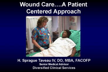
Wound Care'A Patient Centered Approach PowerPoint PPT Presentation
1 / 94
Title: Wound Care'A Patient Centered Approach
1
Wound Care.A Patient Centered Approach
H. Sprague Taveau IV, DO, MBA, FACOFP Senior
Medical Advisor Diversified Clinical Services
2
Faculty Disclosure H. Sprague Taveau IV, DO, MBA
- It is the policy of Diversified Clinical
Services to ensure balance, independence,
objectivity, and scientific rigor in all of its
individually sponsored or jointly sponsored
educational programs. - H. Sprague Taveau IV, DO, MBA, FACOFP is the
Senior Medical Advisor for Diversified Clinical
Services. He owns no stock nor does he have a
financial interest in any products or services
that are being presented in this program. - REGRETABLY!!!
3
- Goals Establish a universally applicable
approach to wound patient assessment - Objectives
- Acquire a systematic plan for wound patient
assessment - Know the assessment steps for wound evaluation,
description and identification of local factors
affecting wound healing - Know the assessment steps for wound patient
evaluation and identification of systemic factors
affecting wound healing - Know the indications for secondary testing in
wound patient evaluation
4
Wound Healing Failure
Infection
Ischemia Hypoxia
Chronic Inflammation
Cellular Failure
Recurrent Injury
5
Philosophy of wound patient assessment
- Identify an ulcer/wound etiology or diagnostic
category - Identify the final common pathways to healing
failure acting in this patient which enables
setting key therapeutic goals for management - Identify the presence of co-morbidities that
might impact the selection of treatment options
or other aspects of patient care.
6
Step 1 Obtain a complete history of the wound
- Date of occurrence, duration
- Etiologytrauma, pressure, surgery, infection,
etc. - Ameliorating and exacerbating factors
- Evaluations previously completed
- Treatments previously administered
- Impact on the patient
7
Step 2 Obtain a complete review of systems
- To elicit other important information about wound
etiology - To identify co-morbidities that might impact
wound healing or selection of treatment
interventions - Define factors that might affect treatment
options - Define general health status, activity level
8
Step 2 Obtain a complete review of systems
- All of the information obtained in Step 2 becomes
vitally important in Step 7.
9
Grading Ambulatory Status
- Grade VI Unlimited community ambulator
- Grade V Limited community ambulator
- Grade IV Unlimited household ambulator
- Grade III Limited household ambulator
- Grade II Supervised household ambulator
- Grade I Wheelchair ambulator
- Grade 0 Bedridden
Volpicelli LJ, Chambers RB, Wagner FW. J BONE
JOINT SURG 1983, 65-A599-605.
10
Step 3 Determine the location of the wound
- Location can point us to specific essential
aspects of treatment independent of the specific
etiology
11
Step 3 Determine the location of the wound
- Is the wound on the lower extremity?
- If yes, can be diabetic, neuropathic, primary
arterial, primary venous, inflammatory,
malignant, infectious, etc. - Location can be an important factor in
differential diagnosis of wound etiology
12
Step 3 Determine the location of the wound
- Is the wound on the lower extremity?
- If yes, be sure to complete a full vascular
assessment - Arterial
- Venous
- If yes, also complete a neurosensory/muscular
status assessment
13
Step 3 Determine the location of the wound
- Is the wound on the lower extremity?
- If no, is the wound over a bony prominence in
contact with a support surface? - If yes, consider the most likely diagnosis is a
pressure ulcer
14
Step 5 Assess the woundMEASURE
M Measure E Exudate A Appearance (wound
bed and surrounding skin) S Suffering U
Undermining R Reevaluation E Edge
Keast DH, et al. MEASURE A proposed assessment
framework for developing best practice
recommendations for wound assessment. Wound Rep
Reg 2004 12S1-S17.
15
Step 5 Assess the wound and wound patient
- Appropriate ancillary testing to assess the
wound - Culture
- Histopathology
- Blood work
- Diagnostic radiology
- Appropriate ancillary testing to define the
significance and impact of co-morbidities - ECG, CXR
- Blood work
- Consultations
16
Assess for Loss of Protective Sensation
17
Step 5 Assess the wound
- Define the impact of the wound on the patient
- Suffering Pain
- Severity and Complexity
18
(No Transcript)
19
(No Transcript)
20
Step 6 Identify the final common pathways acting
to cause wound healing failure
- Infection
- Malperfusion/hypoxia
- Cellular failure
- Unrelieved pressure/trauma
- Pathological inflammation
21
Step 7 Identify the co-morbidities impacting
wound healing and wound care
- Diabetes mellitus
- ESRD/dialysis
- Edema (as secondary etiology)
- CHF
- Chronic arterial insufficiency (as secondary
etiology) - Mobility impairment/CVA/cord injury
- Smoking
- COPD
- Vasculitis, Raynauds, other collagen vascular
disease - Wound contamination including incontinence
- Steroid therapy
- Chemotherapy
- Distant malignancy
- Malnutrition
22
Step 8 Define the key therapeutic objectives
- Resolution of infection and maintenance of
bacterial balance - Enhancement of perfusion and oxygenation
- Resolution of edema
- Relief of pressure
- Ambulatory off loading
- Mechanical stabilization
- Enhancement of tissue growth
- Pain control
- Preservation of function
- Exudate control and wound moisture balance
- Odor control
- Patient/caregiver education
- Patient compliance
23
(No Transcript)
24
Fundamental Questions
- Is the wound healable?
- Is the patient optimized to support healing?
- Nutrition
- Glycemic control
- Metabolic control
- Is the wound optimized to support
healing/response to local interventions? - How important is time?
- DFUrisk of osteomyelitis, progression
- Preserving, restoring function
- How compliant is the patient/family?
- Be cognizant of complications of technology
25
Step 9 Select the most appropriate treatment
plan (CPG) for management and the specific
interventions appropriate for this patient.
26
Wound Diagnosis or Type
- Diabetic ulcer
- Arterial insufficiency ulcer
- Venous leg ulcer
- Pressure ulcer
- Surgical wound/dehiscence/failing flap or graft
- Progressive soft tissue infection/osteomyelitis
- Laceration/acute traumatic injury/crush injury
- Burn
- Abrasion/skin tear
- Contact dermatitis
- Dermatological conditions/rash
- Vasculitis ulcer
- Radiation wound
- Stoma wound
- Other
27
Step 10 Monitor the response to therapy and
adjust as needed
- Follow a subset of the steps used in the initial
evaluation - Failure to make consistent clinical progress
should prompt a thorough re-evaluation and a
change in the treatment plan
28
(No Transcript)
29
Essential step 1 Adequate perfusion?
If you dont get water to the garden, the garden
wont grow!!
30
Essential step 2 Non viable tissue?
Wounds wont heal in a sewer!!
31
Essential step 3 Inflammation or infection?
Wounds with BUGS dont heal!!
32
Essential step 4 Edema?
Wounds dont heal in a swamp!!
33
Essential step 5 Wound microenvironment
conducive to healing?
34
Essential step 6 Tissue growth optimized?
35
Essential step 7 Offloading, pressure relief?
Wounds dont heal under pressure!!
36
Essential step 8 Pain controlled?
37
Essential step 9 Host factors optimized?
Wounds dont heal without building blocks!!
38
Steps in the Evaluation of the Problem
WoundOverview of Assessment
Recap
- Identify the wound etiology
- Define the extent and character of the wound
- Identify the specific pathophysiology of wound
healing failure Final common pathways - Identify co-morbidities impacting wound healing
or treatment options - Develop a treatment plan addressing all of the
above
39
(No Transcript)
40
Goals Objectives
- Goal
- To gain an appreciation for the indications for
treating patients with HBO. - Objectives
- To familiarize you with emergent indications
- To familiarize you with non-emergent indications
- To familiarize you with contra-indications
- Relative
- Absolute
41
Emergency/Acute Indications
- Cerebral Arterial Air or Gas Embolism
- Carbon Monoxide Poisoning
- Cyanide Poisoning
- Hydrogen Sulfide Poisoning
- Clostridial Myositis Myonecrosis
- Acute Traumatic Ischemia
- Crush Injury
- Compartment Syndrome
- Replantation Limb/Digits Etc.
42
Emergency/Acute Indications
- Decompression Sickness
- Exceptional Blood Loss (Anemia)
- Intracranial Abscess
- Necrotizing Soft Tissue Infections
- Thermal Burns
- Combined Synergistic Necrotizing STI
- Compromised Skin Grafts/Flaps
43
Acute Traumatic Ischemia
- 4 year old slipped and fell into a riding lawn
mower, sustaining a mid-calf amputation of his
leg. Leg was successfully replanted. - Ischemic time 10 hours
- Txd aggressively with HBO
44
Acute Traumatic Ischemia
- Appearance of muscle three days after
replantation shows 100 viability as HBO
counteracted reperfusion injury.
45
Acute Traumatic Ischemia
- Three Months after Injury
- HBO _at_ 2.4 ATA x 90 minutes q8h x 6
- Then q12h x 4
46
Acute Traumatic Ischemia
- The result was excellent function of the leg.
The patient regenerated his nerves and ended up
with a sensate foot. He was able to walk and run
with the aid of a brace.
47
Crush Injury
- Crush Injury with avulsion of palmar skin
- Appearance at time of presentation 1 hour after
injury
48
Crush Injury
- Elevation of avulsed palmar skin of crushed right
hand
49
Crush Injury
- Immediate post-op view
- Note vertical blue line through mid-palm
- Area not expected to survive
50
Crush Injury
- 11 weeks post injury
- HBO _at_ 2 ATA x 90 minutes q8h x 3 then q24h x 17
51
Crush Injury
- 11 weeks post injury
- Full range of motion
52
Non-approved Emergent Indications
- Retinal Artery Insufficiency
- Actinomycosis
53
Chronic/Elective Indications
- Problem Wounds
- Diabetic Foot Ulcers (Chronic Wagner III)
- Arteriolar Insufficiency
- Etc.
- Chronic Refractory Osteomyelitis
- Delayed Radiation Injury
- Soft Tissue
- Bony
- Meleny Ulcer (Invasive Group A Strep)
54
Age associated differences in cellular
Proliferation (in vitro)
New born
Young adult
Old adult
(Buras and Buras, Harvard Medical School, MGH,
Boston)
55
Decreased cellular proliferation with diabetes
(Buras and Buras, Harvard Medical School, MGH,
Boston)
56
HBO Dramatically Increases Old Adult Fibroblast
Proliferation
(Buras and Buras, Harvard Medical School, MGH,
Boston)
57
HBO Dramatically Increases Diabetic Fibroblast
Proliferation
(Buras and Buras, Harvard Medical School, MGH,
Boston)
58
TcpO2 As A Predictor of Wound Healing in Diabetic
Foot Wounds
Initial healing success
Initial healing failure
Pecoraro, et al. Diabetes 401305-1313, 1991
59
(No Transcript)
60
(No Transcript)
61
2 hours pre2 hours post
What About Smoking?
62
Smoking Effects on Benefit from HBO
The avg pt with gt 10 pk/yrs who benefitted from
HBOT needed 8-14 more HBO treatments than a non
smoker for the same outcome (Otto Fife, UHM
200027(2)83-89.
63
Wagner Classification Diabetic Foot Ulcers
Wagner FW. Foot Ankle 1981, 64-122
- Grade 0 Intact skin
- Grade I Superficial without penetration deeper
layers - Grade II Deeper reaching tendon, bone, or joint
capsule - Grade III Deeper with abscess, osteomyelitis, or
tendonitis extending to those structures - Grade IV Gangrene of some portion of the toe,
toes, and/or forefoot - Grade V Gangrene involving the whole foot or
enough of the foot that no local
procedures are possible
Grade I or II w/Infection Grade III
64
Problem Wounds
- Achilles tendon rupture repair
- 4 months post-op
- Suture line breakdown 2 weeks post-op
- Multiple failed attempts at secondary closure
65
Problem Wounds
- TCOMs in the periwound area demonstrated soft
tissue hypoxia immediately adjacent to wound edges
66
Problem Wounds
- 5 weeks post-HBO
- HBO _at_ 2 ATA x 90 minutes q24h x 20
- Routine wound care
- Oral antibiotics
67
Problem Wounds
- Posterior view
- Excellent range of motion
- Ambulating without difficulty
68
Problem Wounds
- Non-healing transmetatarsal amputation
- Suture line breakdown
- 3 mos s/p Fem/Tib bypass
- Considering BKA
69
Problem Wounds
- 10 weeks post-HBO
- Complete healing
- No surgical debridement
- No revision
- No BKA
- HBO _at_ 2 ATA x 90 minutes q24h x 20
70
Soft Tissue Radionecrosis
- Malignant Fibro-Histiocytoma
- Wide excision
- Radiation therapy
- 2 months post-op
- Dehiscence
- Radionecrosis
- Purulent drainage
71
Soft Tissue Radionecrosis
- Close-up view
- 9 x 6.5 cm
- Stage III/IV Ulceration
72
Soft Tissue Radionecrosis
- 1 week post-HBO
- 2 ATA
- 90 minutes each
- Q24h
- 20 treatments
- 5 days/week
- Routine wound care
- Oral antibiotics
73
Soft Tissue Radionecrosis
- 10 days post-STSG
- Ambulating without difficulty
- No further procedures required
74
Compromised Flap
- ORIF open fracture right Tibia
- Wound break down
- Exposed plate
- Flap rotated
- Skin graft to donor site
- Distal ischemia
- Impending necrosis
75
Compromised Flap
- Post-HBO x 10 Treatments
76
Compromised Flap
- Complete Healing
- HBO _at_ 2.4 ATA x 90 minutes q12h x 6
- Then 2 ATA x 90 minutes q24h x 14
- No further procedures necessary
77
Old Absolute Contraindications
- Known Malignancies
- Increased Vascularity
- Implanted Pacemakers
- Manufacturing Defects
78
Absolute Contraindications
- Untreated Pneumothorax
- Optic Neuritis
- Pregnancy
- Retrolentil fibroplasia
- Early closure of PDA
Weigh risk/benefit ratio
79
Chemotherapy and HBO Risks
- Oxygen Toxicity (Pulmonary CNS)
- Alkylating Agents
- Plant Alakaloids
- Anthracyclines
- Antineoplastic/Cytotoxic Agents
- Anti-tumor Antibiotics
- Cyto-skeletal disrupters (Taxanes)
- Epipodophyllotoxins
- Epothilones
- Peptide Antibiotics
- Platinum Based Agents
- Topoisomerase II Inhibitors
80
Chemotherapy and HBO Risks
Since there are no RCTs and very few case reports
regarding chemotherapeutic agents and HBO, we can
only extrapolate from information in the
literature as it pertains to mechanism of action.
In our opinion, patients undergoing
chemotherapy with the aforementioned agents
should not be treated with HBO for at least 6
weeks or 6 half lives (whichever is longer) after
their last dose of that agent.
81
Chemotherapy and HBO Risks
- Probably Safe
- Monoclonal antibodies
- Nucleotide analogs and precursor analogs
- Retinoids
82
Medication and HBO Risks
- Oxygen Toxicity (Pulmonary)
- Amiodarone
IV gt than Oral.Irreversible
83
Relative Contraindications
- Upper Respiratory Infections
- Chronic Sinusitis
- Emphysema w/CO2 Retention
- High Fevers
- History of Seizure Disorder
84
Relative Contraindications (Continued)
- History of Surgery for Otosclerosis
- PE tubes
- Viral Infections
- Get worse
- Congenital Spherocytosis
- Hemolysis in presence of increased paO2
- History of Optic Neuritis
- May be associated with blindness
85
Complications Side Effects
- Barotrauma of the Ear
- PE tubes
- CNS Oxygen Toxicity
- Pulmonary Oxygen Toxicity
- Visual Refractive Changes
86
Complications Side Effects (Continued)
- Numb Fingers
- Dental Problems
- Occult abcess
- Claustrophobia
87
Sepsis
- 79 Mortality Untreated
- 65 Mortality Treated
- 90 minutes BID _at_ 2.5 ATA
- Reduction in splenic bacterial CFUs
Buras, J.A. et al
88
UTA Hermann Memorial Multiplace
89
Perry Sigma 40
90
Perry Sigma 34
91
Seachrist 3600E
92
Seachrist 3200
93
Brooks AFB Research Chamber
94
Home Grown HBO Can
95
Wound Care.A Patient Centered Approach
Thanks for your attention! Do you have any
questions?
Diversified Clinical Services

