
Metabolomic Analyses of Lipid and Aqueous Liver Extracts in TCDDTreated Mice
1 / 1
Title: Metabolomic Analyses of Lipid and Aqueous Liver Extracts in TCDDTreated Mice
1
Metabolomic Analyses of Lipid and Aqueous Liver
Extracts in TCDD-Treated Mice Kent, M.N.1, Reo,
N.V.1, Jahns, G.J.2, DelRaso, N.3, Boverhof,
D.R.4,Burgoon, L.D.4,6, Jump, D.5, and
Zacharewski, T.R.4,6 1Department of Biochemistry
Molecular Biology, Boonshoft School of
Medicine, Wright St University, Dayton, OH 2BAE
Systems, San Diego, CA 3AFRL/HEPB,
Wright-Patterson AFB, OH 4Department of
Biochemistry Molecular Biology, 5Department of
Physiology, 6National Food Safety Toxicology
Center, Center Integrative Toxicology, Michigan
St University, East Lansing, MI.
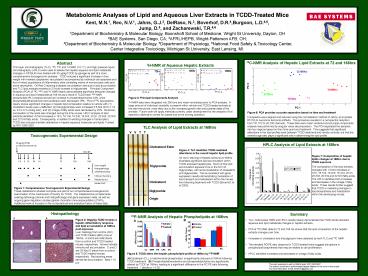
13C-NMR Analysis of Hepatic Lipid Extracts at 72
and 168hrs
Abstract
1H-NMR of Aqueous Hepatic Extracts
Thin layer chromatography (TLC), 13C, 31P, and 1H
NMR (14.1 T), and high pressure liquid
chromatography (HPLC) were used to assess the
hepatic aqueous and lipid metabolite changes in
C57BL/6 mice treated with 30 ug/kg TCDD by gavage
as part of a more comprehensive toxicogenomic
analysis. TCDD induced a significant increase in
liver weight with marked cytoplasmic
vacuolization accompanied by individual cell
apoptosis and foci of mixed populations of
inflammatory cells consisting mainly of
mononuclear cells and some neutrophils. Oil Red
O staining indicated vacuolization was due to
lipid accumulation and TLC lipid analysis
revealed a 2.5-fold increase in triglycerides.
Principal Component Analysis (PCA) of 13C, 31P,
and 1H NMR clearly demonstrates significant
temporal changes in aqueous and lipid metabolites
at 168 hrs as a result of TCDD treat. 31P NMR
phospholipid (PL) analysis showed an increase in
phosphatidylcholine 24, while phosphatidylethanol
amine and cardiolipin each decreased 16. PCA of
13C lipid spectra clearly shows significant
changes in hepatic lipid composition relative to
vehicle with (1) cholesterol levels were
unaffected, (2) triacylglycerides were increased
3.5-fold (20.8 1.9 vs. 6.0 0.3 umol/g liver),
and (3) omega-3 fatty acids were decreased by
22. Moreover, composition of the lipids also
changed during the time course. HPLC analysis of
lipid extracts identified gt3-fold increases in
160, 181n9, 182n6, 183n3, 202n, 203n6,
203n9 and 225n3 fatty acids. Consequently, in
addition to eliciting changes in transcription,
TCDD also induces dramatic alterations in hepatic
aqueous molecules and lipids. Funded by RO1
ES013927.
Figure 3 Principal Components Analysis 1H-NMR
data were integrated into 280 bins and mean
normalized prior to PCA analysis. A large amount
of individual variability is present within
vehicle and TCDD-treated animals at the late
time-points, most likely due to the increased
growth rate and pubertal state of the animals.
Future work will apply methods currently under
development for the analysis of full spectra to
attempt to correct for biases due to the binning
operation.
Figure 6 PCA provides accurate separation based
on time and treatment Full spectra were aligned
and reduced using the normalization method of
Jahns, et al (poster 1219) to overcome technical
artifacts. This procedure resulted in a net
spectral reduction from 131,072 to 43,700
channels. These data were mean centered and
principal components analysis was performed by
singular value decomposition. The PCA separated
the samples into four regions based on their
time-point and treatment. This suggests that
significant alterations in the lipid profiles
exist between TCDD treatment and vehicle
controls, but that the animals age also plays a
significant role in determining the lipid profile.
TLC Analysis of Lipid Extracts at 168hrs
T
V
V
T
HPLC Analysis of Lipid Extracts at 168hrs
Cholesterol Ester
Figure 4 TLC identifies TCDD-mediated
alterations in the overall hepatic lipid
profile Oil red O staining of hepatic sections at
168hrs illustrated significant lipid accumulation
within TCDD-exposed hepatocytes. Much of this
lipid accumulation appears to be in the form of
triglycerides, with some localization of
cholesterol and diglycerides. This is consistent
with gene expression results demonstrating
modulation of lipid transport and metabolism
within the mouse liver following treatment with
TCDD (Boverhof, et al 2005).
Figure 7 Composition of hepatic lipids changes
at 168hrs due to TCDD exposure The composition of
the lipid extracts changed with gt3-fold increases
in 160, 181n9, 182n6, 183n3, 202n, 203n6,
203n9 and 225n3 fatty acids. The HPLC
identifies both increases and decreases in the
omega-3 fatty acids. These results further
suggest that TCDD is mediating changes in lipid
biosynthesis and metabolism within the developing
mouse.
Triglyceride
Cholesterol
Diglyceride
Origin
Histopathology
Summary
31P-NMR Analysis of Hepatic Phospholipids at
168hrs
Figure 2 30µg/kg TCDD invokes a hepatic
inflammatory response and lipid accumulation at
168hrs post exposure. Liver histology from
control (A/C) and TCDD treated (B/D) mice at
168hrs. A and B are HE stains from a control
and TCDD treated mouse, respectively. Arrows
indicate immune cell accumulation. C and D are
Oil Red O stains from a control and TCDD treated
mouse, respectively. Red staining areas denote
fat accumulation. Bars 10 µm
B
- TLC, multinuclear NMR and HPLC results clearly
demonstrate that TCDD elicits dramatic aqueous
and lipid metabolite changes in hepatic extracts. - PCA of 13C NMR data for 72 and 168 hrs shows that
the lipid composition of the hepatic extracts
changes over time. - Increases in cholesterol and triacylglycerol were
detected by both TLC and 13C NMR - The elevated PC/PE ratio observed in TCDD-treated
livers suggest disruptions in phospholipid
biosynthesis that may be related to cell
proliferation. - HPLC identified increases and decreases in
omega-3 fatty acids.
A
5.00
4.00
3.00
2.00
uMol/g tissue
1.00
0.00
CL
SPM
PS
LPC
PI
Figure 5 TCDD alters the hepatic phospholipid
profile at 168hrs by 31P-NMR (A)Cardiolipin (CL),
a mitochondrial phospholipid, is significantly
reduced at 168hrs following TCDD-treatment. (B)
Phosphotidylethanolamine and phosphatidylcholine
are significantly altered by TCDD at 168hrs,
leading to a significant difference in the PCPE
ratio following treatment. denote p lt 0.05.
This work supported in part by NIEHS grant RO1
ES013927. The authors would like to acknowledge
Meghan Makley for her work on this project For
more information, e-mail kentmi11_at_yahoo.com
Recommended
