
THREE MAIN TYPES OF MUSCLE PowerPoint PPT Presentation
1 / 38
Title: THREE MAIN TYPES OF MUSCLE
1
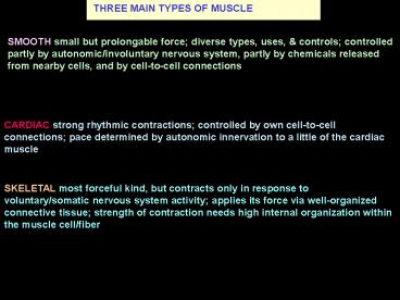
THREE MAIN TYPES OF MUSCLE
SMOOTH small but prolongable force diverse
types, uses, controls controlled partly by
autonomic/involuntary nervous system, partly by
chemicals released from nearby cells, and by
cell-to-cell connections
CARDIAC strong rhythmic contractions controlled
by own cell-to-cell connections pace determined
by autonomic innervation to a little of the
cardiac muscle
SKELETAL most forceful kind, but contracts only
in response to voluntary/somatic nervous system
activity applies its force via well-organized
connective tissue strength of contraction needs
high internal organization within the muscle
cell/fiber
2
THREE MAIN TYPES OF MUSCLE III
SKELETAL most forceful kind
but contracts only in response to
voluntary/somatic nervous system activity
applies its force via well-organized
connective tissue
strength of contraction needs high internal
organization within the muscle cell/fiber
3
SKELETAL MUSCLE INNERVATION
Axons/nerve fibers to motor end-plates to cause
contraction
striated/cross-banded myofiber
TENDON
4
THREE MAIN TYPES OF MUSCLE II
CARDIAC strong rhythmic contractions
controlled by own cell-to-cell connections
pace determined by autonomic innervation to a
little of the cardiac muscle
5
CARDIAC MUSCLE
striated/cross-banded CARDIOMYOCYTES
INTERCALATED DISK
Reticular fiber
central NUCLEUS
Capillary
branching muscle fibers
Sarcolemma external lamina
6
INTERCALATED DISC - electro-mechanical union
ID is a strong myocyte-myocyte attachment
electrical connections
Fascia adherens strength
Maculae adherens strength
Gap junction transmits contraction
7
THREE MAIN TYPES OF MUSCLE I
SMOOTH small but prolongable force diverse
types, uses, controls controlled partly by
autonomic/ involuntary nervous system, partly by
chemicals released from nearby cells, and by
cell-to-cell connections
8
SMOOTH MUSCLE
SMOOTH MUSCLE CELL has same contractile control
machinery as skeletal myocyte, but less
organized
Filaments attach to DENSE BODIES serving the role
of Z-lines
CAVEOLAE for stimulus-contraction coupling serve
role of T-tubule SR system
9
SKELETAL MUSCLE
striated/cross-banded myofiber
capillary
sarcolemma
TENDON
endomysium CT
Myofiber in cross-section
10
SKELETAL MUSCLE Connective Tissue Organization
MYOCYTE
PERIMYSIUM
creates
FASCICLE/ bundle
endomysium
EPIMYSIUM
11
Myofiber in cross-section
myofibrils
Each myofibril consists of bundled myofilaments
thick MYOSIN
But, at regular intervals along the relaxed
fiber, only thin or only thick filaments are
found. Why?
thin ACTIN
12
ACTIN MYOSIN FILAMENTS IN MUSCLE
Z line/disc
thick MYOSIN filament
thin ACTIN filament
In muscle, for strong shortening (contractile)
force the actin filaments are stabilized and
interdigitated with thicker myosin filaments,
which pull them in deeper
13
BANDING-PATTERN CHANGES IN CONTRACTION
A band
I band
I band
actin
myosin
M line but no H zone
Z line
1
Sarcomere shortens
A band unchanged
3
2
I band shortens
4
H zone disappears
14
MUSCLES II
Cardiac muscle the heart are described under
Muscle as a tissue, in the Cardiovascular
section
Smooth muscle is described under Muscle, the
tissue, and as each tubular organ system is
covered.
Skeletal muscle is the principal component of the
Muscles of the body, which are the topic of
this section
Since these muscles attach to move bones, the
outline will go regionally through the two major
divisions of the skeleton axial appendicular
15
MUSCLES VII
Having reached the skeletal muscles, the plan is
as follows.
Well explain the naming of the movements that
muscles cause at joints
We make deliberate movements, mostly of the limbs
and hands, but there is a background
Trunk muscles have to make respiratory movements
to provide oxygen
Head trunk muscles have to hold or change
postures so that the limbs are oriented right
So axial muscles will be covered first, then
the muscles that link axial appendicular
skeletons, then the limbs
16
(No Transcript)
17
(No Transcript)
18
Right ARM Deltoid muscle I
Anterior view
Acromion spine
DELTOID m
SCAPULA
Glenoid fossa
HUMERUS
The deltoids wide origins are the clavicle,
scapular acromion spine
It narrows to the insertion on the deltoid
tuberosity, giving it a fan shape
Deltoid tuberosity
Lateral view
19
MUSCLE ACTIONS IV Multiple actions
More than one motion is possible around a joint
Thus, around the shoulder, the arm can be
abducted, adducted, flexed, extended, rotated
medially, rotated laterally, circumducted
Marieb Fig 6.13, p. 169
We speak of particular muscles as having a
certain action as abductors, flexors, extensors,
etc
ABDUCTION
Harmonious action requires coordination in the CNS
20
For example, this deltoid muscle can be easily
felt coming over the tip of the shoulder, as you
pull your arm away from your side
MUSCLE ACTIONS I Abduction
The action of pulling the arm away from the
midline
Absent - away from class
21
MUSCLE ACTIONS II Adduction
Adduction - action of pulling the arm toward the
midline
This motion requires relaxation of the deltoid
muscle contraction of other muscles from the
ribs scapula to the humerus
22
Rotator-cuff muscles I
to tighten shoulder joint (Glenohumeral)
23
Rotator-cuff muscles III
The arrangement allows the humerus great freedom
of movement
but, is vulnerable to painful tears of the cuff
structures to dislocation of
the head out of the shallow fossa
The four cuff muscles of the scapula are
Supraspinatus
Infraspinatus
Subscapularis
Teres minor
24
(No Transcript)
25
ARM MOVEMENT II Pectoralis major
Large muscles from the axial skeleton - spine
ribs ( pelvis) are needed
Tendon inserts into antero-medial side of humerus
Pectoralis major
ACTIONS - adduction, flexion medial rotation of
the humerus
Origin - medial ribs, sternum clavicle
26
MUSCLE ACTIONS V
Thus, around the shoulder, the arm can be
abducted, adducted, flexed, extended, rotated
medially, etc
CAUTION
Arm here has its technical meaning - the piece
above the elbow, which is another joint with its
own ranges of motion
Thus, one can flex the shoulder , but extend
the elbow
27
MUSCLE ACTIONS VII WRIST PRONATION
SUPINATION SHOULDER ELBOW ACTIONS
Having closed ones fingers over the food, one
rotates the wrist and lower forearm to have the
food facing ones mouth. Then, flexing the elbow
shoulder joints brings the food to the mouth.
Turning the wrist so that the palm faces
downwards (platewards) is PRONATION.
Note crossing of forearm bones
The motion of turning the wrist so that the palm
faces upwards (mouthwards) is SUPINATION.
Followed by elbow shoulder flexion
28
MUSCLE ACTIONS X Another classification scheme
The deltoid muscle is the PRIME MOVER in
abduction of the shoulder
Any muscle helping it in this action is its
SYNERGIST, e.g., supraspinatus
The opposing adductor muscles that had to relax
are the ANTAGONISTS
The spinal muscles holding the scapula in place
are FIXATORS
ABDUCTION
Harmonious action requires coordination in the CNS
29
ARM CROSS-SECTION Compartments
The elbow region has far fewer movements open to
it than the wrist and digits.
The muscles are generally bulky, but few, so that
no subdivision of the two compartments is needed
30
Lets return to
Biceps brachii for refining movements
2
Glenoid fossa
1
SCAPULA
Bone protrudes to give wanted direction of pull,
while tendon stays straight
Coracoid process
1
LONG HEAD
2
SHORT HEAD
Tendon curves, guided along a groove to alter
angle of pull
HUMERUS
3
Tendon overshoots fastened to bone, so that it
rotates the bone
RADIUS
3
Radial tuberosity
31
POSTERIOR Right ARM Triceps muscle medial head
SCAPULA
Glenoid fossa
HUMERUS
MEDIAL
Insertion onto
Olecranon
ULNA
32
POSTERIOR Right ARM Triceps lateral long heads
SCAPULA
Glenoid fossa
LONG
LATERAL
HUMERUS
Insertion onto
Olecranon
ULNA
33
UPPER EXTREMITY Innervation pattern I
Upper extemity is the collective name for arm,
forearm, hand
Musculocutaneous N
Shoulder
FLEXORS
Radial N
ARM
Elbow
EXTENSOR
FOREARM
Thumb
Wrist
HAND
Fingers
34
UPPER EXTREMITY Innervation pattern II
Median N
Ulnar N
Shoulder
Radial N
ARM
Elbow
FLEXORS
FOREARM
Thumb
Wrist
EXTENSORS
Fingers
35
UPPER EXTREMITY Innervation pattern III
Median N
Ulnar N
Shoulder
ARM
Elbow
FOREARM
Thumb
Wrist
HAND
Fingers
36
UPPER EXTREMITY Innervation pattern IV
Median N
Ulnar N
Musculocutaneous N
Shoulder
Radial N
ARM
FLEXORS
Elbow
FOREARM
EXTENSORS
Thumb
Wrist
Fingers
37
(No Transcript)
38
SENSORY INNERVATION OF PALMAR HAND
Ulnar nerve
is most important for using the hand
Superficial Radial nerve more
for back of the hand
Recommended
