Microscopy - PowerPoint PPT Presentation
1 / 44
Title: Microscopy
1
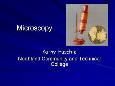
Microscopy
- Kathy Huschle
- Northland Community and Technical College
2
Microscopy
- microscope
- produces enlarged images
- studies the morphology (what it looks like) of a
microorganism
3
Microscopy
- microscopy includes
- magnification
- resolution
- contrast
4
Magnification
- magnification
- a result of refraction or bending of light rays
5
Resolution
- resolution
- ability to distinguish detail
- will allow you to observe one layer at a time
6
Contrast
- contrast
- visible shades in a specimen
- contrast is often enhanced by staining
7
Light Microscopes
- Bright-field Microscope
- field of view is brightly illuminated with
visible light - also known as compound microscope
8
Fluorescence Microscope
- Fluorescence Microscope
- use with fluorescent stains
- emit light at a different wavelength
- different wavelengths have different colors
9
Fluorescence Microscope
- dyes can be chemically linked specific chemical
targets - target can be visualized and quickly and
positively identified - allows for rapid diagnosis
Click icon to use a virtual fluorescence
scope. Click Try the Simulator
10
Fluorescence Microscope
Image of cell division taken by fluorescence
microscope
11
Dark Field Microscope
- Dark-field Microscope
- enhance contrast without staining
- most staining will kill the microorganism
- allows for the viewing of live specimens
- special condenseris used
- focuses light at an angle
- reflected off specimen
Treponema pallidum image from Dark Field
Microscope
12
Phase Contrast Microscope
- Phase Contrast Microscope
- contrast without staining
- difference in density produces difference in
light - brings direct and reflected light rays together
to form an image of the specimen
image of cell division taken by phase contrast
microscope
13
Phase Contrast Microscope
- allows for observation of living organisms
Click icon to use a virtual phase contrast
scope. Click Try the Simulator
14
Interference Microscope
- Interference Microscope
- two light waves combined
- beam is split
- one part through the specimen
- one part around
- recombined
15
Interference Microscope
- specimen looks three dimensional
16
Confocal Scanning Laser Microscope
- constructs a 3-dimensional image
- specimen is stained with fluorescent dye
17
Confocal Scanning Laser Microscope
- specimen is illuminated one plane at a time
- computer processes the images into 2 and 3
dimensional images
18
Electron Microscopy
- electron beam is used in the place of a beam of
light - electromagnets control focus, illumination and
magnification - higher magnification up to 100,000X
19
Electron Microscopy
- electron beam must be contained in a vacuum to
eliminate disturbance from air molecules - large, bulky unit
- specimen must be killed
- special preparation process
- problem with artifacts
20
Transmission Electron Microscope
- TEM
- used for observing layers of cells
21
Transmission Electron Microscope
- 10,000 100,000 X magnification
rubber particle magnified 100,000 times
22
Transmission Electron Microscope
Click icon to use a virtual Transmission
electron scope. Click Try the Simulator
23
Scanning Electron Microscope
- SEM
- used for surface features, not internal features
- knocks electrons out of specimen surface
- collected for image
24
Scanning Electron Microscope
- SEM
- produces a 3-D image
- 1,000 10,000 X magnification
image of table salt taken with an SEM
25
Scanning Electron Microscope
Click icon to use a virtual Scanning electron
scope. Click Try the Simulator
26
Contrast Staining
- stain is a salt with a negative and positive
charged ion - color portion is a positive charge and will be
attracted to the negative charged microbial cell
Enterobacter
27
Contrast Staining
- stain
- increases contrast between specimen and
background - most microorganisms are colorless and difficult
to see
Unstained Halophile
Stained Halophile
28
Contrast Staining
- simple stains
- positive staining
- cells are dark against light background
- makes cell shapes and arrangements visible
yeast stained with methylene blue stain
29
Contrast Staining
- negative staining
- clear microorganisms against a dark background
- better view of microbial shape due to lack of
distortion - stain is not taken up by the microorganism
- very useful with microorganisms with capsules
negatively stained mixed bacteria culture
30
Differential Staining
- differential staining
- multiple stains
- differentiate bacteria according to their
reaction to the staining process - differentiation is based on differences in cell
structure - Gram stain
- acid fast stain
31
Differential Staining
- Gram Stain
- most widely used procedure
- process uses a stain, wash, counterstain
32
Differential Staining
- Gram Stain
- Gram positive bacteria retain the purple stain
- Gram negative bacteria appear pink after the
counterstain
Gram - Gram
33
Differential Staining
- acid-fast staining
- waxy chemical in the cell wall allows the
bacteria to retain the stain
Cryptosporidum muris
34
Differential Staining
- acid-fast staining
- makes Mycobacterium ( causative agent for
tuberculosis) easy to recognize in sputum, amidst
numerous organisms
sputum sample containing M. tuberculosis
35
Special Stains
- stains that color only certain parts of bacteria
- endospore staining
- reveals the presence of endospores
- produced by few bacteria
- Clostridium and Bacillus
A is the microorganism, B indicates the endospore
36
Special Stains
- flagella staining
- adheres to an enlarges the flagella for ease of
observation
flagella
37
Bacteria MorphologyShape
- 3 basic shapes
- coccus
- bacilus
- spiral
spiral
coccus
bacillus
38
Bacteria Morphologyshape
- coccus
- primarily spherical in shape
- can be oval, elongated or flattened on one side
- plural cocci
Enterococcus sp.
39
Bacteria Morphologyshape
- bacillus
- rod shaped
- plural bacilli
Bacillus anthracis
40
Bacteria Morphologyshape
- spiral
- one or more twists
- never straight
Treponema pallidum
41
Bacterial MorphologyArrangement
- single
yeast cells
42
Bacterial Morphologyarrangement
- diplococci or diplobacilli
Neisseria gonorrhoeae
43
Bacterial Morphologyarrangement
- chains of coccus or bacillus
Streptococcus sp.
44
Bacterial Morphologyarrangement
- clusters


