In Vitro Culture Methods PowerPoint PPT Presentation
1 / 81
Title: In Vitro Culture Methods
1
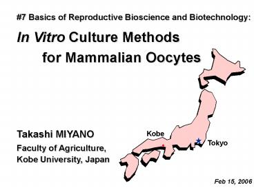
7 Basics of Reproductive Bioscience and
Biotechnology
In Vitro Culture Methods for Mammalian Oocytes
Takashi MIYANO
Faculty of Agriculture, Kobe University, Japan
Feb 15, 2006
2
Kobe
Kobe Beef
Japanese Black
3
In Vitro Culture Methods for Mammalian Oocytes
1) Oogenesis in Mammals - Oocyte Growth -
Oocyte Maturation - Oocyte Meiosis 2) In Vitro
Maturation (IVM) of Oocytes 3) In Vitro Growth
(IVG) of Oocytes
4
zona pellucida
first polar body
secondary oocyte
mouse mature oocytes
5
Embryo
Primordial germ cells
Oogonia
Spermatogonia
Oocytes
Spermatocytes
Eggs
Spermatozoa
Gametes
Zygotes
6
Pig Reproductive Organs
ovary
uterus
oviduct
7
Oocyte Growth
fully grown oocytes
growing oocytes
non-growing oocytes
8
ovaries
pituitary
9
Oocyte Maturation
Gonadotropins (FSH, LH)
fully grown oocytes
eggs mature oocytes
10
Gametes
Spermatozoon (pl. Spermatozoa)
Egg
11
oviduct
2-cell embryo
morula
uterus
zygote
blastocyst
Fertilization
implantation
Ovulation
ovary
12
1) Oogenesis in Mammals
1) Oocytes are produced in the ovary. 2) Oocytes
grow with the follicular development. 3)
Fully-grown oocytes start to mature after the
stimulation of the gonadotropic hormones (FSH
and LH). 4) Mature oocytes are ovulated to the
oviduct where oocytes are fertilized with
the spermatozoa.
13
1) Oogenesis in Mammals - Oocyte Growth -
Oocyte Maturation - Oocyte Meiosis 2) In Vitro
Maturation (IVM) of Oocytes 3) In Vitro Growth
(IVG) of Oocytes
14
Meiosis
Mitosis
DNA replication
meiosis I
cell division
meiosis II
cell division
cell division
Molecular Biology of THE CELL 4th Ed. (2002)
daughter cells
gametes
15
puberty
birth
fetus
?
undifferentiated gonad
?
germ cells in mitosis (PGC, spermatogonia,
oogonia)
germ cells in meiosis (spermatoctyes, oocytes)
16
homologous chromosomes
sister chromatids
17
Mitosis and Oocyte Meiosis
DNA
18
Oocyte Meiosis
meiosis I
prophase I (GV)
metaphase I
anaphase I - telophase I
emission of 1st polar body
meiosis II
metaphase II
anaphase II - telophase II
Sperm penetration emission of 2nd polar body
pronucleus stage
19
1) Oogenesis in Mammals - Oocyte Meiosis
1) Oocyte meiosis starts in the fetal gonads. 2)
Oocytes enter prophase I and arrest at this stage
before birth. 3) Oocytes grow to the full
size during the arresting period of prophase
I. 4) Fully-grown oocytes resume meiosis and
mature to metaphase II after the
gonadotropic stimulation.
20
1) Oogenesis in Mammals - Oocyte Growth -
Oocyte Maturation - Oocyte Meiosis 2) In Vitro
Maturation (IVM) of Oocytes 3) In Vitro Growth
(IVG) of Oocytes
21
Objectives of In Vitro Experiments
1) Mechanisms in Vivo 2) Applications Animal
production Assisted Reproductive Technology (ART)
22
In Vitro Maturation of Oocytes
Pincus, G. and Enzmann, E.V. (1935) The
comparative behaviour of mammalian eggs in vivo
and in vitro. I. The activation of ovarian eggs.
J. Exp. Med., 62, 655-675. Edwards, R.G.
(1965) Maturation in vitro of mouse, sheep, cow,
pig, Rhesus monkey and human ovarian oocytes.
Nature, 208, 349-351.
23
Pig ovaries
24
Pituitary
Gonadotropins FSH, LH
Ovary
Oocyte Maturation Cumulus expansion
25
Pig oocyte at prophase I (GV stage)
26
Chromosome condensation starts
GVI
GVIII
GVII
0 h
GVIV
LD
MI
18 h
27 h
27
Gonadotropins (GTH)
Germinal vesicle (GV, Prophase I)
Diakinesis
Metaphase I (MI)
Metaphase II (MII)
0 hr
18-24 hr
27 hr
36 hr
- Chromosome condensation
- Nucleolus disassembly
- GVBD (Nuclear membrane breakdown)
- Spindle formation
28
Somatic cell
Oocyte
DNA red tubulin green
29
Collection of pig follicles 1
30
Collection of pig follicles 2
31
cumulus granulosa cells
antrum
oocytes
mural granulosa cells
theca cells
32
Oocyte-cumulus complexes
Oocyte-cumulus-granulosa cell complexes
33
theca cells
34
In Vitro Maturation of Pig Oocytes in Kobe
- Materials
- Medium
- Hormone
- Temperature
- Gas phase
- Dish
- Culture
Oocyte-Cumulus-Granulosa Cell Complexes from
large antral follicles (4 -6 mm) TCM199 (10 FCS,
0.1 mg/ml Na-pyruvate, 0.08 mg/ml kanamycin) 0.1
IU/ml hMG (human menopausal gonadotropin)
Follicle Shell 38.5 - 39.0ºC 5 CO2 in air Falcon
1008 Rocking
35
(No Transcript)
36
Cdc2 kinase (MPF) induces Oocyte Maturation
Fertilization
Cdc2
active form
37
Sperm penetration Electro-stimulation
GTH
GV
MII
MI
Zygote
cyclinB1
cyclinB1
ERK1
ERK1
ERK2
ERK2
Miyano Lee, 2003
38
Micro-dissection of pig follicles
5 mm
1 mm
0.5 mm
39
100
80
of GVBD oocytes
60
40
M II
20
AI-TI
D-M I
0
105
110
115
120
100
Oocyte diameter (µm)
Follicle diameter (mm)
1.0-1.5
4-6
0.5-0.7
Meiotic competence of pig oocytes
40
Acquisition of Maturational Competence
100 µm 0.5 - 0.7 mm
Incompetent Oocytes
110 µm 1.0 - 1.5 mm
120 µm 4.0 - 6.0 mm
Competent Oocytes
41
2) In Vitro Maturation of Oocytes
1) Oocyte at the GV stage mature to metaphase II
(Oocyte Maturation). 2) Oocyte maturation
can be induced in vitro (IVM). 3) Oocytes
maturation is accompanied with cumulus
expansion. 4) Small oocytes have no ability to
resume meiosis or no ability to mature to
metaphase II. 5) Oocyte maturation is induced by
Cdc2 kinase.
42
1) Oogenesis in Mammals - Oocyte Growth -
Oocyte Maturation - Oocyte Meiosis 2) In Vitro
Maturation (IVM) of Oocytes 3) In Vitro Growth
(IVG) of Oocytes
43
fully grown oocytes
Oocyte Maturation
growing oocytes
Oocyte Growth Follicular Development
44
Oocyte Growth Follicular Development
fully grown oocytes
Secondary follicle
Antral follicle
Primary follicle
growing oocytes
Primordial follicle
45
Oocytes in bovine ovary
46
Oocytes in pig ovary
47
Oocytes increase in size
Mouse
15 - 20 µm
75 µm
30 µm
120 - 125 µm
Cow
48
Numbers of ovarian follicles in mammals
Primordial follicles
Developing follicles
Species
Mouse
4,270
676
Sheep
105,450
475
Cow
120,000
300
Pig
420,000
84,000
Human
302,000
12,090
Mean number per pair of ovaries (Gosden and
Telfer, 1987 Erickson, 1966)
49
Experiments of Oocyte Growth
Applications
- Production of a huge number of eggs
- Assisted Reproductive Technology (ART)
- Oocyte Banking (with cryopreservation)
Basic Questions
- Initiation of oocyte growth ?
- Follicle/Oocyte selection ?
- Interaction between oocyte and somatic cells ?
50
(No Transcript)
51
(No Transcript)
52
(No Transcript)
53
Growth of Mouse Oocytes in vitro
Newborn ovary
Organ-cultured ovary
54
Organ Culture
Oocyte-granulosa cell complex Culture
Follicle Culture
agar
collagen
collagen gel
55
Granulosa cells connect with the oocytes via the
projections.
56
Bovine Oocytes in Early Antral Follicles
(0.5 - 0.7 mm)
90 - 99 µm
120 - 125 µm
57
cow
Bovine ovary
Embedding in collagen gel
Oocyte-cumulus-granulosa cell complexes
Early antral follicles (0.5 - 0.7 mm)
Culture for 14 - 16 days in TCM199 containing
10 FCS (or 4 mg/ml BSA), 0.1 mg/ml Na pyruvate,
4 mM hypoxanthine (38.5 ºC, 5CO2 - 95 air)
58
cow
collagen gels
4 polyvinylpyrrolidone
Hirao et al. (2004)
Harada et al. (1997)
59
Development of in vitro grown bovine oocytes
After 14 days of growth culture, recovered
granulosa cell-enclosed oocytes were matured for
24 hr, and subsequently inseminated with bovine
spermatozoa.
Yamamoto et al. (1999)
60
IVG of Mammalian Oocytes
IVG
IVM
Species
Baby
IVF
oocyte ø
days
Mouse
20 µm
8 14
Yes
Yes
Yes
Eppig O'Brien, 1996
Pig
80 µm
16
Yes
No
Yes
Hirao et al., 1994
Cow
Yes
95 µm
14
Yes
Yes
Yamamoto et al., 1999
Cow
Yes
95 µm
14
Yes
Yes
Hirao et al., 2004
Ovarian organ-culture for 8 days followed by
Oocyte-Granulosa cell Complex culture for 14
days.
61
Bovine Oocytes in Secondary Follicles
(0.15 - 0.2 mm)
50 µm
120 - 125 µm
62
Ovarian Oocytes
In Vitro Growth Culture
Xenotransplantation to Immuno-deficient Mice
63
Auto-transplantation (auto-grafting) Allo-transpl
antation (allo-grafting) Xeno-transplantation
(xeno-grafting)
64
Live birth after ovarian tissue transplant
D. M. Lee et al.
Nature, vol. 428, 11 March, 2004
65
Applications of xenografting into
immuno-deficient mice
Honaramooz A, et al., Nature 418 778-781 (2002)
SCID mice have few circulating lymphocytes and
little or no serum immunoglobulin. Homozygosity
for the severe combined immune deficiency (scid)
mutation results in a block in T and B lymphocyte
development.
66
500 µm
bovine preantral follicles
67
SCID mouse
68
Bovine secondary follicles developed to antral
follicles 6 weeks after transplantation to SCID
mice
200 µm
69
In vitro maturation and fertilization of bovine
oocytes grown in SCID mice
2 mm
70
Bovine Oocytes in Primordial Follicles
(0.04 mm)
30 µm
120 - 125 µm
71
Bovine primordial follicles
72
100 µm
Bovine primordial and primary follicles 6 weeks
after transplantation into SCID mice
73
Growth of bovine oocytes in SCID mice
Primordial follicle
Secondary follicle
74
Pig Oocytes in Primordial Follicles
(0.04 mm)
6 months old
30 µm
120 - 125 µm
10 days old
75
Pig primordial follicle (6 months old)
100 µm
Pig primordial follicles 2 months after
transplantation into SCID mice
76
Pig primordial follicle (10 days old)
400 µm
Pig primordial follicles developed to antral
follicles 2 months after transplantation into
SCID mice
77
Newborn
Activated
Oocytes in primordial follicles in the adult
animals take a longer time before starting their
growth.
78
3) In Vitro Growth of Oocytes
1) Contact with the surrounding granulosa cells
is important for the oocyte growth. 2) Mouse
oocytes in the primordial follicles are able to
grow to the final size in vitro. 3) Pig and
bovine oocytes in the mid-growth stage are
able to grow in vitro. 4) In xenotransplantation
to immuno-deficient mice, oocytes in much
smaller follicles from domestic species grow
to the final size.
79
Ovarian small oocytes
IVG
Xenotransplantation
IVM ? IVF ? ET
80
21st Century COE Program The Japan Society for
the Promotion of Science, Creative Scientific
Research (13GS0008)
81
7 Basics of Reproductive Bioscience and
Biotechnology In Vitro Culture Methods for
Mammalian Oocytes Takashi MIYANO
Question Improvement of culture methods is
important for the IVG and IVM. But why the basic
study on the mechanisms (for example molecular
mechanisms) in oocyte growth and maturation is
required?

