Mikrowiazka protonowa i akcelerator Van de Graaffa
Title:
Mikrowiazka protonowa i akcelerator Van de Graaffa
Description:
Mikrowiazka protonowa i akcelerator Van de Graaffa –
Number of Views:61
Avg rating:3.0/5.0
Title: Mikrowiazka protonowa i akcelerator Van de Graaffa
1
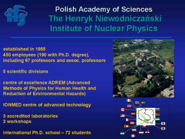
Polish Academy of Sciences The Henryk
Niewodniczanski Institute of Nuclear Physics
established in 1955 450 employees (190 with
Ph.D. degree), including 67 professors and
assoc. professors 5 scientific divisions
centre of excellence ADREM (Advanced Methods of
Physics for Human Health and Reduction of
Environmental Hazards) IONMED centre of
advanced technology 3 accredited laboratories
2 workshops international Ph.D. school 72
students
2
Interdisciplinary and applied research at the IFJ
PAN - examples
Equipment for proton eye therapy optical line
with beam formation and monitoring system
Proton microprobe experimental chamber with
external beam, used for biological experiments
Geophysical probe used in exploration of gas and
oil fields
Thermoluminescent detectors and their
installation in a phantom for the study of
radiation dose distribution in humans during
space flights
UHV system for preparation of multilayers and for
surface analysis
3
Department of Applied Spectroscopy
Atomic Force Microscopy
Surface and Thin Films techniques and PAC / MS
Perturbed Angular Correlations and Mossbauer
Spectroscopy
MicroProbe and the Single Hit Ion Facility
Head Assoc. Prof. Wojciech M. Kwiatek 8
researchers, 6 technicians, 10 PhD students The
research activity of DAS ? bio-medical
applications and condensed matter studies using
nuclear methods - ion beam techniques PIXE,
PIGE, RBS with home facilities - SR-XRF,
XANES, and EXAFS with synchrotron radiation. ?
AFM / SFM technique applied to both biological
and material science research. ? the structure of
intermetallic compounds, alloys, minerals and
metals investigated using the PAC and
Mössbauer effect techniques.
4
Nuclear Microprobe at the IFJ
- VdG HVEC KN-3000
- accelerating voltage up to 2.5 MV
- ion beam H, He
- focusing - down to the size of 7 µm
- beam current 100 pA and more
- unique construction (short 230 cm)
Main ion beam techniques used ? PIXE ? RBS
? STIM
5
Studies of sediments in the Dobczyce lake
source of water for Krakow. Broad
collaboration AGH, UJ, IFJ, PP, gov. and city
authorities Accumulation of toxic substances in
sediments Depth profiling with the use of
nuclear methods.
Fe Ka
6
Compozite structure of wood cells in petrified
wood(collaboration with the University of
Lublin)
7
Elemental composition of soil and dust
microparticles by the µPIXE technique.
8
Microprobe applications in geology rocks dating
basing on Pb/Th/U contentin monazite
crystals(collaboration with the Geological
Institute of PAN)
Monazite grains (dia 30100 µm), fixed in Epoxy
resin and polished with diamond paste to remove
upper layers of material. Monazite, a phosphate
of rare earth elements, (Ce, La, Th) PO4 is
frequently used in geological sciences to date
rocks
9
(No Transcript)
10
Studies of skin as a protective barrier (NANODERM
project, finished 2006)
Stratum corneum
Stratum granulosum
Stratum spinosum
Dermis
400 µm 200 µm
? formulations containig TiO2 have been
applied to animal and human skin ? after
incubation, formulations have been washed out
and the depth distribution of Ti was
measured.
11
Autoradiography image of a hair follicle (image
size 300 x 220 µm2). Dark pixels correspond to
positron halo due to radiolabelled TiO2.
Outer layer of epidermis with a visible follicle.
Skin was treated with a formulation containing
TiO2 (application time 45)
12
Oxidation state studied by XANES
Laboratori Nationali di Frascati HASYLAB, Hamburg
The XANES spectra of sulphur and iron edge in
cancerous tissues are shifted in relation to
non-cancerous ones. It proves the domination of
iron appearance on 3rd oxidation state in
cancerous tissues while in healthy part of
tissues iron appears on 2nd oxidation state. The
sulphur state is still not well understood.
AFM as the early detection technique revealing
changes in cells
13
Cells damage due to natural backgroundDNA
damage due to cosmic radiation and ionizing
radiation of the background are MILLION times
less numerous than damage due to normal
metabolism 102 vs 108 /
dayBackground radiation 1 electron / cell /
year 1 mGy 1 a particle / cell / 100 years
200 mGy 1 Gy 1J/1kgBackground
radiation effects are due to single tracks in
cells, separated in time and space
14
Random beam applied to cells population, averag
e dose
15
Microbeam, SIH individual cells targeted, exact
dose
16
The IFJ PAN single ion hit facility
2.5 MV VdG
beam off
online imaging
single proton detection
XY stage control
deflection plates
slits
optical system
focusing
meas. chamber exit window
Control
cell dish XY stage
200 p/cell
17
External beam at the SIH experimental chamber
6th FP MC-RTN CELLION 10 European institutions,
over 20 recruited young researchers cellion.ifj.e
du.pl
18
X-ray microprobe at the IFJ PAN
Rigaku Innovative Technologies (RIT)
ML optics as Zone Plate alternative for Ti
K-alpha line (4.5 keV) Multilayer mirrors,
design KB cylindical optics, roughness lt 3
A Intensity increase over zone plates gt 7,
magnification 11 1.5 micron focal spot size at
300 mm distance from the source 4 micro
attenuators, software controlled
Hamamatsu L 9191 X-ray lamp point source (1 µm
diameter) Max 140 kV, 200 µA Mutiple targets
Ti, Mo, Ag


