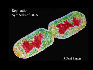
Replication: Synthesis of DNA - PowerPoint PPT Presentation
1 / 31
Title:
Replication: Synthesis of DNA
Description:
Relieves torsional strain generated by DNA unwinding. DNA gyrase (DNA topoisomerase II) ... the leading strand proceeds as DNA is unwound by the DnaB helicase. ... – PowerPoint PPT presentation
Number of Views:206
Avg rating:3.0/5.0
Title: Replication: Synthesis of DNA
1
Replication Synthesis of DNA
J. Paul Simon
2
Learning Objectives
Define the following terms as they apply to the
process of DNA replication Leading
strand Lagging strand Okazaki fragments RNA
primer Continuous and discontinuous
synthesis Processivity Proofreading Replication
fork Template Bidirectional Semi-conservative
3
Describe the primary events of DNA
replication with regard to the following enzymes,
proteins, and substrates DNA helicase DNA
topoisomerase (Type I and II) Single-stranded
DNA binding proteins DNA primase DNA
polymerase DNA ligase Nucleotide triphosphates
Identify when DNA replication occurs in
eukaryotes.
4
Describe the mechanism by which ddI prevents
DNA synthesis. Describe how AZT,
5-fluorouracil, and doxorubicin inhibit DNA
replication.
5
Map of the E. coli chromosome showing the
relative positions of genes encoding many of the
proteins involved in DNA synthesis, repair and
recombination.
6
Abbreviations used in naming genes and proteins
Bacterial genes are named using three lowercase,
italicized letters that reflect their apparent
function. dna DNA replication uvr resistance
to damaging effects of UV
radiation rec recombination pol DNA
polymerase Where several genes effect the same
process, capital letters (A, B, C, etc.) are
added dnaB, uvrD.
7
When referring to the protein, italic type is not
used and the first letter is capitalized. dnaA
gene product DnaA recQ gene product RecQ xpC
gene product xpC (XPC) XPC is involved in
Xeroderma pigmentosum. The Blooms Syndrome
gene, blM, produces the protein BLM. The
Werners Syndrome gene, wrN, produces the protein
WRN.
8
When does DNA synthesis occur?
In prokaryotes, cell division is relatively
simple. Once the DNA of the cell is replicated,
each copy moves toward an opposite side of the
cell by attaching to the cell membrane. The cell
elongates until it is approximately double its
original size. Finally, the cell membrane
pinches inward and forms two new cells. As long
as nutrients are available, prokaryotes continue
to replicate DNA and divide.
9
Eukaryotic cell cycle
10
Prokaryotes (E. coli)
The single chromosome is a circular duplex of DNA.
Single origin of replication (oriC)
Synthesis is bidirectional two replication forks
Replication forks dynamic points where parental
DNA is being unwound and the separated strands
quickly replicated.
Replication is semi-conservative each strands
serves as a template for the synthesis of a new
strand.
11
DNA synthesis proceeds in a 5 3 direction.
DNA synthesis is semidiscontinuous synthesis is
continuous on the leading strand, and
discontinuous on the lagging strand.
12
Elongation of a DNA chain by DNA polymerase a
single unpaired strand is required as template,
and a primer strand (RNA) is required to provide
a free 3 hydroxyl group to which a new
nucleotide unit is added.
13
(No Transcript)
14
These characteristics make the different DNA
polymerases suitable for different synthetic
functions. Two key properties proofreading and
processivity.
15
Tautomeric forms of uracil
Proofreading
Error correction by the 3 5 exonuclease
activity.
16
Proofreading (continued)
17
DNA polymerase III is the principal replication
enzyme (or replicase) in E. coli.
DNA polymerase III (pol III) has very high
processivity.
18
Two b subunits of E. coli polymerase III form a
circular clamp that surrounds DNA. The clamp
slides along the DNA, enhancing the processivity
of the enzyme by preventing its dissociation from
the DNA.
19
Proteins required to initiate replication at E.
coli origin
20
Model for initiation of replication at the E.
coli origin, oriC
21
Elongation
Leading strand synthesis is straightforward
Primase makes a 10 to 60 nucleotide RNA primer
at the origin. Deoxyribonucleotides are added
to the primer by DNA polymerase III. Leading
strand synthesis proceeds continuously, keeping
pace with the unwinding of DNA at the
replication fork. Lagging strand synthesis is
more complex Okazaki fragment synthesis is in
the opposite direction from leading strand
synthesis. Yet both strands are synthesized by
a single DNA polymerase III dimer in a highly
coordinated manner.
22
Okazaki fragment synthesis
(a) At intervals, primase synthesizes an RNA
primer for a new Okazaki fragment.
(b) Each primer is extended by DNA polymerase.
(c) DNA synthesis continues until the fragment
extends as far as the primer of the previously
added Okazaki fragment. A new primer is
synthesized near the replication fork to begin
the process again.
23
Coordination of leading and lagging strand
synthesis
(a) Continuous synthesis on the leading strand
proceeds as DNA is unwound by the DnaB
helicase. (b) DNA primase binds to DnaB,
synthesizes a new primer, then dissociates. (c)
The DNA polymerase clamp-loading complex
catalyzes the loading of a new b sliding clamp at
the new RNA primer. Meanwhile, the Okazaki
fragment that was being synthesized is completed.
When synthesis of an Okazaki fragment is
complete, replication halts and the core subunits
of DNA polymerase III dissociate from their b
sliding clamp (and the completed Okazaki
fragment) and re-associate with the new one. (d)
This initiates the synthesis of a new Okazaki
fragment, and synthesis on both leading and
lagging strands resumes. This complex of
proteins is likely associated with the plasma
membrane and does not move. The DNA moves
through the complex.
24
Coordination of leading and lagging strand
synthesis
25
RNA primer
RNA primers in the lagging strand are removed by
the 5 to 3 exonuclease activity of DNA
polymerase I and replaced with DNA by the same
enzyme. The remaining nick or gap is sealed by
DNA ligase.
26
Termination of replication
Completion of synthesis leaves the circular
chromosomes catenated. In E. coli, a type II
topoisomerase (topoisomerase IV) cuts both
strands of DNA, allowing the circles to separate.
27
Replication in eukaryotes
Multiple origins of replication autonomously
replicating sequences (ARS) or replicators. Origin
recognition complex (ORC) binds to
replicator. Replication of human chromosomes
proceeds bidirectionally from multiple
replicators (30,000 to 300,000 base pairs apart).
28
Eukaryotic DNA polymerases
DNA polymerase alpha (a) a multisubunit enzyme
with primase activity but no 3-5 exonuclease
activity (proofreading). Thought to synthesize
short primers for Okazaki fragments. DNA
polymerase delta (d) multisubunit enzyme
associated with proliferating cell nuclear
antigen (PCNA), analogous to beta subunit of pol
IIIa circular clampthat enhances processivity
has 3-5 exonuclease activity (proofreading)
appears to carry out both leading and lagging
strand synthesis of DNA.
29
Mechanism of action of 5-fluorouracil
5-Fluorouracil is converted by nucleotide salvage
pathways to both ribosyl- and deoxyribosyl-derivat
ives. One of these is FdUMP. This compound is a
suicide inhibitor of thymidylate synthase.
Thymidylate synthase converts dUMP to dTMP.
Inhibition of this enzyme stops the production of
dTTP, one of the substrates required for DNA
synthesis. The ribosyl derivatives are
incorporated into RNA and interfere with RNA
function.
30
Mechanism of action of doxorubicin
Derived from Streptomyces, doxorubicin
intercalcates or slips in between the stacked
bases in the DNA. This inhibits the type II
topoisomerase II which relieves torsional strain
as DNA is synthesized. Without relief of this
torsional strain, the replication fork halts, and
DNA synthesis cannot continue.
Type I topoisomerase acts by transiently
breaking one strand of DNA, rotating one of the
ends about the unbroken strand, and then
rejoining the broken ends. Type II topoisomerase
breaks both DNA strands to relieve torsional
strain, and then rejoin the breaks.
31
Mechanism of action of AZT and DDI
AZT is taken up by T lymphocytes and converted to
AZT triphosphate. HIVs reverse transcriptase
has a higher affinity for AZT triphosphate than
the normal substrate, dTTP,
and therefore inhibits dTTP binding. When AZT is
incorporated into the DNA, the lack of a 3
hydroxyl group halts virial DNA synthesis. AZT
triphosphate is not as toxic to the T lymphocytes
themselves because cellular DNA polymerases have
a lower affinity for this compound than
dTTP. Dideoxyinosine acts in a similar manner.
The lack of a 3 hydroxyl on this compound causes
termination of DNA chain growth.


