
Medical Imaging - PowerPoint PPT Presentation
1 / 21
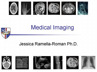
Title:
Medical Imaging
Description:
images from: http://nobelprize.org/medicine/laureates/2003/press.html. X-Ray imaging ... (X-Ray or Gamma-Ray) of a isotope that was injected in a patient to image ... – PowerPoint PPT presentation
Number of Views:774
Avg rating:5.0/5.0
Title: Medical Imaging
1
Medical Imaging
- Jessica Ramella-Roman Ph.D.
2
Instructor
- Office ---gt Pangborn Hall -105B
- Email ---gt ramella_at_cua.edu
- Office phone ----gt 202 319 6247
- Office hours ? By appointment
- Research ---gt Biomedical Optics
- Lab ---gt Pangborn Hall, 118
3
Textbook
- The essential physics of medical imaging, second
edition, J.T. Bushberg, J.A. Seibert,
E.M.Leidholdt, JR. J.M. Boone, Editor Lippincott
WilliamsWilkins - ISBN 0-683-30118-7
- CLASS SLIDES WILL BE DISTRIBUTED
4
More suggested reading
- Naked to the Bone Medical Imaging in the
Twentieth Century (Paperback)by Bettyann Kevles - Emc2 A Biography of the Worlds Most Famous
Equation by David Bodanis
5
Grading
- 30 Homework and class participation
- 30 Midterm Exam
- 40 Final Exam
http//policies.cua.edu/academicgrad/gradesfull.cf
miii
6
CUA Grading System
- http//policies.cua.edu/academicgrad/gradesfull.cf
miii
7
Class Structure
- 1 h 15 min - lecture
- 20 min break
- 1 h 15min lecture and discussion
- I will ask you to read the chapter we are dealing
with before hand.
8
Class calendar
9
Class calendar
10
Class calendar
11
Class calendar
12
WHAT IS MEDICAL IMAGING?
13
Medical imaging
- Includes many imaging modalities
- X-ray, Ultrasound, CT, PET, MRI, and many others
- Uses physics, math, engineering tools, biology,
(...) to image different part of the interior of
the human body.
14
Radiation is
- Energy that travels through space and matter
- We are interested in electromagnetic radiation
- Xray
- Visible Waves
- Gamma rays ()
15
Electromagnetic spectrum
wavelength (nm)
1015 1012 109 106 103 100 10-3 10-6
frequency (Hz)
60 16 1012 1018 1024
UV
Radio, TV, Radar, MRI
IR
g-Rays
X-Rays
Radiant Heat
Cosmic Rays
10-12 10-9 10-6 10-3 100 103 106 109
energy (eV)
visible
16
Magnetic resonance Imaging (MRI)
- Paul Lauterbur and Peter Mansfield 1970-1980
- Uses very powerful magnets (1.5 Tesla) and the
nuclear magnetic - resonance properties of the proton
- Detects the radio frequency emitted by the
protons - (Proton spin flip)
- Tomographic imaging modality
- 10 minutes for a complete scan
images from http//nobelprize.org/medicine/laur
eates/2003/press.html
17
X-Ray imaging
- Discovered by Wilhelm Roentgen Nov 8, 1895
- Published December 12, 1895
- Uses X-Rays (0.01-0.1 nm)
- Ionizing radiation
- 10-100 KeV
- Resolution mm
- Penetration all body
- Mammography is done with
- low power X-rays
images from http//imagers.gsfc.nasa.gov/ems/xr
ays.html
18
Computer Tomography (CT)
- Full body CT inventor 1970-2 Robert Lendley
- Uses X-Rays, rotates the source detector around
the body - Very quick (10 sec)
- Image soft tissue and vascular network
images from http//www.radiologyinfo.org
19
Ultrasound
- Inventor Donald 1957, used on pregnant woman in
1958 - Uses sound waves
- Sound enters the tissues and is reflected by
internal structures - Echoes
- Its a scanning process
- Much less harmful than ionizing mechanism
- Doppler ultrasound is used to measure flow
images from http///www.medical.philips.com
20
Nuclear Imaging
- Inventor Many over the years John Ernest
Lawrence, Ager ... - Uses the decay (X-Ray or Gamma-Ray) of a isotope
that was injected in a patient to image internal
organs (emission images) - Gives information on the physiology of the
patient - PET-gt Positron Emission Tomography
- Radioactive isotopes Fluorine18 and Oxygen 15
emits positrons (e) - e combined to e- produces annihilation radiation
- Annihilation radiation similar to gamma-ray but
produces 2 photons ( in opposite directions) - Uses a ring of detector to record the photon
- Produces a tomographic image
- SPECT-gt Single Photon Emission Computed
Tomography - Gamma ray or Xray emission from the patient is
recorder at several - different angle. (tomography)
- Radioactive isotopes Fluorine18 and Oxygen 15
emits positrons (e)
images from http///www.medical.philips.com
21
- PLEASE READ CHAPTER 1 and 2 of the text book by
next week,
Recommended
CrystalGraphics Presentations


