
Anoxia tolerance and expression of heat shock proteins in myocardium of animals
1 / 1
Title: Anoxia tolerance and expression of heat shock proteins in myocardium of animals
1
Anoxia tolerance and expression of heat shock
proteins in myocardium of animals Xxxx
Xxxx Southwestern Oklahoma State University
ABSTRACT Heat shock proteins are proteins
produced in the cell in response to different
stresses such as greatly increased or decreased
temperature, hypoxia, anoxia, ischemia, and heavy
metals. These proteins not only protect the cell
from the current stress, but also impart a
tolerance for that stress upon subsequent
exposures. The cardiac cells of animals are one
type of cell that produces these heat shock
proteins (Hsps). Mammalian myocardium is
particularly sensitive to hypoxia or ischemia.
Turtles, on the other hand, are quite tolerant of
anoxic environments, surviving up to 12 weeks
while being submerged in oxygen-free water.
Intermittent hypoxia has been shown to increase
the degree of anoxia tolerance while decreasing
the amount of heat shock proteins expressed.
Studies have also been done on the possibility of
conferring on a cell the over-expression of
particular Hsps without having to go through the
adaptation stage. This has been tested by
transfecting myocardial cells with plasmids
containing genes which, once transcribed, produce
Hsps. The goal of this paper is to investigate
the differences in Hsps expression in the
myocardium of painted turtles and softshell
turtles compared to that of rabbits and rats to
assess the role of intermittent hypoxia in
decreasing Hsps production and to examine the
possibility of gene therapy in Hsps production.
Hsp60 Hsp60 is a constitutive stress protein
that is found in the matrix of mitochondrion
(Chang, et al., 2000 Snoeckx, et al., 2001.)
Hsp60 presence seems to be critical for normal
protein import into mitochondria, and thus is
fundamental for mitochondrial function (Snoeckx,
2001). Myocardial expression of Hsp60 under
normoxic conditions appeared to correlate with in
vivo anoxia tolerance (Chang, et al., 2000). The
most anoxic-tolerant animal, the painted turtle,
had myocardial Hsp60 levels five times higher
than rat heart, three times higher than rabbit,
and nearly twice as high as softshell turtle
heart. The softshell had levels much higher
(Plt0.05) than observed in both mammalian species.
Thus, constitutive levels of Hsp60 maybe one
evolutionary advantage turtles use to survive
periods of anoxia (Chang, et al., 2000). Hsp70,
Hsp72/73 The HSP70 family mainly functions as
chaperone proteins (Chang, et al., 2000 Snoeckx,
et al., 2001). Hsp72 is the inducible form,
while Hsp73 is the constitutive form (Chang, et
al., 2000). A high degree of tolerance to
ischemic injury and thermal stress exists in rat
myogenic cells that highly express Hsp70 (Chang,
et al., 2001 Heads, et al., 1994). Chang, et
al., (2000) reported all four species had
absolute concentrations of Hsp72/73 lower than
the concentration levels of Hsp60. The rapid
return (within 12 hours) of Hsp72/73 to control
levels in painted turtles seemed to be an
indicator of the species having a more finely
turned stress protein response than the softshell
turtle (Chang, et al., 2000). Mohan, et al.,
(2001) reported intermittent hypoxia (IH) reduces
the amount of Hsp70 in atria of guinea pigs, but
remained unchanged in the ventricles. Hsp90 and
to a lesser extent Hsp70 remained suppressed
following in-vitro hypoxia (IV) in IH animals.
Thus, Hsp expression in atria following IH may
act as a cellular marker for long-term adaptation
to hypoxic stress rather than a direct modulator
of the cellular mechanism underlying the
pre-conditioned response of the atria
itself. Other than pre-conditioning a cell to a
stress and causing the expression of Hsps, it has
been shown that transfecting a cell with the
needed gene can confer protective measures on the
cell. Heads, et al., (1994) reported the
transfection of a myocyte cell line with a vector
containing a single human Hsp70 gene, which is
capable of driving high levels of expression of
the inducible form of Hsp70 protein, can confer
thermotolerance to these cells. Over-expression
Hsp70 alone can confer at least the same degree
of protection as pre-conditioning with a
sublethal stress. This is important because the
approach can be further developed in terms of
determining the role of individual Hsps in
adaptation to different stress and in terms of
developing a strategy in which resistant
phenotypes can be exogenously conferred on
myocytes (Heads, et al., 1994). Future
implications As of yet, few studies have been
done on heat shock protein production in human
cells. It is known that Hsps usually serve as
chaperone proteins in human cells, but not much
is known about any protective measures of human
Hsps. CONCLUSIONS While heat shock proteins
usually serve as chaperones for protein folding
in animal cells, they do confer cardioprotection
in cardiac cells following sublethal stresses.
Specifically, heat shock proteins are important
in the toleration of environments with low
oxygen. Some animals have shown that
pretreatment with bouts of intermittent hypoxia
confer the same degree of anoxia tolerance while
reducing the amount of heat shock proteins
produced. Other studies have shown that it is
possible to impart heat shock protein production,
thereby improving the tolerance for anoxia, while
avoiding the preconditioning stage usually
needed.
Introduction It has been shown that heat shock,
defined as body temperature being raised 5C
above normal body temperature, elicits a heat
shock response characterized by the synthesis of
new heat shock proteins (Hsps) normally absent in
tissues of adult animals and by an increased
synthesis of constitutively present heat shock
proteins (Snoeckx, et al. 2001 Tanonaka, et al.,
2001). This event is followed by an increased
tolerance to normally lethal temperatures and
resistance toward other events like hypoxia,
ischemia, and inflammation. The production of
Hsps following a particular sublethal stress is
dependent on the presence of a transcription
factor known as the heat shock factor (HSF).
Several factors can lead to HSF activation (Fig.
1). The basic event leading to the transcription
of an hsp-gene is the unfolding of proteins. A
balance exists between the binding of cognate
Hsps to HSF and to stress-mediated unfolding of
another protein. When proteins unfold due to
stress, Hsps detach from HSF and attach to the
unfolded protein. This frees HSF for activation
which leads to the transcription of hsp-genes.
(Morimoto, et al., 1992) As with other organs,
the cells of the heart produce heat shock
proteins. Specific sublethal stresses of hypoxia
and ischemia elicit the production of heat shock
proteins in the myocardium (Chang, et al., 2000).
Mammalian cardiac cells are particularly
sensitive to hypoxia or ischemia. During these
stresses, a rapid depletion of high energy
phosphate compounds occurs along with
intracellular acidosis and decreased cardiac
function (Chang, et al., 2000). Differences
occur among mammalian species as to how tolerant
a species is to anoxia, but all mammals
inevitably die after prolonged bouts without
oxygen under normal conditions. In contrast,
cardiac muscle cells of freshwater turtles are
extremely tolerant of anoxia. Some species of
turtles can maintain cardiac function for 12
weeks of submergence in O2-free water at 3C and
recover to control level of contraction rate and
pressure generation after air breathing (Herbert
Jackson, 1985). The goal of this paper is to
determine if Hsps play a role in the anoxic
tolerance exhibited by turtles and if it is
possible to increase anoxia tolerance while
decreasing the amount of Hsps produced.
References Cited Change, J., A.A. Knowlton, and
J.S. Wasser. 2000. Expression of heat shock
proteins in turtle and mammals hearts
relationship to anoxia tolerance. Am. J. Physiol.
Regulatory Integrative Comp. Physiol.
278R209-R214. Heads, R.J., D.S. Latchman, and
D.M. Yellon. 1994. Stable high level expression
of a transfected human Hsp70 gene protects a
heart derived muscle cell line against thermal
stress. J. Mol. Cell Cardiol. 26695-699. Herbert
, C.V., and D.C. Jackson. 1985. Temperature
effects of the responses to prolonged
submergence in the turtle Chrysemys picta
bellii. II. metabolic rate, blood acid-base and
ionic changes, and cardiovascular function in
aerated and anoxic water. Physiol. Zool.
58670-681. Mohan, R.M., S. Golding, and J.
Paterson. 2001. Intermittent hypoxia improves
atrial tolerance to subsequent anoxia and
reduces stress protein expression. Acta Physiol.
Scand. 17289-95. Morimoto, R.I., K.D. Sarge,
and K. Abravaya. 1992. Transcriptional regulation
of heat shock genes. J. Biol. Chem.
26721987-21990. Snoeckx, L.H., R.N. Cornelussen,
F.A. Van Nieuwenhoven, R.S. Reneman, and J. Van
Der Vusse. 2001. Heat shock proteins and
cardiovascular pathophysiology. Physiological
Reviews. 811461-1490.
METHODS Chang, et al. (2000) tested the
difference in the expression of Hsps in western
painted turtles, softshell turtles, rats and
white rabbits. Turtles undergoing anoxia were
compared to the rest of the animals kept in
normal or normoxic conditions. Normoxic animals
were allowed free access to air while anoxic
turtles underwent forced submergence. Blood
samples from the animals were tests for the
presence of Hsp60 and Hsp72/73. Mohan, et al.
(2001) tested whether intermittent hypoxia
improved tolerance to anoxia and reduced
expression Hsps in guinea-pigs. The researchers
used difference combinations of intermittent
hypoxia, in-vitro hypoxia, and control on the
animals. The amount of heat shock protein
present in the cardiac tissue was measured by
monoclonal anti-Hsp70 and anti-Hsp90
antibodies. Heads, et al. (1994) tested whether
cells from a clonal muscle cell line called
myocytes, which retain certain features of
cardiac cells, can be protected against sublethal
stresses by transfection with an Hsp70 gene
thereby directly conferring thermoresistance
while bypassing the adaptation process. Control
cells were pre-conditioned with sublethal heat,
allowed to recover, then subjected to a severe
heat stress to determine the time needed for
normal accumulation of Hsp70 and development of
thermotolerance in normal cells. A second group
of cells were transfected with either a control
vector or a vector carrying a full-length DNA
coding for human Hsp70. These transfectants were
then subjected to a normally lethal heat stress.
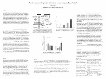
RESULTS During normoxic treatment, there was a
significant difference in the amount of Hsp60
expressed in the two turtle species, rabbits, and
mice. The most anoxic-tolerant animals, the
painted turtle, expressed the highest levels of
Hsp60, followed by softshell turtles, rabbits,
and mice (Fig. 2). Hsp72/73 expression amount
the species was not significantly different.
Following anoxic treatment, Hsp60 levels in
either turtle species did not deviate
significantly from control values after 12 h of
anoxia or 12 or 24 h of recovery. The changes in
the expression of Hsp72/73 were quite different
between the painted and softshell turtles (Fig.
3). Animals tested for a change in heat shock
proteins expression following intermittent
hypoxia showed typical adaptations to hypoxic
exposure when compared to the control group,
which were allowed free access to air. IH groups
also had significantly (Plt0.05) higher absolute
heart weights as well as ventricular weight/body
ratios (Mohan, et al., 2001). No expression of
Hsp70 was observed in cells transfected with the
control plasmid therefore, the process of
transfection was not stressful to the cell. The
magnitude of thermoresistance of the experimental
cells was similar to, or slightly greater than,
that observed in cells which had adapted to heat
stress by producing Hsp70 endogenously (Heads, et
al., 1994).
Recommended
