Pseudoseptic Arthritis in Diffuse Systemic Sclerosis PowerPoint PPT Presentation
1 / 1
Title: Pseudoseptic Arthritis in Diffuse Systemic Sclerosis
1
Pseudo-septic Arthritis in Diffuse Systemic
Sclerosis
Elise Martin1 Dr. Sean Whelton, MD2
1Georgetown University School of Medicine,
Washington, DC 2Department of Medicine,
Georgetown University Hospital, Washington, DC
Georgetown University
erosions and/or digital ulcers as well? In the
reported cases of intra-articular calcification
in SSc, 4 cases specifically note the presence of
ulcers.8-10 2 cases specifically noted
erosions.6,8 The remaining cases did not discuss
whether these factors were present or not. While
this information can not positively correlate
either erosions or ulcers with intra-articular
calcinosis, it does provide reason to consider
evaluating for them in a case series.
Another important possible risk factor that
should be examined is the presence of
subcutaneous calcinosis, which is common in SSc.
Our patient had soft tissue calcinosis before she
developed the intra-articular calcification.
This was also the case in several of the patients
described in the literature with acute
intra-articular calcification.6,7,9,10 The
presence of soft tissue calcinosis was not
discussed in the remaining case reports. While
this information may not help in identifying at
risk patients, since calcinosis is much more
common, it may aid in treatment and prevention
strategies once effective treatments for
subcutaneous calcinosis are identified.
An important concern for our patient is the risk
of recurrence of acute joint calcification. In
the study by Fam and Pritzker, two of the
patients discussed with SSc and intra-articular
calcification had had more than one attack at the
same joint.9 They explained that multiple
attacks can occur and are most likely to recur at
the same joint. Based on their research, the
patient discussed in this report is at risk for
future recurrences. It would be difficult to
estimate her actual risk of recurrence, however,
since so few cases of intra-articular
calcifications have been documented and long term
follow up has not been reported. Thus
far, no specific treatments have been tested to
treat intra-articular calcification or prevent
possible recurrences. At discharge, this patient
was only sent home with narcotic pain control and
close follow up. In the study by Fam and
Pritzker, the patients were sent home on a 7-21
day course of NSAIDS, which consisted of either
Indomethacin or Naproxen.9 While specific
treatments for intra-articular calcification have
not been identified, several treatments for
subcutaneous calcinosis in SSc are currently
being evaluated. Case reports have been
published with mixed reviews for various
treatments for subcutaneous calcinosis and
calcinosis universalis, including diltiazem,12-13
warfarin,14-17 minocycline,18 and colchicine.19
None of these studies were able to demonstrate
reduction of subcutaneous calcinosis in a large
enough sample size to conclusively demonstrate
effectiveness, but some of the other effects of
these medications, including improvement in blood
pressure or antibiotic properties may provide
additional reasons to attempt this medications.
While these medications have not been studied in
intra-articular calcifications in SSc, they
appear to be reasonable options if this patients
calcifications should return. The
mechanism behind intra-articular calcification
has not been established. Devogelear, Huaux, and
Maldague identified two possible theories to
explain this process.8 Their first theory
explained that the deposition of the calcium and
hydroxyapatite was primary and lead to
destruction of the joint and the patients
symptoms. In their second theory, primary joint
destruction occurs and that destruction leads to
the calcium deposition in the joint. They
believe that the second theory is more likely and
we agree. SSc is known to cause significant
joint damage,5 even without intra-articular
calcification, and it is reasonable that this
damage can lead to the presence of calcium
phosphate and its deposition in the joint space.
In February 2009, the patient presented
for the worsening left elbow pain and swelling.
The pain extended proximally into the mid
proximal arm and distally to the proximal
forearm. She denied any trauma to the joint.
She also denied fevers, but reported subjective
chills and night sweats. On physical exam, the
left elbow was warm and swollen, with no
erythema. The joint was fluctuant and
exquisitely tender to palpation. It was
partially flexed and she was unable to extend or
further flex the joint. The non-healing ulcers
were still present on her right hand. The
lesion was initially concerning for septic
arthritis, with the presumed source being the
non-healing digital ulcers. The joint was
aspirated and a white, chalky material was
removed from the joint. Cytopathology of the
synovial fluid demonstrated microcalcification
and scattered acute inflammatory cells in a
background of a fibrinous material. A grams
stain of the fluid did not demonstrate any
bacteria and only a few WBCs were present. The
culture of the fluid did not grow out any
organisms. The sample was evaluated for the
presence of crystals and none were noted in the
sample. The elbow joint was x-rayed to
assess for trauma figure 1 after the initial
aspiration. X-rays revealed degenerative changes
at the elbow joint with calcific lesions adjacent
to the joint. There was a posterior displacement
of the posterior fat pad, consistent with an
effusion. There was irregularity and destruction
of the articular surface of the distal humorus at
the elbow. The pain and swelling
continued after the aspiration. An irrigation
and debridement of the left elbow was preformed
two days after the initial aspiration and an
additional 20 ml of milky white aspirate was
removed. The culture and crystal examination of
the fluid were also negative. The patient
was discharged on her home medications and pain
control with Percocet. At 1 week follow
up, the patient noted significant improvement in
both range of motion and pain. 1 month later,
the patient experienced a recurrence of symptoms.
Summary
Joint involvement is a common feature in systemic
sclerosis. Acute calcification of the joints,
however, has only been reported in few cases of
scleroderma and it has not specifically been
reported in diffuse scleroderma. Here we report
a case of acute intra-articular calcinosis of the
elbow joint in a patient with diffuse
scleroderma. She presented with an acute
pseudo-septic arthritis. Her elbow joint was
aspirated and a chalky, white fluid was removed.
Evaluation of synovial fluid demonstrated the
presence calcium phosphate particles. This case
report discusses the need for further evaluation
of the incidence of intra-articular calcinosis in
scleroderma, possible correlations with potential
risk factors, the likelihood of recurrence, and
the need for effective treatment options.
Introduction
Systemic sclerosis (SSc) is a connective
tissue disorder characterized by autoimmunity,
inflammation, vasculopathy, and extensive
fibrosis. The etiology behind this disorder is
still unknown.1 SSc is typically
divided into two categories based on the extent
of skin involvement limited cutaneous and
diffuse cutaneous.2 In limited scleroderma, the
skin fibrosis is limited to the distal
extremities. It is sometimes referred to as
CREST syndrome (calcinosis, Raynaud phenomenon,
esophageal dysmotility, sclerodactyly, and
telangiectasia).2,3 There is an association with
other internal organ disease, including pulmonary
arterial hypertension and gastrointestinal
dysmotility.2,3 Limited scleroderma is typically
associated with the presence of anti-centromere
antibodies.3 Diffuse scleroderma is
characterized by widespread skin fibrosis that
includes the distal and proximal extremities, the
face, and the trunk. Patients with diffuse
scleroderma may also have arthritis, myopathy,
joint contractures, interstitial lung disease,
pulmonary arterial hypertension, and various
autoantibodies, including anti-Scl 70
(anti-topoisomerase I). The skin changes
progress rapidly in these patients and they have
an early onset of internal organ involvement.2
The two classifications of SSc also carry
prognostic value. Limited scleroderma carries a
better prognosis and the onset of internal organ
involvement is delayed in comparison to diffuse
scleroderma. Joint involvement is a
common feature in SSc and has been reported in up
to 46-97 of patients with SSc.4 A study of 38
patients by Baron and Keystone found that 66 of
patients had joint pain, 61 had joint
inflammation, 42 had periarticular osteoporosis,
34 had joint space narrowing, and 40 had
erosions.5 50 of these patients also had
calcinosis of soft tissues.5 Intra-articular
calcification, however, has only been reported in
few cases in SSc and is not a common feature in
this disease.6-11 In a review of the
literature for cases of SSc and intra-articular
calcification, only a handful of cases have been
reported in the English literature. Here we
report a case of a woman with a pseudo-septic
joint that further testing of the aspirated
synovial fluid demonstrated the presence of acute
intra-articular calcification.
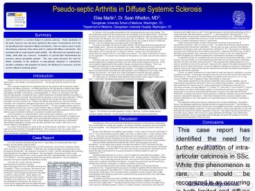
A
B
Figure 1 The left elbow joint after aspiration
of chalky, white fluid. A) Elbow partially
flexed. B) Elbow partially extended. Circles
indicate location of the intra-articular
calcifications.
Discussion
Conclusions
Cytopathology of the synovial fluid
demonstrated the presence of intra-articular
calcifications. Since examination for crystals
was negative, the sample most likely represents
calcium phosphate and hyproxyapatitie. The
radiographs of her elbow support the presence of
calcium in the joint space. Review of the
literature has identified approximately 11 other
cases of acute joint calcification in SSc.6-11
The involved joints include the digits, wrist,
knee, and elbow joints. In each of these cases,
a chalky, white fluid was aspirated from the
affected joint and analyzed for crystals and
bacterial. Each case identified calcium
phosphate in the synovium and was believed to be
hydroxyapatite. Radiographic analysis further
confirmed the presence of calcification in the
joints of these patients. Fam and
Pritzker identified three patients with CREST
syndrome and acute calcific periarthritis.9 In
their article, they propose a possible link
between CREST syndrome and intra-articular
calcification. This was supported by another
case report of acute intra-articular
calcification in a patient identified as having
CRST.6 The other case reports of intra-articular
calcification did not specify the type of
scleroderma or include the presence of antibody
patterns to allow for classification.7,8,10,11
The patient described above, however, is
Scl-70 positive and anticentromere negative.
This information, combined with her clinical
presentation of SSc, strongly supports a
diagnosis of diffuse scleroderma. This is
interesting because the other case reports of
intra-articular calcification were mainly seen in
patients with limited scleroderma. No cases have
previously reported specifically identified
intra-articular calcification in a patient with
diffuse scleroderma. This new information
points to the need for further recognition of
acute intra-articular calcification in SSc. It
appears that this process is not limited to
patients with the limited form of SSc and can
also be present in the diffuse form. Further
evaluation of the actual incidence of acute
intra-articular calcification in SSc should be
further evaluated to provide clinicians with a
better understanding of the disease process when
a patient with SSc presents to them with what
initially appears to be a septic joint.
No specific risk factors have been correlated
with intra-articular calcinosis in SSc.
Subcutaneous calcinosis, however, has been noted
more frequently in patients with articular
erosions5 and/or digital ulcers.4 The patient
described above has both erosions and digital
ulcers, which raises an interesting question is
there a correlation between intra-articular
calcinosis and
This case report has identified the need for
further evaluation of intra-articular calcinosis
in SSc. While this phenomenon is rare, it should
be recognized is as occurring in both limited and
diffuse scleroderma. Further evaluation of the
actual frequency, risk factors, recurrence risk,
and possible treatment options should be
evaluated.
Case Report
A 51 year old African American female
with a 1 week history of worsening left elbow
pain, swelling, and decreased range of motion.
The patient has a past medical history of
mild hypertension, but had been otherwise healthy
until 5 years prior to her presentation. At that
time, she developed fatigue and polyarthritis of
both her large and small joints. Her symptoms
continued for 2 years. She then developed skin
thickening that extended from her fingers to her
elbows. She was tested for the presence of
anti-nuclear antibodies and was noted to have a
both a positive ANA with a speckled pattern and a
positive anti-Scl-70 Table 1. She was
diagnosed at that time with diffuse scleroderma.
She was started on prednisone because of
significant inflammatory arthritis.
References
1. Varga, J., Abraham, D. Systemic sclerosis
a prototypic multisystem fibrotic disorder. J
Clin Investigation 2007117557-567. 2. Chung,
L., Lin, J., Furst, D.E., Fiorentino, D. Systemic
and localized scleroderma. Clinics in Dermatology
200624374-392. 3. Fritzler, M.J., Kinsella,
T.D. The CREST syndrome a distinct serologic
entity with anticentromere antibodies. Am J Med
198069520-6. 4. Avouac, J., Geurini, H.,
Assous, N., Chevrot, A., Kahan, A., Allanore, Y.
Radiological hand involvement in systemic
sclerosis. Ann Rheum Dis 2006651088-1092 5.
Baron, M., Lee, P., Keystone, E.C. The articular
manifestations of progressive systemic sclerosis
(scleroderma). Ann Rheum Dis 198241147-152. 6.
Albert, J.j Ott, H. Unusual articular
abnormalities in scleroderma. Clin Rheumotol
19843323-327. 7. Brant, K.D., Krey P.R.
Chalky Joint Effusion. The result of massive
synovial deposition of calcium apatite in
progressive systemic sclerosis. Arthritis Rhuem
197720792-6. 8. Devogelaer, J.P.,
Huaux, J.P., Maldague, B., Malghem, J., Noel, H.,
Nagant De Deuxchaisnes, C. Intra-articular
calcification in progressive systemic sclerosis.
Clin Rheumatol 19865262-267. 9.
Fam, A.G., Pritzker, K.P. Acute calcific
periarthritis in scleroderma. J Rheumatol
1992191580-5. 10. Resnick, D., Scavulli, J.F.,
Georgen, T.G., Genant, H.K., Niwayama, G.
Intra-articular calcification in scleroderma.
Radiology 1977124685-688. 11. Resnick, D.
Scleroderma with intra-articular calcification.
J Rheumatol 19785114-115. 12. Dolan, A.L.,
Kassinom, D., Gibson, T., Kingsley, G.H. Case
Report. Diltiazem induces remission of calcinosis
in scleroderma. Brit J Rheumatol 199534
576-578. 13. Vayssairat, M., Hidouche, D.,
Abdoucheli-Baudot, N., Gaitz, J.P. Clinical
significance of subcutaneous calcinosis patients
with systemic sclerosis. Does diltiazem
induce regression? Ann Rheum Dis
199857252-254. 14. Berger, R.G., Featherstone,
G.L., Raasch, R.H., McCartney, W.H., Hadler, N.M.
Treatment of calcinosis universalisis with
low-dose warfarin. Am J Med 19878372-76. 15.
Cukierman, T., Elinav, E., Korem, M.,
Chajek-Shaul, T. Low dose warfarin treatment for
calcinosis in patients with systemic sclerosis.
Ann Rheum Dis 2004631341- 1343. 16.
Lassoued, K., Saiag, P., Anglade, M.C., Roujeau,
J.C., Touraine, R.L. Failure of warfarin in
treatment of calcinosis universalis. Am J Med
198884795-796. 17. Yoshida, S. and Torikai, K.
The effect of warfarin on calcinosis in a patient
with systemic sclerosis. J Rheumatol
1993201233-1235. 18. Robertson, L.P., Marshall,
R.W., Hickling, P. Treatment of cutaneous
calcinosis in limited systemic sclerosis with
minocycline. Ann Rheum Dis 200362267-269. 19.
Fuchs, D., Fruchter, L., Fishel, B., Holtzman,
M., Yaron, M. Colchicine suppression of local
inflammation due to calcinosis in dermatomyositis
and progressive systemic sclerosis.
Clin Rheumatol 19865527-530.
Table 1 Patients antibody pattern at
diagnosis.
Over the next 3 years, she also developed
gastrointestinal involvement, including
gastroesophageal reflux and intermittent diarrhea
and constipation. While on prednisone, the
extent of skin thickening decreased and only
involved her hands and fingers. She developed
contractures of the digits on both her hands and
feet. Small, painful, non-healing ulcers
developed on the tips of the 2nd and 3rd fingers
of her right hand. In the months before her
presentation for elbow pain, the ulcers were
concerning for osteomyelitis and the patient had
been on and off oral antibiotics to treat
non-healing ulcers.
Acknowledgments
I would like to thank our patient, TD, for not
only sharing her medical information, but also
her story.

