Development and Aging PowerPoint PPT Presentation
1 / 54
Title: Development and Aging
1
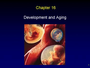
Chapter 16
- Development and Aging
2
Outline
- Fertilization
- Development before Birth
- Fetal Circulation
- Embryonic Development
- Fetal Development
- Birth
- Development after Birth
- Aging
3
Fertilization
- requires the collective action of many sperm
- Several sperm must work to penetrate the corona
radiata - Several sperm must work to penetrate zona
pellucida
4
Fertilization
- enzymes in acrosome of sperm dissolve away the
corona and zona
5
Fertilization membrane
- As soon as sperm penetrates, the eggs plasma
membrane and zona pellucida change to prevent
polyspermy
before
after
6
Fertilization
- One sperm enters egg and nuclei fuse, producing a
zygote
7
Zygote
8
Processes of pre-natal development
- Cleavage - Cell division without growth
- begins with zygote formation
- Morphogenesis - Shaping of embryo
- begins shortly before implantation
- Differentiation - Cells take on specific
structure and function - immediately follows morphogenesis
- Growth - Increase in number and size of cells
9
Embryonic Development
- When a zygote begins dividing, it is termed an
embryo. - Developing embryo travels down oviduct and
eventually implants in endometrium - human chorionic gonadotropic hormone (hCG)
produced by embryo confirms pregnancy - If implantation does not occur, a woman never
knows fertilization took place
10
Development before Birth
- Stages of development.
- Morula - Solid mass of cells resulting from
cleavage. - Blastocyst - Ball of cells formed from morula.
- Embryonic disk - Inner mass of cells of
blastocyst. - Gastrula - Embryo composed of three tissues.
- Ectoderm, mesoderm, endoderm.
11
Fertilization - Implantation
- approximately 7 days
- cleavage leads to formation of morula (mulberry)
- beginning of morphogenesis causes morula to
become a blastocyst - inner cell mass embryo
- trophoblast secretes hCG
12
Pre-implantation Development
13
Post-Implantation Development
- inner cell mass becomes two layers of
cellsembryonic disk - cavity above disk becomes amniotic cavity
- cavity below becomes yolk sac
- gastrulation third layer of cells forms
(mesoderm)
14
Primitive Node and Primitive Streak
- Marks extent of gastrulation
- resulting mesoderm will
- line future coelom
- form somites
15
Neurula Stage
- Mesoderm becomes notochord
- ectoderm located just above the notochord
develops into beginning of nervous system by
induction - induction the process where one tissue induces
another to differentiate
16
Neural Folds
- mark differentiation of ectoderm into neural
tissue
17
Neural Tube
- Neural folds fuse to form neural tube
18
Primitive Streak and Neurula
19
Extraembryonic Membranes
- Membranes external to the embryo
- Amnion - Provides fluid environment for
developing embryo and fetus - Yolk sac - First site of red blood cell formation
- Allantois - Contributes to cardiovascular system
20
Extraembryonic Membranes
21
Placenta
- fetal contribution from the chorion
- complete at 2 months
22
Fetal Circulation
- The umbilical cord stretches between the placenta
and the fetus and contains the umbilical arteries
and veins - Exchange of gases and nutrients between maternal
and fetal blood takes place in the umbilical
arteries - Umbilical vein carries blood and oxygen away from
the placenta to the fetus
23
fetal circulation
24
Embryonic Development
- Embryonic development occurs from the second week
to the eighth week. - Fetal development occurs from the third month
through the ninth month.
25
Summary of Embryonic Development
- blastocyst first week
- Implantation by end of the second week
- gastrulation by end of the third week
- Placenta forming by end of fourth week, complete
by end of second month
26
Day 14 Implantation Complete
27
Day 21 Gastrulation
28
Day 35 Placenta Forming
29
Five Week Embryo
30
End of the Second Month
- all organs have appeared and the placenta is
fully functioning - Embryonic development complete
31
Fetal Development
- At the beginning of the third month, head growth
begins to slow and the body increases in length - Ossification centers appear in bones
- Sex can be determined sometime in the third month
32
Three-Four Month Fetus
33
Fifth through Seventh Months
- Mother begins to feel fetal movement
- Wrinkled skin covered by fine hair, lanugo, is
covered by a greasy substance vernix caseosa - Lungs lack surfactant
34
Six Month Fetus
35
Eighth and Ninth Months
- Fetus usually rotates so head is pointed down
toward cervix - Fetus is now about 530 mm in length and weighs
about 3,400 g - Full-term babies have the best chance of survival
36
Development of Male and Female Sex Organs
- Sex of an individual is determined at the moment
of fertilization? - Gonads arise form indifferent tissue that can
develop into ovaries or testes, depending on the
action of hormones - In the absence of a Y chromosome and in the
presence of two X chromosomes, ovaries develop
instead of testes
37
At 6 Weeks Indifferent Gonads
- Y chromosome causes gonads to become testes
- testosterone secreted by testes causes male
pattern of development - without Y chromosome and/or testosterone, female
(default) pattern of development occurs
38
Development of Male Duct Systems
- mesonephric ducts develop into the male duct
system (epididymis and vas) because of
Testosterone (androgens) - androgens (anti Mullerian hormone) cause
paramesonephric ducts to degenerate
39
Development of Female Duct Systems
- paramesonephric ducts persist and become uterine
tubes and uterus in the absence of testosterone
40
Male versus Female Duct Development
41
At 6 Weeks -- Indifferent Genitalia
- glans and urogenital groove can develop into
either sex
42
Development of Male Genitalia
- by 14 weeks testosterone causes urogenital groove
to close - glans becomes glans and shaft of penis
- labioscrotal folds become scrotum
43
Development of Male Genitalia
- without testosterone urogenital groove does not
close - glans becomes shaft and glans of clitoris
- labioscrotal folds become labia majora
44
Male versus Female Genital Development
45
Birth
- True labor is marked by uterine contractions that
occur regularly every 15-20 minutes and last for
40 seconds or more. - Positive feedback control.
- Parturition.
- Stage 1.
- Mucous plug may be expelled from cervical canal.
- Cervix dilates completely.
46
Birth
- Stage 2.
- Babys head descends into the vagina.
- Baby is delivered.
- Stage 3.
- Placenta delivered.
47
Stages of Parturition
48
Female Breast and Lactation
- Female breast contains 15-20 lobules, each with a
milk duct beginning at the nipple and ending in
alveoli. - In pregnancy, breasts enlarge as ducts and
alveoli increase in number and size. - Milk usually not produced during pregnancy.
- Prolactin suppressed due to increase in estrogen
and progesterone. - Suckling stimulates release of oxytocin.
49
Female Breast Anatomy
50
Development after Birth
- Aging encompasses progressive changes that
contribute to an increased risk of infirmity,
disease, and death. - Theories.
- Genetic in Origin.
- Whole-Body Process.
- Extrinsic Factors.
51
Effect of Age on Body Systems
- Skin.
- Skin becomes less elastic due to changes in
elastic fibers. - Processing and transporting.
- Heart shrinks due to a reduction in cardiac
muscle. - Blood pressure gradually increases.
- Liver not as efficient in metabolizing drugs.
- Blood supply to kidneys reduced.
52
Effect of Age on Body Systems
- Integration and coordination.
- Few neural cells of the cerebral cortex are lost
during the aging process. - Reaction time slows.
- Loss of skeletal muscle mass not uncommon.
- Reproductive system.
- Females undergo menopause.
- Male androgen levels fall between ages 50-90, but
sperm produced until death.
53
Review
- Fertilization
- Development before Birth
- Fetal Circulation
- Embryonic Development
- Fetal Development
- Birth
- Development after Birth
- Aging
54
(No Transcript)

