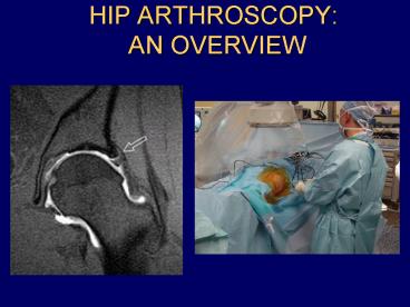
HIP ARTHROSCOPY: AN OVERVIEW PowerPoint PPT Presentation
1 / 55
Title: HIP ARTHROSCOPY: AN OVERVIEW
1
HIP ARTHROSCOPY AN OVERVIEW
2
Purpose
- Review causes of hip and groin pain in athlete
- Discuss indications for hip arthroscopy
- Review, if any, history physical findings of a
patient who may benefit from hip arthroscopy - Review portal placement and anatomy
- Review literature on outcomes of hip arthroscopy
3
AAOS OKU Sports Med 2 Groin Pain in the Athlete
- Athletic Pubalgia
- Rectus abdominus insertion with pain in inguinal
canal - Adductor longus inflammation
- Adductor (Groin) Strain
- Piriformis Syndrome
- Hamstring Syndrome
- Pain overlying ischial tubersosity
4
AAOS OKU Sports Med 2 Groin Pain in the Athlete
- Snapping Hip
- Iliopsoas gliding over iliopectineal eminence or
femoral head - IT band over greater troch
- Biceps over ischial tuberosity
- Iliofemoral ligaments over femoral head
5
AAOS OKU Sports Med 2 Groin Pain in the Athlete
- Iliopsoas tendonitis
- Iliotibial band syndrome
- Osteitis Pubis
- R/O infx, frx, neoplasm, prostatitis,
endometriosis, tendonitis - Primary (noninfectious inflammatory condition
secondary to repetative micro trauma) vs.
secondary
6
AAOS OKU Sports Med 2 Groin Pain in the Athlete
- Contusion
- Hip pointer (ASIS)
- Bursitis
- Fractures
- Stress
- Pelvis
- Femoral neck
- Apophyseal avulsion (ASIS, AIIS, Ischial
tuberosity - Traumatic
- SCFE
7
AAOS OKU Sports Med 2 Groin Pain in the Athlete
- Intra-articular pathology Synovitis
Loose bodies Labral tears AVN DJD
8
Hip Arthroscopy
- Not frequently performed
- Difficult because
- Highly constrained joint
- Deeply constrained by muscular capsular
attachments - Surrounding neurovascular structures at risk
- Equipment is improving
9
Diagnostic Applications of Hip Arthroscopy
- Evaluation of hip pain
- Use as a diagnostic tool when have intractable
hip pain with reproducible physical findings and
functional limitations which fail to respond to
traditional conservative measures - Intra-articular pathology often not evident on
plain x-ray, CT, or MRI - The most common physical finding suggestive of an
intra-articular disorder is a painful inguinal
click when hip is extended from a flexed position.
10
Symptoms of loose bodies
- Locking
- Anterior inguinal pain
11
Symptoms of Acetabular Labral tears
- Anterior inguinal pain
- Painful clicking
- Transient locking
- Giving way
- Positive Thomas extension test
12
Symptoms of a Chondral defect
- Anterior inguinal pain
- Hip arthroscopy should not be performed for
nonspecific pain
13
Therapeutic Applications of Hip Arthroscopy
- Synovitis
- Difficult to diagnose
- Yield biopsy specimen
- Synovectomy
14
Therapeutic Applications of Hip Arthroscopy
- ?efficacy of synovectomy in hip arthroscopically
- Septic Arthritis
- Culture specimens
- Debridement
- Placement of suction drains
- Loose bodies
- Arthroscopic removal
15
Therapeutic Applications of Hip Arthroscopy
- Osteoarthritis
- Aid in staging
- Indicated in young patient with residual joint
space who has failed traditional conservative
therapy - Recent acute change in symptomatology
- Debridement of chondral flaps
16
Therapeutic Applications of Hip Arthroscopy
- Torn Labrum
- Role of acetabular dysplasia
- Lack of lateral and anterior coverage
- Higher incidence of labral tears
- Ligamentum Teres defect and Synovial Folds
- Pediatric Infections
17
Therapeutic Applications of Hip Arthroscopy
- Avascular Necrosis of the Femoral Head
- Diagnostic purposes
- Assess for possible vascularized fibula
- R/O chondral flap tears
- Total hip arthroplasty
- Debris removal
- Loose cement
18
Anatomic Structures at Risk
- Femoral artery
- Femoral nerve
- Lateral femoral cutaneous nerve (LFCN)
- Sciatic nerve
- Gluteal vessels
19
Distance from portal to anatomic structures Byrd,
Arthroscopy, 1995, 11(4)
- Anterior
- ASIS 6.3 cm
- LFCN 0.3 cm
- Femoral nerve at level of sartorius 4.3 cm
- Femoral nerve at level of rectus femoris 3.8 cm
- Femoral nerve at level of capsule 3.7 cm
- Ascending branch of lat circumflex art. 3.7 cm
20
Distance from portal to anatomic structures Byrd,
Arthroscopy, 1995, 11(4)
- Anterolateral
- Superior Gluteal nerve 4.4 cm
- Posterolateral
- Sciatic Nerve 2.9 cm
21
Anterior (Anterolateral) Portal
- Junction between horizontal line at pubic
symphysis and vertical line from ASIS - Angle 45 degrees medially cephalad
- Very close to LFCN, avoid by minimizing skin
incision - Scope visualization of anterior neck, superior
retinacular fold, and ligamentum teres - 70 scope necessary for visualization of anterior
labrum
22
Anterior Paratrochanteric Portal (Anterolateral)
- 2 to 3 cm anterior 1 cm proximal or distal to
the greater trochanter - Visualization of anterior neck and head, capsular
folds, and labrum - If too anterior on approach can damage NV bundle
- Superior gluteal nerve at risk in its course
through the gluteus medius
23
Proximal Trochanteric Portal
- 2 to 3 cm proximal to greater troch
- Directed medially slightly superiorly (aim
toward center of hip) - Visualization of labrum, femoral head, and fovea.
24
Posterior Paratrochanteric Portal (Posterolateral)
- 2 to 3 cm posterior to the greater trochanter
- Sciatic nerve at risk. Especially if leg is
externally rotated - Visualization of posterior capsule
25
Joint Distraction
- Forces can be very high (25 200lb)
- Contribution of physiologic negative
intra-articular pressure - Good anesthesia
- Hip flexion and internal rotation can increase
anterior capsular space (but draws sciatic nerve
closer posteriorly) - Lateral vector should also be used to obtain some
lateral subluxation
26
Positioning
- Supine vs. Lateral
- Some of the laterally based portals allow better
access to labrum anteriorly
27
Supine Position
- Position on table
- Peroneal post positioned for some lateralization
with distraction - Goal of appx 1 cm distraction
- Inject joint to insufflate joint capsule and
release vaccum. This will enhance ability for
distraction - Anterolateral portal is made first
- Anterior portal is then made under direct
visualization - Make posterolateral portal
28
Arthroscopic Anatomy
- From Anterolateral portal
- Anterior wall and anterior labrum
- From Posterolateral portal
- Posterior wall and posterior labrum
- From Anterior portal
- Lateral labrum and its capsular reflection
- Articular surface visualization enhanced by IR
ER of leg - Difficult to see inferior capsule, inferior
acetabulum, and transverse acetabular ligament
29
Contraindications
- Conditions that limit joint distraction
- Protrusio acetabuli
- End-stage DJD
- Ankylosing spondylitis
- AVN pressure changes may effect already
compromised femoral head blood supply
30
Complications
- Traction injuries
- Transient neuropraxia to pudendal and sciatic
nerves - Pressure necrosis to foot, scrotum, or perineum
- Direct neurovascular injury
- Iatrogenic chondral injury
- Iatrogenic labral injury
- Instrument breakage
31
Labral Tears
- Difficult to diagnose
- May not be seen on MRI or double contrast
CT-arthrography - Fluoro guided diagnostic injection often helpful
in differentiating b/w intra- vs. extra-articular
pathology - Despite ineffectiveness in diagnosing labral
pathology, MRI is necessary to r/o Stage I AVN
32
Byrd Jones, Prospective Analysis of Hip
Arthroscopy with 2-Year Follow-up, Arthroscopy,
Vol. 16, No. 6, 2000, 578-587.
- Outcome study of heterogenous patient population
with hip pain. - 38 procedures on 35 patients with minimum of
2-year follow-up - Harris Hip scores pre-op 1, 3, 6, 12, 24 mo.
post-op or until subsequent procedure - Variables studied Age, sex, duration of
symptoms, onset of symptoms, CE angle, diagnosis,
workers comp, and pending litigation.
33
Byrd Jones, Prospective Analysis of Hip
Arthroscopy with 2-Year Follow-up, Arthroscopy,
Vol. 16, No. 6, 2000, 578-587.
- Median Harris Hip scores improved from 57 to 85
- 10 cases ( 9 patients) underwent second procedure
at avg of 10 mo. - Diagnoses
- Labral pathology (23)
34
Byrd Jones, Prospective Analysis of Hip
Arthroscopy with 2-Year Follow-up, Arthroscopy,
Vol. 16, No. 6, 2000, 578-587.
- without chondral injury 31 point improvement
- with chondral injury 18 point improvement
- Chondral damage (15) 18 point improvement
- Arthritic disorder (9) 14 point improvement
- Synovitis (9) 26 point improvement
- Loose bodies (6) greatest improvement 34
points - AVN (4)
35
Byrd Jones, Prospective Analysis of Hip
Arthroscopy with 2-Year Follow-up, Arthroscopy,
Vol. 16, No. 6, 2000, 578-587.
- Poor results of arthroscopy as a palliative
procedure - Cont to question role of arthroscopy in staging
- Perthes (2)
- Synovial Chondromatosis 1
- Ligamentum Teres damage 1
36
Byrd Jones, Prospective Analysis of Hip
Arthroscopy with 2-Year Follow-up, Arthroscopy,
Vol. 16, No. 6, 2000, 578-587.
- No significant difference in results based on CE
angle (only one patient with dysplasia, i.e. CE
angle lt 20), work comp, or pending litigation.
However, anecdotally work comp and litigation
seemed to do better.
37
Onset duration of symptoms
- patients with acute or traumatic onset of
symptoms with greater improvement than those with
insidious onset of symptoms - Longer duration of symptoms especially in male
counterparts correlated with less successful
outcomes
38
Complications
- LFCN neuropraxia resolved
- Myositis of anterior quad following removal of
loose bodies for synovial chondromatosis-
responded to exc.
39
Conclusion
- Hip arthroscopy can be performed for a variety of
conditions (except end-stage AVN) with reasonable
expectations of success.
40
Dorfmann and Boyer, Arthroscopy of the Hip 12
Years of Experience, Arthroscopy, Vol. 15, No.
1, 1999, 67-72.
- Review of 413 patients over 12 years
- 68 for diagnostic purposes
- 32 for operative purposes
- Arthroscopy performed with and without traction
41
Dorfmann and Boyer, Arthroscopy of the Hip 12
Years of Experience, Arthroscopy, Vol. 15, No.
1, 1999, 67-72.
- Labral lesions commonly overestimated at
arthrography. Only 18 cases of 413 confirmed
arthroscopically (4.4) - 93 of 103 arthroscopies for chondromatosis were
therapeutic (90.3) - 55 normal hip scopes 13.3 too high
42
Dorfmann and Boyer, Arthroscopy of the Hip 12
Years of Experience, Arthroscopy, Vol. 15, No.
1, 1999, 67-72.
- Mixed traction technique
- Indications
- Undiagnosed hip pain despite complete work-up
- Undiagnosed catching or locking of the hip
- Diagnostic arthroscopy especially beneficial for
biopsy specimens in inflammatory synovitis, etc. - Removal of loose bodies is main therapeutic
indication
43
Lage, Patel, and Villar, The Acetabular Labral
Tear An Arthroscopic Classification,
Arthroscopy, Vol. 12, No. 3, 1996, 269-272.
- 267 hip scopes
- 37 labral tears
- 4 Etiologies
- Traumatic (7) clear history with no degen
cartilage changes - Degenerative (18) if degenerative changes
present in cartilage or labrum - Idiopathic (10)
- Congenital (2) - two subluxing labra which were
functionally abnormal
44
Lage, Patel, and Villar, The Acetabular Labral
Tear An Arthroscopic Classification,
Arthroscopy, Vol. 12, No. 3, 1996, 269-272.
- Morphological Classification
- Radial Flap (21)
- Radial Fibrillated (8)
- Longitudinal Peripheral (6)
- Unstable (2)
- 62 tears on anterior labrum
- No correlation of tear type and location
associated with etiology - No mention of indications, history, or PE
findings - No mention of outcomes
45
Farjo, Glick, Sampson, Hip Arthroscopy for
Acetabular Labral Tears, Arthroscopy, Vol 15,
No. 2, 1999, 132-137.
- Attempt to define clinical presentation,
diagnosis, and outcome of arthroscopic
debridement of acetabular labral tears. - Retrospective review of 28 labral tears with min.
of one year of follow-up with subjective outcome
analysis.
46
Farjo, Glick, Sampson, Hip Arthroscopy for
Acetabular Labral Tears, Arthroscopy, Vol 15,
No. 2, 1999, 132-137.
- Presenting symptoms
- 36 recalled a specific event
- 64 with mechanical symptoms
- 57 described clicking
- 18 described locking
- 14 giving way
47
Farjo, Glick, Sampson, Hip Arthroscopy for
Acetabular Labral Tears, Arthroscopy, Vol 15,
No. 2, 1999, 132-137.
- Physical exam - no specific reproducible pattern
- provocative positioning ranged from flex/IR to
ext/ER - provocative position did not correlate with
location of labral tear
48
Farjo, Glick, Sampson, Hip Arthroscopy for
Acetabular Labral Tears, Arthroscopy, Vol 15,
No. 2, 1999, 132-137.
- Radiography
- 50 DJD
- MRI pos. in 5 of 21
- Arthrography pos. in 1 of 8
49
Farjo, Glick, Sampson, Hip Arthroscopy for
Acetabular Labral Tears, Arthroscopy, Vol 15,
No. 2, 1999, 132-137.
- Arthroscopic Findings
- 17 tears of anterior labrum
- 7 tears of posterior labrum
- 4 tears of superior labrum
50
Farjo, Glick, Sampson, Hip Arthroscopy for
Acetabular Labral Tears, Arthroscopy, Vol 15,
No. 2, 1999, 132-137.
- Subjective outcome scores
- 13 good results
- 15 poor results
- correlation present between radiographic presence
of arthritis, femoral chondromalacia, acetabular
chondromalacia, and poor result - 10 of 14 (71) with good result in patients
without radiographic evidence of arthritis
51
Farjo, Glick, Sampson, Hip Arthroscopy for
Acetabular Labral Tears, Arthroscopy, Vol 15,
No. 2, 1999, 132-137.
- Complications
- 2 Sciatic nerve palsies
- 1 Pudendal nerve palsy
- All resolved sponteously without sequelae
52
Farjo, Glick, Sampson, Hip Arthroscopy for
Acetabular Labral Tears, Arthroscopy, Vol 15,
No. 2, 1999, 132-137.
- Conclusion
- Good result of labral tear debridement if no
evidence of arthritis - Poor result of debridement if radiographic
evidence of arthritis or arthroscopic evidence of
chondromalacia - Questions the efficacy of Hip arthroscopy for DJD
- Difficult to diagnose labral pathology without
arthroscopy.
53
Byrd, Avoiding the Labrum in Hip Arthroscopy,
Arthroscopy, Vol. 16, No. 7, 2000, 770-773.
- Iatrogenic intra-articular damage to the joint is
likely the most common complication associated
with hip arthroscopy. - Use of cannulated instrumentation
- Anterolateral portal established first blind
under fluoro
54
Byrd, Avoiding the Labrum in Hip Arthroscopy,
Arthroscopy, Vol. 16, No. 7, 2000, 770-773.
- Reposition the needle after breaking the negative
intra-articular vacuum if any concern about
position of needle and guide wire - Use 70 degree arthroscope for direct
visualization of anterior and posterolateral
portals - After making accessory portals look at
anterolateral portal to ensure no labral damage.
55
ThankYou

