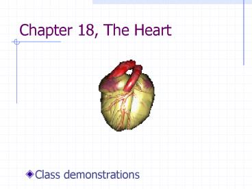
Chapter 18, The Heart PowerPoint PPT Presentation
1 / 36
Title: Chapter 18, The Heart
1
Chapter 18, The Heart
- Class demonstrations
2
Two main functions
- Pulmonary circulation
- Superior inferior vena cava
- R atrium
- R ventricle
- Pulmonary arteries
- Lungs oxygenation
- Pulmonary veins
- Return to heart
18.1
3
Two main functions
- Systemic circulation
- L atrium
- L ventricle
- Aorta
- Arteries
- Capillaries
- Tissues
- Veins
- Vena cava
18.1
4
Location of heart
- Thorax
- Posterior to sternum costal cartilages
- Mediastinum (between lungs)
- Superior to diaphragm
- Anterior to vertebrae and large vessels
18.2abc
5
Location of heart
- Apex of heart
- Left side
- Between ribs 5-6
- Below nipple
- Base of heart
- Posterior surface
5
6
18.2a
6
Pericardium
18.2c
- 3 layered sac
- Fibrous pericardium
- Protective layer
- Attached to great vessels (aorta, pulmonary
artery veins) - Attached to diaphragm
- Anchors heart in place
18.3
7
Pericardium
- Serous pericardium
- Parietal layer
- Visceral layer
- Pericardial cavity
- Serous fluid
- Permits movement
18.3
8
Heart wall layers
18.3
- Epicardium
- Visceral layer of serous pericardium
- Myocardium
- Cardiac muscle fibers
- Arranged in bundles
- Squeezes blood out of heart
- Endocardium
- Continuous with endothelium
18.4
9
Heart chambers
- R-L atria
- R-L ventricles
- Interatrial septum
- Interventricular septum
18.5be
10
External landmarks
- Coronary sulcus
- Anterior interventricular sulcus
- Posterior interventricular sulcus
18.5bd
11
R Atrium
- External landmarks
- R auricle (not labeled)
18.5ad
12
R atrium
not in text
- Internal landmarks
- Superior inferior vena cava
- Coronary sinus
- Fossa ovalis
- Former foramen ovale
18.5e
13
Foramen ovale
- In fetus
- Shunts 1/2 blood from R to L
- Closes at birth (fossa ovale)
19.26a
14
Prenatal Heart Animation
- http//www.heartcenteronline.com/myheartdr/home/sh
ow_animations.cfm?cmbtopics174 - this is slow sometimes
15
R atrium
- Tricuspid valve
- R atrioventricular valve
18.5e
16
R ventricle
- External landmarks
- Coronary sulcus
- Anterior interventricular sulcus
- Posterior interventricular sulcus
18.5b
17
R ventricle
- Internal landmarks
- Trabeculae carnae
- Tricuspid valve
- Valve flaps
- Chordae tendineae
- Papillary muscles
- Pulmonary trunk
- Pulmonary semilunar valve
18.5e
18
L atrium
- External landmarks
- 2R 2L pulmonary veins
- L auricle
18.5bd
19
L atrium
- Internal landmarks
- 4 pulmonary veins
- Mitral valve
- L atrioventricular valve
- Bicuspid valve
18.5e
Episcopal Bishop Persell of Chicago wearing a
miter.
20
L ventricle
- External landmarks
- Coronary sulcus
- Anterior-posterior interventricular sulci
- Apex
18.5bd
21
L ventricle
18.5e
- Internal landmarks
- Mitral valve
- Chordae tendineae
- Papillary muscles
- Trabeculae carnae
- Ascending aorta
- Aortic semilunar valve
- Thicker myocardium
18.7
22
Path of blood through the heart
- Superior inferior vena cava and coronary sinus
- R atrium
- Tricuspid valve
- R ventricle
- Pulmonary semilunar valve
- Pulmonary trunk
- (lungs)
18.17a
23
Path of blood through the heart
- 4 pulmonary veins
- L atrium
- Mitral valve
- L ventricle
- Aortic semilunar valve
- Ascending aorta
18.17a
24
Heartbeat
- Two atria contract
- Two ventricles contract
- 70-80 heartbeats / min, average
- Systole contraction
- Diastole relaxation
25
AV valves
- Prevent backflow from ventricles to atria
- Diastole of ventricles, systole of atria
- Valve open
- Systole of ventricles, diastole of atria
- ? BP in ventricles
- AV valves close
- Papillary muscles contract
- ? tension on chordae tendineae
18.9ab
26
Semilunar valves
- Three cusps
- Prevent backflow from aorta pulmonary trunk
into ventricles - Passive action
- Systole of ventricles opens valves
- Diastole of ventricles
- ? BP
- Backflow fills cusps and closes valve
18.10ab
27
Heart sounds
- lub dub
- lub closing of AV valves
- dub closing of semilunar valves
(Synapse Publishing,Inc.)
http//www.medlib.com/spi/coolstuff2.htm
28
Heart conducting system
- Cardiac muscle fibers form 2 networks via gap
junctions at intercalated discs - Atrial network
- Ventricular network
4.14b
18.4
29
Heart conducting system
- Sinoatrial node
- Internodal fibers
- Atrioventricular node
- Atrioventricular bundle
- Bundle branches
- Purkinje fibers
18.12
30
Heart conducting system
- Sinoatrial (SA) node
- In R atrial wall
- Pacemaker
- Generates AP
- Triggers contraction of atria
- Sends AP along internodal path
18.12
31
Heart conducting system
- Atrioventricular (AV) node
- Triggers ventricular contraction
- Relays AP to AV bundle
- RL bundle branches
- Interventricular septum
- Purkinje fibers
- Near apex
- Turn superiorly
18.12
32
Parasympathetic innervation
- Slows heart rate
- Pathway
- Reticular formation in medulla
- Cardioinhibitory center
- Vagus nerve (CN X)
- To SA AV nodes
- ACh
18.13
33
Sympathetic innervation
- ? heart rate
- ? force of contraction
- Cardioacceleratory center
- Medullary reticular formation
- Preganglionic sympathetic neurons in thoracic
spinal cord - Postganglionic sympathetic neurons to SA AV
nodes - NE, NE ? ? R
18.13
34
Coronary circulation
- L coronary artery
- From ascending aorta
- Two branches
- Anterior interventricular artery (LAD)
- Branches to RL ventricles, interventricular
septum - Circumflex artery
- Supplies L atrium
- Coronary sulcus
18.14a
35
Coronary circulation
- R coronary artery
- From ascending aorta
- In coronary sulcus
- Two branches
- Marginal artery
- Posterior interventricular artery
- Supplies R atrium and ventricle
18.14
36
Coronary Art. Bypass Surgery
- http//www.heartcenteronline.com/myheartdr/home/sh
ow_animations.cfm?cmbtopics183

