Chemical Signals PowerPoint PPT Presentation
1 / 31
Title: Chemical Signals
1
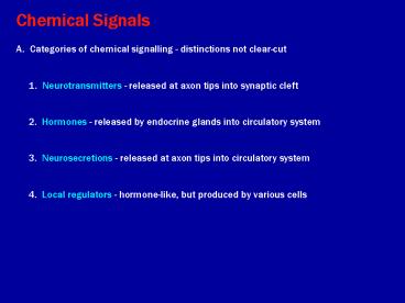
Chemical Signals A. Categories of chemical
signalling - distinctions not clear-cut 1.
Neurotransmitters - released at axon tips into
synaptic cleft 2. Hormones - released by
endocrine glands into circulatory system
3. Neurosecretions - released at axon tips into
circulatory system 4. Local regulators -
hormone-like, but produced by various cells
2
B. Neurotransmitters revisited. 1.
Integral to function of nervous system
2. Various molecules (Ach, norepinephrine,
glutamate, etc.) 3. Synthesized in cell
nucleus, transported to axon tip in vesicles
4. Released into the synaptic cleft
5. Function determined by gated channels
affected
3
Figure 45.4 One chemical signal, different
effects
4
C. Endocrine System 1. Endocrine glands
secrete hormones into circulatory system
2. Hormones circulated everywhere
3. Target specific cell populations. 4.
Only cells that have receptor molecules are
affected
5
Two mechanisms 1. Hormone docks with
receptors on cell surface initiates signal
transduction pathway w/in the cell a.
activates enzymes in cytoplasm b. activates or
inhibits gene transcription 2. Hormone
enters cell and nucleus a. unites with receptor
molecule and b. acceptor site on DNA c.
initiates transcription of mRNA, resulting in
protein synthesis by ribosomes E.g., steroid
hormones
6
Figure 45.5 Human endocrine glands surveyed in
this chapter
7
Figure 45.6b Hormones of the hypothalamus and
pituitary glands
8
Figure 45.3 Mechanisms of chemical signaling a
review
9
Table 45.1 Major Vertebrate Endocrine Glands and
Some of Their Hormones (HypothalamusParathyroid
glands)
10
Table 45.1 Major Vertebrate Endocrine Glands and
Some of Their Hormones (PancreasThymus)
11
D. Neurosecretory cells of nervous system
1. Typical neurons (cell body, axon,
neurotransmitters) 2. Transmitter
synthesized in nucleus and exported to axon
tip a. Released by neurons in posterior
pituitary, taken into
capillaries and circulated as hormones.
Example ADH, oxytocin
produced in neurons originating in hypothalamus
of brain transported down axons to
posterior pituitary released into
capillaries and circulate as hormones
12
Figure 45.6a Hormones of the hypothalamus and
pituitary glands
13
b. Neurons in hypothalamus release hormone into
pituitary portal blood
vessels. Example Gonadotropic Releasing
Hormone (GnRH) Released into portal
capillaries Causes release of Follicle
stimulating hormone (FSH) and
luteinizing hormone (LH) into general circulation
(stimulates testes or ovaries)
14
Figure 45.6b Hormones of the hypothalamus and
pituitary glands
15
- Local Regulators
- 1. Growth factors
- increase rate of development of various tissues
- (including nerve growth)
- b. may affect synaptic transmission in brain
- 2. Nitric oxide (NO) gas
- a. released by many types of cells
- b. toxic to bacteria and certain cancer cells
- c. secreted by endothelial cells of
capillaries - causes dilation - 3. Prostaglandins
- in semen causes contraction of smooth muscles of
uterus, - helping to convey sperm to fallopian tubes
- b. in lungs diff prostaglandins contract or relax
blood vessels
16
- Chemical Signals Case Studies
- Molt and metamorphosis in insects
- Calcium regulation in mammals
- III. Glucose regulation in mammals
- Stress Syndrome in mammals
- Pineal and Biorhythms in mammals
17
Figure 45.0 A monarch butterfly just after
emerging from its cocoon
18
Figure 45.2 Hormonal regulation of insect
development (Layer 1)
19
Figure 45.2 Hormonal regulation of insect
development (Layer 2)
20
Figure 45.2 Hormonal regulation of insect
development (Layer 3)
21
Figure 45.x1 Pupa
22
Figure 45.1 An example of how feedback
regulation maintains homeostasis
23
Figure 45.9 Hormonal control of calcium
homeostasis in mammals
24
Figure 45.10 Glucose homeostasis maintained by
insulin and glucagon
25
Figure 45.11 Derivation of endocrine cells of
the adrenal medulla and neurons from neural crest
cells
26
Figure 45.14 Stress and the adrenal gland
27
- Pineal Gland and Daily and Annual Cycles in
Vertebrates - Pineal gland on top of brain stem, between
cerebral hemispheres - In most vertebrates, contains photoreceptors (not
in mammals) - In mammals receives input from suprachiasmatic
nucleus - and the SCN is the probable biological
clock in mammals - (associated with the optic nerve - hence
light cycle data). - Secretes hormone melatonin during dark period -
affected by SCN - input, but has endogenous rhythym.
- Melatonin affects activity cycles, and
reproductive cycles - (and skin pigmentation in some lower
vertebrates).
28
Figure 45.8 Feedback control loops regulating
the secretion of thyroid hormones T3 and T4
29
Figure 45.12 The synthesis of catecholamine
hormones
30
Figure 45.13 Steroid hormones from the adrenal
cortex and gonads
31
Figure 45.7 Two thyroid hormones

