
PH3MI Medical Imaging - PowerPoint PPT Presentation
1 / 253
Title:
PH3MI Medical Imaging
Description:
Medical imaging has come a long way since 1895 when R ntgen first described a new kind of ray' ... the heart of radiography, angiography, fluoroscopy and ... – PowerPoint PPT presentation
Number of Views:580
Avg rating:3.0/5.0
Title: PH3MI Medical Imaging
1
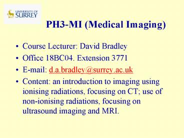
PH3-MI (Medical Imaging)
- Course Lecturer David Bradley
- Office 18BC04. Extension 3771
- E-mail d.a.bradley_at_surrey.ac.uk
- Content an introduction to imaging using
ionising radiations, focusing on CT use of
non-ionising radiations, focusing on ultrasound
imaging and MRI.
2
Medical imaging using ionising radiations
- Medical imaging has come a long way since 1895
when Röntgen first described a new kind of ray. - That X-rays could be used to display anatomical
features on a photographic plate was of immediate
interest to the medical community at the time. - Today a scan can refer to any one of a number of
medical-imaging techniques used for diagnosis and
treatment.
3
Digital Systems
- The transmission and detection of X-rays still
lies at the heart of radiography, angiography,
fluoroscopy and conventional mammography
examinations. - However, traditional film-based scanners are
gradually being replaced by digital systems - The end result is the data can be viewed, moved
and stored without a single piece of film ever
being exposed.
4
Detail detectability test phantoms
5
Detail-detectability
6
The Physical Probe
- X-rays also form basis of computed tomography
(CT) systems, which can obtain a series of 2D
"slices" through body. - Other physical techniques also used single
photon emission CT (SPECT) and positron emission
tomography (PET) rely on use and properties of
radionuclides,while MRI exploits well known
principles of NMR, and is starting point for
fMRI. - Last but not least, ultrasound uses
high-frequency sound in similar manner to
submarine sonar to produce images of tissue and
blood vessels.
7
Prospects
- Even if patients remain absolutely still while
being scanned, a beating heart or the movements
associated with breathing can sometimes distort
the final images. - We therefore look forward to "intelligent
acquisition" systems that allow for the effects
of patient motion.
8
Live-time imaging fluoroscopy
- Used for obtaining cine x-ray images of a patient
in functional studies. - Radiologist uses switch to limit x-ray beam
transmitted through patient. Transmitted beam
falls upon fluorescent screen coupled to an
image intensifier coupled to a TV camera. - Fluoroscopy often used to observe digestive
tract. Also used during diagnostic and
therapeutic procedures, observing action of
instrument used to diagnose or treat patient.
9
X-ray Image Intensifiers (II) for Fluoroscopy
- X-ray II converts transmitted x rays into
brightened, visible light image. - Within II, input phosphor converts the x-ray
photons to light photons, which are then
converted to photoelectrons within photocathode. - The electrons are accelerated and focused by
series of electrodes striking output phosphor,
which converts accelerated electrons into light
photons that may be captured by various imaging
devices.
10
Image Intensifier
11
- Through this process, several thousand light
photons are produced for each x-ray photon
reaching the input phosphor. - Most modern image intensifiers use CsI2 for input
phosphor because it has high absorption
efficiency and thus decreases patient dose. - Image intensifiers come in various sizes, most
having more than one input image size or
magnification mode.
12
Magnification Modes and Spatial Resolution
- Changing voltage to electronic lenses of II will
change magnification of II. - In magnification, smaller area of the input
phosphor is used, giving effect of zooming. - Because input field size is reduced, exposure to
input phosphor must be increased to maintain
constant brightness level at output phosphor. - To maintain same noise level, dose quadruples
when magnification doubled. - The smaller the field size, the larger the
magnification the higher the patient dose. - Higher magnification modes produce increased
spatial resolution. Spatial resolution of II in
range 46 lp/mm. Spatial resolution of system
depends on other imaging components in imaging
chain.
13
Fate of a 50 keV x-ray photon totally absorbed in
input phosphor
- Absorption will result in 2 x 10³ light
photons. Half of these might reach
photocathode. - If efficiency of photocathode 15, 150
electrons released. - If acceleration voltage 25 kV, efficiency of
electron optics is 90 and each 25 keV electron
releases 2 x 10³ light photons in output
phosphor, then 2.7 x 10⁵ light photons
produced. - If 70 of these transmitted through output
window, outcome is light pulse of 2 x 10⁵
photons produced following absorption of a single
50 keV x-ray.
14
Performance Characteristics
- Brightness Gain The gain in image brightness
results from combined effects of image
minification and acceleration of the electrons - Minification Gain Obtained because electrons from
relatively large photocathode focused down to
smaller area of output phosphor. Gives rise to an
increase in number of electrons/mm2. Gain is
given by ratio of areas of input and output
phosphors, expressed as
15
- Thus, for input phosphors with diameters between
15 and 40 cm and output phosphor of 2.5 cm
diameter, minification gain is between 36 and
256. - Flux Gain Results from acceleration given to
electrons as they are attracted from photocathode
to output phosphor. Dependent on applied voltage
and typically between 50 and 100. - Brightness Gain Overall brightness gain given by
- Brightness Gain (Minification Gain) x (Flux
Gain) - Thus, when Minification Gain 100 and Flux
Gain 50 then Brightness Gain 5,000.
Brightness Gains to more than 10,000 are
achievable
16
Limiting Spatial Resolution
- This can be assessed using a Pb bar test pattern,
determining highest spatial frequency that can be
resolved and given in line pairs per mm (lp/mm). - Images of a test pattern are shown in figure
below, where a 23 cm II is operated in 23 cm
(left), 15 cm (middle) and 11 cm (right) mode.
The parameter is generally expressed for centre
of field of view, since it decreases towards
image periphery depending on quality of electron
optics.
17
(No Transcript)
18
General Principles
- Resolution
Ability to discern two points close together
Unresolved
Resolved
19
General Principles
- Contrast
Ability to discern interesting object from noise
or other tissues
Good Contrast
Poor Contrast
20
CT scanning and reconstruction
- As in MRI, computed tomography is a method that
can be used to create cross-sectional images. - CT refers to a generalised methodology for image
reconstruction, the practical techniques of which
can be sub-divided into attenuation CT and
emission CT. - Examples of latter include PET and SPECT. In this
course, we will consider attenuation CT and, in
particular, x-ray CT.
21
CT imaging
- Goal of x-ray CT is to reconstruct an image whose
signal intensity at every point in region imaged
is proportional to µ (x, y, z), where µ is linear
attenuation coefficient for x-rays. - In practice, µ is a function of x-ray energy as
well as position and this introduces a number of
complications that we will not investigate here. - X-ray CT is now a mature (though still rapidly
developing) technology and a vital component of
hospital diagnosis.
22
Principles of x-ray attenuation
- Attenuation CT imaging based on Beers Law,
describing how an x-ray beam is reduced in
intensity as it passes through a medium. - In a uniform substance of linear attenuation
coefficient µ, x-ray intensity, as measured by
a detector placed at depth d is - 1
- where I0 is intensity measured at depth zero.
23
- Suppose we now consider a set of blocks of
different material, each of width ?y. The x-ray
intensity measured at the exit of the set of
blocks is 2 - For the limit ?y ? 0, N ? ?, this becomes
- 3
24
Fig 1 Schematic diagram of a 1st generation CT
scanner
(a) X-ray source projects a thin pencil beam of
x-rays through sample, detected on the other
side of the sample. Source and detector move in
tandem along a gantry. (b) Whole gantry rotates,
allowing projection data to be acquired at
different angles.
25
First-generation CT apparatus
- As shown in Fig 1a, the first-generation CT
apparatus consisted of a source and detector,
placed on either side of object to be imaged.
These slide along in tandem. - Consider intensity of x-ray signal received by
detector when source-detector assembly is at
position x - 4
26
- EMI CT head scanner (Mayo Clinic, Rochester,
Minn, circa 1973)
and an 80 x 80-matrix head CT image obtained with
it.
27
General Principles
- Image Display - Pixels and voxels
28
The Radon transformation
- In a first-generation scanner, the
source-detector track can rotate around the
sample, as shown in Fig 1. We will denote the
x-axis along which the assembly slides when the
assembly is at angle f by xf and the
perpendicular axis by yf. - Clearly, we may relate our (xf, yf) coordinates
to the coordinates in the un-rotated lab frame by
5
29
Figure 2 Relationship between Real Space and
Radon Space
Highlighted point on right shows where the value
?f (xf) created by passing the x-ray beam through
the sample at angle f and point xf is placed.
Note that, as is conventional, the range of f is
-p / 2, p / 2, since the remaining values of f
simply duplicate this range in the ideal case.
30
- Hence, the projection signal when the gantry is
at angle f is - 6
- We define the Radon transform as
- 7
31
Radon Space
- We define a new space, called Radon space, in
much the same way as one defines reciprocal
domains in a 2-D Fourier transform. Radon space
has two dimensions xf and f . At the general
point (xf, f), we store the result of the
projection ?f(xf). - Taking lots of projections at a complete range of
xf and f fills Radon space with data, in much
the same way that we filled Fourier space with
our 2-D MRI data.
32
Fig 3. Sinograms for sample consisting of a small
number of isolated objects.
In this diagram, the full range of f is -p, p
is displayed.
33
Relationship between real space and Radon space
- Consider how the sinogram for a sample consisting
of a single point in real (image) space will
manifest in Radon space. - For a given angle f, all locations xf lead to
?f(xf) 0, except the one coinciding with the
projection that goes through point (x0, y0) in
real space. From Equation 5, this will be the
projection where xf x0 cos f y0 sin f.
34
- Thus, all points in the Radon space corresponding
to the single-point object are zero, except along
the track - 8
- where R (x2 y2)1/2 and f0 tan-1 ( y / x).
- If we have a composite object, then the filled
Radon space is simply the sum of all the
individual points making up the object (i.e.
multiple sinusoids, with different values of R
and f0). See Fig 3 for an illustration of this.
35
Reconstruction of CT images
- This is performed by a process known as
back-projection, for which the procedure is as
follows - Consider one row of the sinogram, corresponding
to angle f. Note how in Fig 3, the value of the
Radon transform ?f(xf) is represented by the
grey level of the pixel. When we look at a single
row (i.e., a 1-D set of data), we can draw this
as a graph see Fig 4(a). Fig 4(b) shows a
typical set of such line profiles at different
projection angles.
36
Fig 4a. Relationship of 1-D projection through
the sample and row in sinogram
37
Fig 4b. Projections at different angles
correspond to different rows of the sinogram
38
Fig 4c. Back-projection of sinogram rows to form
an image. The high-intensity areas of image
correspond to crossing points of all three
back-projections of profiles.
39
Reconstruction of CT images (continued)
- Place the sinogram row at an angle f in real
space. Then smear it out evenly all the way
along the yf -direction. - Repeat the two steps above for all the lines in
the sinogram see Fig 4c. Where the
back-projections overlap, the signal adds
constructively to give high-intensity image
regions.
40
Blurring
- This is not quite the whole story. It turns out
that the image that is produced by this method is
blurred, as shown in Fig 5. - To get exactly the right representation of the
object, we need an additional mathematical
trick called filtering.
41
Fig 5. Blurring problem with non-filtered
back-projection.
A point like object reconstructs to a blurred
object. Since all objects may be regarded as the
sums of a number of point-like objects, every
image will be blurred unless the projections are
first filtered.
42
General Principles- Filtered BP
Forward Projection
Back- Projection
Filtered Back-Projection
Filtered Projection
43
Further generations of CT scanner
- The first-generation scanner described earlier is
capable of producing high-quality images.
However, since the x-ray beam must be translated
across the sample for each projection, the method
is intrinsically slow. - Many refinements have been made over the years,
the main function of which is to dramatically
increase the speed of data acquisition.
44
- Scanner using different types of radiation (e.g.,
fan beam) and different detection (e.g., many
parallel strips of detectors) are known as
different generations of X-ray CT scanner. We
will not go into details here but provide only an
overview of the key developments.
45
Four generations of CT scanner
46
X-rays CT - 1st Generation (1975)
- Single X-ray Pencil Beam
- Single (1-D) Detector set at 180 degrees opposed
- Simplest and cheapest scanner type but very slow
due to - Translate(160 steps)
- Rotate (1 degree)
- 5minutes (EMI CT1000)
- Higher dose than fan-beam scanners
- Scanners required head to be surrounded by water
bag
47
X-rays CT - 2nd Generation (1980)
- Narrow Fan Beam X-Ray
- Small area (2-D) detector
- Fan beam does not cover full body, so limited
translation still required - Fan beam increases rotation step to 10 degrees
- Faster (20 secs/slice) and lower dose
- Stability ensured by each detector seeing
non-attenuated x-ray beam during scan
48
X-rays CT - 3rd Generation (1985)
The General Electric CTi CT scanner (1999)
Scott White Texas A M University College of
Medicine
49
X-rays CT - 3rd Generation
50
X-rays CT - 3rd Generation
- Wide-Angle Fan-Beam X-Ray
- Large area (2-D) detector
- Rotation Only - beam covers entire scan area
- Even faster (5 sec/slice) and even lower dose
- Need very stable detectors, as some detectors are
always attenuated - Xenon gas detectors offer best stability (and are
inherently focussed, reducing scatter) - Solid State Detectors are more sensitive - can
lead to dose savings of up to 40 - but at the
risk of ring artefacts
51
X-rays CT - 3rd Generation Spiral
- Schematic diagram of the operation of a helical
CT scanner - The patient couch translates as the x-ray source
is rotated
http//dolphin.radiology.uiowa.edu/ge/
52
X-rays CT - Multi Slice
Latest Developments - Spiral, multislice CT?
Cardiac CT
53
X-rays CT - 4th Generation (1990)
- Wide-Angle Fan-Beam X-Ray Rotation Only
- Complete 360 degree detector ring (Solid State -
non-focussed, so scatter is removed
post-acquisition mathematically) - Even faster (1 sec/slice) and even lower dose
- Not widely used difficult to stabilise rotation
expensive detector - Fastest scanner employs electron beam, fired at
ring of anode targets around patient to generate
x-rays. - Slice acquired in 10ms - excellent for cardiac
work
X-rays CT - Electron Beam 4th Generation
54
CT Numbers
- Linear attenuation coefficient of each tissue
pixel is compared with that of water
55
- Example values of µt
- At 80 keV µbone 0.38 cm-1
- µwater 0.19 cm-1
- The multiplier 1000 ensures that the CT (or
Hounsfield) numbers are whole numbers.
56
Windowing in CT
57
General Principles
- Image Display - Look up tables
Intensity
159
128
255
0
Pixel Value
Int
Int
Int
Pixel Value
Pixel Value
Pixel Value
Logarithmic
Exponential
Inverse Linear
58
General Principles
- Image Display - Look up tables
Same image windowed to different levels
Colour Look-up table
Brain image
59
Windowing and window level
60
Ring artefact in a third-generation scanner
61
Detail-contrast diagram for a CT scanner
62
Patient Dose in CT
63
Radiation Risks
Risk of fatal cancer - 1 in 20,000 per mSv per
year
64
CT and corresponding pixels in image
65
- Simple numerical
- example
66
The decision/confusion matrix
67
(No Transcript)
68
Sensitivity and specificity
- The test results can be
- True positive (TP) A positive test result in
the presence of the disease - True negative (TN) A negative test result in
the absence of the disease - False positive (FP) A positive test result in
the absence of the disease - False negative (FN) A negative test result in
the presence of the disease
69
sensitivity and specificity 2 x 2 table below
labeled with test result on lhs, disease status
on top
70
Sensitivity vs false-positive rate line D is
worthless, B is good, A is ideal
71
(No Transcript)
72
(No Transcript)
73
(No Transcript)
74
(No Transcript)
75
(No Transcript)
76
sensitivity and specificity
- Sensitivity the ability of the test to identify
the disease in the presence of the disease - TP / (TP FN)
- Specificity the ability of the test to exclude
the disease in the absence of the disease - TN / (TN FP)
77
Ultrasound for imaging
- Basic principle same as used in radar and sonar
and similar to echo-location method of bats. - Emitter sends out pulses of sound. These bounce
off objects and returned echoes provide
information about object, in particular location,
size and reflectional properties. - Gases and liquids support only longitudinal
waves solids support transverse waves as well,
but these are rapidly attenuated for non-rigid,
soft solids.
78
Basic principles
Fig 1. Longitudinal waves in gas
79
- The fundamental equation of ultrasound is
- 1
- where d distance of the reflecting object
from the source/detector of ultrasound - c speed of the ultrasound
- t round-trip time of the pulse, from
emission to reception.
80
What do we mean by ultrasound?
- Acoustic waves with frequencies above those which
can be detected by the human ear. In practice, 20
kHz lt f lt 200 MHz. - An acoustic wave is a propagating disturbance in
a medium, caused by local pressure changes at a
transducer. - The molecules of the medium oscillate about their
resting (equilibrium) positions, giving rise to a
longitudinal waves. - c ? 1540 m/s ? 6.5 µs/cm in most body tissues
- ? c / f 1.5 mm at 1 MHz.
81
Speed of sound
- The speed of sound is a constant in a given
material (at a given temperature), but varies in
different materials - Material Velocity ( m/s)Air 330Water 1497Me
tal 3000 - 6000Fat 1440Blood 1570Soft
tissue 1540
82
Uses of ultrasound imaging
- Most widespread use is in medical imaging
- Non-invasive, low risk
- Obstetrics, abdominal problems, measurement of
blood flow and detection of constrictions in
arteries and veins. - Also used in non-destructive testing in industry
e.g., cracks in structures. - Sonar, underwater imaging (e.g., in submarine
echo-location devices).
Fig 2 Typical obstetric ultrasound scan
83
A-Mode
- Simplest form of ultrasound instrument
- Pulses of ultrasound in a thin beam are emitted
from a transducer into the body and encounter
interfaces between different organs. - Some of the sound energy is reflected at each
stage and some continues through to be reflected
in turn by deeper organs. - The returning pulses are detected by the
transducer and the amplitude of the signal is
displayed on an oscilloscope. If the time-base of
the scope is constant, then the distance across
the screen corresponds to the depth of the object
producing the echo, in accord with Eq. 1.
84
- A-mode imaging gives information very quickly and
involves a minimum of sophisticated apparatus - Weakness is that this information is
one-dimensional i.e., along the line of the
beam propagation. - Nowadays, this mode has been largely superseded
by the brightness B-mode (see later).
85
M-mode first u/s modality to record display
moving echoes from the heart
Typical M-mode images. a. from left ventricle, b
from the mitral valve and c from the aortic valve
86
- A-mode still finds uses in ophthalmology, where
the simple structure of the eye makes it
relatively easy to interpret the echoes and where
what is required are straightforward but accurate
measurements of, for example, distance from the
lens to the retina. - Even this very primitive instrument is not as
straightforward as it might seem. To understand
why, we need to look at a number of principles of
physics, engineering and signal processing.
87
Reflection coefficients
- Reflections occur when the incident wave
encounters a boundary between two materials with
different acoustic impedances. - Acoustic impedance Z is the material property
which relates pressure changes p (in excess of
atmospheric) to the vibrational velocity u of the
particles in the medium. - 2
88
- If we are looking at a single plane wave through
a substance with density ? and speed of sound c,
then Z ?c. - When an incident plane wave, with amplitude pi,
travelling through a medium with acoustic
impedance Z1 hits a boundary with a second
material of impedance Z2 at normal incidence,
there is in general both a reflected wave pr and
a transmitted wave pt - 3
89
Significance of reflection coefficients
- (i) Too little reflection is bad. pr / pi ? 0
- Useful images occur only where there is a
difference in acoustic impedance. Tissues with
strikingly different properties in other respects
may have similar acoustic impedances. From Fig.
3, observe that there is virtually no reflection
at a transition from liver to spleen hence the
two tissues will not be delineated from each
other. - (ii) Too much reflection is bad. pr / pi ? ?1
- If difference in acoustic impedance is too high,
then virtually all the incident ultrasound will
be reflected. This means that the boundary is
opaque to ultrasound. The organ in question will
show up very brightly, but there is an inability
to see through it to find out what is underneath
90
Reflection coefficients at various tissue
boundaries
Figure 4
Note that these are power reflection coefficients
(see later).
91
Implications
- No ultrasound images of brain in vivo skull
reflects ultrasound. - Images of the heart have to be taken round the
ribs, which are also opaque. - Finding the right window into the body is
important.
The ultrasound transducer must be coupled to
the body using a special gel. Before an
ultrasound scan, a thin layer of gel is smeared
onto the skin. Why?
92
Answer
- The material from which transducers are made has
a very different acoustic impedance Ztransducer
to that of the body Ztissue and more importantly
that of air Zair. - These large mis-matches between Ztransducer
and Ztissue and between Ztransducer and Zair
mean that the reflection coefficients at these
interfaces are close to -1. - Little of the signal gets through at a
transducer-tissue boundary (pr/pi ? -0.86) and
virtually none at a transducer-air boundary
(pt/pi ? - 0.9997).
93
- By applying the coupling gel, we exclude all air
from region between probe and body and the worst
case scenario of reflection from a transducer-air
boundary is avoided. The reflection coefficient
is still high (0.86), but imaging is possible. - Some manufacturers use impedance matching to
increase amount of transmitted radiation through
transducer-tissue interface. Inside the probe,
there is a matching layer of thickness ?/4
between transducer and tissue. The acoustic
impedance of the matching material is
approximately - 4
94
- The technique has analogues in optics (blooming
of lenses), electronics (coaxial transmission
lines) and quantum mechanics (scattering of
particles by potential wells). - Note that this technique is not suitable in all
cases and, in particular, a ?/4 layer will match
completely only a single frequency of ultrasound.
95
Fig 5 (a) A large degree of reflection occurs at
the interface between the ultrasound transducer
and soft tissue. (b) If the correct thickness of
an appropriate material is built into the probe,
much improved transmission can be obtained. Note
that there is still a thin gel layer (not shown)
between the matching layer inside the probe and
the tissue. This has approximately the same
acoustic impedance as the soft tissue and is used
to exclude air.
96
What other aspects of wave propagation are
important?
- The formulae above are only strictly valid for
an infinite plane reflecting surface. - In the body, there are many structures which are
much smaller than this (e.g., lung tissue is a
fine network of air-filled tubes). These give
rise to a whole series of interfaces, at random
orientations, and the reflections from these
scatter the incident wave.
97
- At a smaller scale where d ltlt ? (e.g., red blood
cells), Rayleigh Scattering occurs and the degree
of scattering varies as f 4 ? 1/?4. - This means that low frequency ultrasound
penetrates tissue better.
98
Absorption
- This is a phenomenon by which organised
vibrations of molecules (i.e. ultrasound) are
transformed into disorganised, random motion.
Acoustic energy ? Heat - The mechanisms for this transfer include fluid
viscosity, molecular excitations and chemical
changes. It is difficult to measure the
proportion of energy loss which occurs by
scattering and the proportion lost by absorption.
99
- The combined effect of absorption and scattering
may be written as 5 - This also applies to the peak oscillation
velocity u0 and the amplitude of displacement a0
of the particles.
100
- Attenuation is approximately proportional to
frequency, so that the depth of penetration goes
down as f rises. - Instead of using amplitude, attenuation is often
measured in terms of a reduction in the power
density transported by the wave. Consider the
units of pu, where u is the particle vibration
velocity - pressure ? velocity N/m2 ? m/s (Nm)s-1/ m2
W/m2 Power/unit area
101
- i.e., pu represents the power being transported
by the ultrasound through a unit area of the
tissue normal to the direction of propagation. It
is often also called the intensity of the
ultrasound and is represented by the symbol I. - If we look at the power (intensity) attenuation,
we see that
6
102
- Now p0(x) p0(0) e- ax and similarly for u0.
- Hence 7
- Attenuation is often measured on a decibel scale,
where - 8
The power density transported decays twice as
quickly as the vibration amplitude.
103
Diffraction
- Huygens Principle states that each point on a
wavefront can be regarded as a secondary source,
emitting spherical wavelets. The new overall wave
is found by summing the contributions from all
the individual wavelets. - Thus, in an ultrasound imager, all points on the
surface of the transducer producing the
ultrasound act as a source of spherical wavelets. - Also, when the ultrasound passes through an
aperture, each point on that aperture is like a
source of secondary wavelets interference
between these wavelets gives rise to diffraction
effects.
104
- Diffraction becomes significant when the
apparatus dimensions and objects examined become
comparable with the radiation wavelength. Thus
acoustic diffraction (? ? 0.1mm) is a much more
significant effect than optical diffraction (? ?
500nm) for biological tissues.
105
Practical ultrasound imaging
- Fig. 6 shows the block diagram of a practical
A-mode scanner. - The new additions, when compared with the simple
diagram of Fig. 1, are concerned with the
practical problems in trying to use reflected
ultrasound, including - how the same probe both transmit pulses and
receive the echoes - how one deals with the signal attenuation by
tissue and - how the signal is displayed.
106
Fig 6. Block diagram of practical A-scanner. Not
all A-mode scanners include a demodulator. At
each stage, the dynamic range values are
approximate and refer to the power range in the
signal. Take the square root (i.e., halve the dB
value) for the corresponding amplitude ranges.
107
Master Clock (PRF Generator)
- This synchronises the various parts of the
scanner (e.g., transmitter, receiver,
oscilloscope time-base) so that each is triggered
to act at the correct time. - PRF stands for pulse repetition frequency, the
frequency at which clock pulses occur and at
which ultrasound pulses are sent out into the
sample.
108
Transmitter/Transducer/Receiver
- On the leading edge of each clock pulse, either a
momentary voltage step, or a short sinusoidal
burst of voltage is applied to the transducer. - The transmitter which performs this must have a
short rise time, i.e., it must be able to go from
zero to its maximum voltage (100200 V) very
quickly (typically lt 25ns), in order to produce
ultrasound pulses with very high frequency
components.
109
Time Gain Compensation (TGC)
- Problem ultrasound is attenuated as it passes
through tissue. - Thus, even for the same type of reflector, the
signal is less for deeper objects. - This effect is very significant. Worked examples
show a typical a value of 0.15 cm1, so on a
typical return trip of 10 cm, the signal is
reduced by exp ( 0.15?10) 0.22 compared with
reflections coming straight from the skin surface.
110
Solution to differential attenuation
- Amplify the later-arriving signals (i.e. the ones
from deeper in the tissue) more than those from
superficial reflections. i.e., change the
receiver gain with time to compensate for the
echo attenuation. - This is achieved by making the gain of the
amplifier dependent on a control voltage.
Specifically, the input voltage is changed by the
TGC unit. - Because of the logarithmic nature of the decrease
in signal, the TGC should increase the gain a
certain number of dB each ms.
111
Worked Example
- An ultrasound beam propagates in uniform liver
tissue with a 0.15 cm1. - If the speed of sound in the tissue is c 1540
m/s, what should be the rate of gain increase by
the TGC? - Since a 0.15 cm1 is the attenuation
coefficient for amplitude the power attenuation
is double this. - Amplifiers are specified in terms of power, so 2a
0.30 cm1 is what we want. - In terms of dB, we have 10 log10 (e 0.3)
1.3 dB cm1 - So we want a gain increase of 1.3 dB for each cm
of travel to cancel out the differential
attenuation. - Now
- This means that required TGC rate is
- Clearly, a specialised amplifier is needed.
112
Principle of operation of time gain compensation
(TGC)
Fig 7
113
TGC Compensation and pitfalls
- In practice, tissue type varies with depth and
the situation is more complicated. - The user is given a range of controls to vary the
TGC. The rate of increase of gain (i.e., d2G/dt2
) varies with time and hence depth. This is not
an exact science! - Notice, too, that by tweaking the time-gain
controls to get a better images, we lose the
information provided by the attenuation
coefficient. - By using this compensation we are ignoring the
physics of the situation. The fact that one might
not be able to see a particular boundary tells us
something about the properties of that boundary.
114
Demodulator
- At the output of the compression amplifier, the
echo signal mirrors that of the pulse, i.e., it
oscillates at the ultrasound frequency of several
MHz. The display is much easier to understand if
this high frequency modulation is removed. - Another way of describing demodulation is to say
we want to change a signal oscillating at a high
frequency to a lower frequency. This is what we
saw in MRI.
115
The B-mode imager
- This is the commonest form of ultrasound imaging,
resembling radar images. - A thin beam of ultrasound is scanned across the
object and the strength of the returned echoes is
displayed on the monitor. - Notice that whilst in radar, full 360? coverage
is required, in medical ultrasound, where only
the body in front of the transducer is of
interest, we look at a limited pie-shaped
sector.
116
2-dimensional imaging
- Fire beam vertically, wait for echoes, store
information then fire new line from
neighbouring transducer etc in a sequence of
B-mode lines. - In linear crystal array, electronic phased array
shoots parallel beams, with field as wide as
probe length. - Curved array creates a wider field than lateral
probe dimension, making possible creation of
smaller footprint for easier access through small
windows at cost of reduced lateral resolution as
scan lines diverge.
117
Principle of B-scanning
Fig 8
118
- To achieve footprint sufficiently small to get
access to heart between ribs, with sufficiently
wide far field, beams have to diverge from
virtually same point. Hence image has to be
generated by single beam from same point,
deflected in different angles to build a sector
image.
119
- B-scan is simply an A-scan in which the
ultrasound beam is moved and the results are
spatially displayed. The ultrasound signal
changes the brightness of a spot on an
oscilloscope screen instead of amplitude of the
trace in A-mode. - What do we need to add to an A-scanner to turn it
into a B-scanner? As soon as we try to turn the
idea into a working system, we find a number of
problems lurking! How do we display the data
received? How do we make the beam sweep across
the sample? What do our data mean?
120
- Fig. 9 is a block diagram of a generic B-scanner.
Only three new items have been added the
co-ordinate generator, the video amplifier and
the beam-steering device.
121
Fig 9. Block diagram of a B-mode scanner
122
The Co-ordinate Generator
- This device is often also called the scan
converter. - It takes information about the instantaneous
orientation of the beam and turns it into the
co-ordinates of a line on the display monitor. - In simple systems, the CRT electron beam is
physically scanned up and down the desired line
(i.e., the co-ordinate generator acts as a
variable voltage source to the scope x- and y-
plates). - On more modern systems, the co-ordinate generator
gives the memory location in which signal
information is stored. The data is then displayed
on a monitor by a computer program.
123
Compression and Amplifier
- Even after passing through the TGC, the range of
signals in the data is still large. - This is due to the range of reflector strengths
in the body see Fig. 4. - The compression amplifier transforms the data by
some rule Vout f (Vin), which reduces the
dynamic range of the data (i.e., compresses the
scale).
124
- Typically, a 4050 dB dynamic range for Iin
(i.e., the ratio Iin max/Iin min ? 104105) is
transformed to an output dynamic range of 10 20
dB (10 100). Remember take the square root of
these values to get the corresponding voltage
amplitude ranges. - This allows low intensity echoes to be seen on
the same display as high intensity ones, i.e.,
strongly reflecting organ boundaries and weakly
reflecting internal structure can be seen on the
same image. A video monitor can display only
about 256 values simultaneously. - This means that
- (i) a huge amount of information is lost as in
the case of the TGC - (ii) one should not normally interpret B-mode
image intensities quantitatively.
125
The Beam-Steering Device
- This is what distinguishes the different types of
scanner. - There are various levels of distinction. The most
basic is between static and real-time scanners.
126
Static B-Scanners
- The transducer is moved manually by the operator.
- The probe slides backwards and forwards over the
patient, changing its angle. - The image is built up line by line. Each time,
the co-ordinates ?1, ?2 and ?3 tell the display
where on the screen to show the results. See Fig.
10 - The advantage of the system is that the operator
can choose which bits of the picture to update
most often and to tailor the scanning motion to
view the feature of interest from several
different directions - It is also very cheap.
127
Co-ordinate generator for static B-scanner
Fig. 10
128
Various types of beam steering device for
real-time scanners
Fig 11
129
- However ...
- The scans take several seconds to build up and
form a complete picture. This is a problem if the
object in question moves in the meantime. - Static B-scanners are not suitable for imaging
of, for example, a beating heart.
130
Real-Time Mechanical Scanners
- Real time scanners acquire anything from a few
frames (images) per second up to several hundred.
They are ideal for imaging motion. - In a motorised scanner, the transducer is moved
mechanically by a motor. - Because of the difficulties of maintaining
contact between the skin and a moving transducer,
a larger probe is used, which contains the
transducer suspended in a bath of oil, with a
window to allow the pulses to leave.
131
- There are several different designs, as shown in
Fig. 11. In all cases, the final device will
depend on obvious mechanical engineering
questions like - How do you make a probe rock backwards and
forwards very fast? Can you make it do so
uniformly? How do you get leads to three
transducers on a ring without everything getting
tangled up when they rotate? - The major disadvantage of this type of device is
that mechanical systems have an inherent speed
limit. - The advantage is that there is no complicated
(and expensive) electronics.
132
Temporal resolution
- To image moving objects frame rate is important,
related to speed of motion of object. Eye can
generally only see 25 FPS (video frame rate),
giving temporal resolution of 40 ms. Higher
frame rate and new equipment offers possibility
of replay at lower rate, eg 50 FPS played at 25
FPS, doubling effective resolution of eye. - In quantitative measurement, whether based on
Doppler effect or 2D B-mode data, sufficient
frame rate is important to avoid undersampling
(if one undersamples at a certain frequency, then
direction of motion becomes ambiguous more
frequent sampling will give correct direction).
133
- Temporal resolution limited by sweep speed, which
in turn is limited by speed of sound, echo from
deepest part of image having to return before
next pulse sent out. Sweep speed can be increased
by reducing number of beams in sector, or
decreasing sector angle. 1st option decreases
lateral resolution, 2nd decreases image field, so
temporal resolution cannot be increased without a
trade off.
134
Electronic Steering Transducer Arrays
- We shall not go into any detail here, but the
basic principle is that a number of very small
transducer are placed into a line and are then
fired separately. - By firing (i.e., sending out a pulse from) the
transducers at different times, one can make
composite wave-fronts (Huygens Principle again!)
which mimic that given out from one of the moving
transducers above. - The beam is scanned in a sector with a frame rate
of at least 20 Hz to minimise flicker. The probe
has no moving parts.
135
(No Transcript)
136
(No Transcript)
137
(No Transcript)
138
- Electronic beam steering is potentially much
faster than mechanical steering and also has the
advantage that the order of sampling of the
different lines is much more flexible. - All modern scanners work this way.
139
Doppler Effect
- As velocity of sound in any medium constant, wave
propagates outwards in all directions with same
velocity, with centre at point of emission. - As source moves, next wave is emitted from a
point further forward. Thus distance between wave
crests decreased in direction of motion/increased
in opposite direction. - As distance between wave crests is equal to
wavelength, wavelength decreases (sound frequency
increases) in front of source/ increases (sound
frequency decreases) behind it. If source
stationary, effect on moving observer similar.
140
- In u/s, wave sent from stationary transducer,
moving blood or muscle firstly moving towards
transducer and then away thus Doppler shift
approximately twice as great. In case of
reflected ultrasound, Doppler shift is
where ? is the angle between the direction of
the motion and the ultrasound beam, v is the
blood or tissue velocity, c is the sound velocity
in tissue, f0 is the transmitted frequency, fD
is the Doppler shift of reflected ultrasound.
141
- Basically, the Doppler effect can be used to
measure blood and tissue velocities from the
Doppler shift of reflected ultrasound
142
Pulsed and continuous wave Doppler
- Can use pulsed Doppler, where pulse sent out, and
frequency shift in reflected pulse measured at a
certain time. This will correspond to a certain
depth, i.e. velocity is measured at a specific
depth, which can be adjusted. The width is the
same as the beam width, and the length of the
sample volume is equal to length of the pulse.
143
- A problem in this is that Doppler shift is very
small compared to u/s frequency. This makes it
problematic to estimate the Doppler shift from a
single pulse, without increasing the pulse length
too far. A velocity of 100 cm/s with a ultrasound
frequency of 3.5 MHz results in a maximum Doppler
shift of 2.3 kHz. - The solution is to shoot multiple pulses in the
same direction and produce a new signal with one
sample from each pulse.
144
- The pulsed modus results in a practical limit on
the maximum velocity that can be measured. In
order to measure velocity at a certain depth, the
next pulse cannot be sent out before the signal
is returned. The Doppler shift is thus sampled
once for every pulse that is transmitted, and the
sampling frequency is thus equal to the pulse
repetition frequency (PRF). Frequency aliasing
occurs at a Doppler shift that is equal to half
of the PRF. fD ½ PRF
145
Tissue Doppler
- The Doppler principle can be used both for blood
flow and tissue velocities. - Main principle is that blood has high velocity
(typically above 50 cm/s, although also all
velocities down to zero), but low density,
resulting in low intensity (amplitude) reflected
signals. - Tissue has high density, resulting in high
intensity signals, but low velocity (typically
below 20 cm/s). - The difference in the applications used for the
two sets of signals is mainly differences in
filtering, applying a high pass filter in Doppler
flow, and low pass filter in tissue Doppler
(although latter not absolutely necessary).
146
Magnetic Resonance Imaging (MRI)
- Introduction
- Basic MR Physics
- Advanced MR Physics
- MR Techniques
- Artefacts
- Advanced Techniques
- Instrumentation
- MR Safety
147
MRI Introduction
- In 1970s Lauterbur introduced concept of magnetic
field gradients, leading to images based on
magnetic resonance. - By 1980s whole body magnets produced in UK,
permitting first in vivo images of human anatomy.
- An estimated 20 million scans now performed
worldwide annually. - Provides excellent soft-tissue contrast can be
acquired in any imaging plane unlike CT, does
not involve ionising radiation. - Imaging modality of choice in brain and spinal
cord routinely used in many other clinical
settings.
148
The Nobel Prize in Physiology or Medicine 2003
Paul C. Lauterbur
Sir Peter Mansfield
149
- In 1971 Raymond Damadian showed that the nuclear
magnetic relaxation times of tissues and tumours
differed, thus motivating scientists to consider
magnetic resonance for the detection of disease. - In 1973 the x-ray-based computerized tomography
(CT) was introduced by Hounsfield. - This date is important to the MRI timeline
because it showed hospitals were willing to spend
large amounts of money for medical imaging
hardware. - Magnetic resonance imaging was first demonstrated
on small test tube samples that same year by Paul
Lauterbur. - He used a back projection technique similar to
that used in CT.
150
- In 1975 Richard Ernst proposed magnetic resonance
imaging using phase and frequency encoding, and
the Fourier Transform. - This technique is the basis of current MRI
techniques. - In 1991, Richard Ernst was rewarded for his
achievements in pulsed Fourier Transform NMR and
MRI with the Nobel Prize in Chemistry. - A few years later, in 1977, Raymond Damadian
demonstrated MRI called field-focusing nuclear
magnetic resonance. - In this same year, Peter Mansfield developed the
echo-planar imaging (EPI) technique. - This technique was to be developed in later years
to produce images at video rates (30 ms / image).
151
- Edelstein and coworkers demonstrated imaging of
the body using Ernst's technique in 1980. A
single image could be acquired in approximately
five minutes by this technique. - By 1986, the imaging time was reduced to about
five seconds, without sacrificing too much image
quality. - The same year people were developing the NMR
microscope, which allowed approximately 10 mm
resolution on approximately one cm samples. - In 1987 echo-planar imaging was used to perform
real-time movie imaging of a single cardiac
cycle. - In this same year Charles Dumoulin was perfecting
magnetic resonance angiography (MRA), which
allowed imaging of flowing blood without the use
of contrast agents.
152
fMRI
- In 1992 functional MRI (fMRI) was developed.
- This technique allows the mapping of the function
of the various regions of the human brain. - Five years earlier many clinicians thought
echo-planar imaging's primary applications was to
be in real-time cardiac imaging. - The development of fMRI opened up a new
application for EPI in mapping the regions of the
brain responsible for thought and motor control. - In 1994, researchers at the State University of
New York at Stony Brook and Princeton University
demonstrated the imaging of hyperpolarized 129Xe
gas for respiration studies.
153
NMR
- Felix Bloch and Edward Purcell, both of whom were
awarded the Nobel Prize in 1952, discovered the
magnetic resonance phenomenon independently in
1946. - In the period between 1950 and 1970, NMR was
developed and used for chemical and physical
molecular analysis. - For years major application in field of
spectroscopy discerning chemical species from
inherent shift in resonant frequency exhibited by
nuclei depends on chemical environment.
154
NMR
- NMR has become the preeminent technique for
determining the structure of organic compounds. - Of all the spectroscopic methods, it is the only
one for which a complete analysis and
interpretation of the entire spectrum is normally
expected. - Although larger amounts of sample are needed than
for mass spectroscopy, NMR is non-destructive,
and with modern instruments good data may be
obtained from samples weighing less than a
milligram.
155
NMR
- The nuclei of many elemental isotopes have a
characteristic spin (I). - Some nuclei have integral spins (e.g. I 1, 2, 3
....), some have fractional spins (e.g. I 1/2,
3/2, 5/2 ....), and a few have zero spin, I 0
(e.g. 12C, 16O, 32S, ....). - Isotopes of particular interest and use to
organic chemists are 1H, 13C, 19F and 31P, all of
which have I 1/2. - Since the analysis of this spin state is fairly
straight forward, a general introductory
discussion of NMR is usually limited to these and
other I 1/2 nuclei.
156
Basic MR Physics Nuclear Spin Behaviour in a
Magnetic Field
- EM tells us that a current carrying conductor
e.g. a piece of wire, produces a magnetic field
encircling it. - When wire formed into a loop, field acts
perpendicular to surface area of loop. - Analogous to this is field produced by negatively
charged electrons orbiting nucleus in an atom, or
spinning charge of nucleus itself. - Spinning momentum of nuclear charge ('the spin')
produces small magnetic field referred to as
magnetic moment. - Under normal circumstances these moments have no
fixed orientation so no overall magnetic field. - However, when nuclei placed in external magnetic
field, for example patient placed in MRI scanner,
they begin to align in given directions dictated
by laws of QM.
157
Nuclear Spin Behaviour in a Magnetic Field
- In case of hydrogen nucleus (single proton with
spin quantum number, I ½), two discrete energy
levels (2I 1) created - (i) a higher energy level where magnetic moments
oppose the external magnetic field, (ii) a
lower energy level in which the nuclei aligned
with magnetic field. - Tiny majority of spins in latter energy state
thereby creating net magnetisation in direction
of main magnetic field. - Population difference therefore sensitivity of
technique, can be altered by reducing temperature
or increasing field, hence need for strong
magnetic field for modern clinical scanners,
between 0.5 and 3.0 Tesla.
158
Behaviour in a Magnetic Field
- Field referred to as B0 to distinguish from
second field described later. - To put into context, 1 Tesla 10,000 Gauss
Earth's magnetic field varies between 0.3 - 0.7
Gauss. - In terms of classical physics, when spin placed
in a magnetic field it precesses about that field
in a motion analogous to a spinning top. - Frequency of precession governed by the Larmor
equation, ?0 ?B0. - Constant of proportionality in equation is
magnetogyric ratio (or gyromagnetic ratio) with
every 'MR visible' nucleus having its own
specific value in units of Hz/T. - For proton, in field strength of 1.5 T, the
associated frequency is about 63.8 MHz, which is
in radio-frequency (RF) range.
159
Figure (left) A net magnetisation is produced
following the application of an external magnetic
field causing a small majority of spins to align
in the direction of the applied field. (right)
Each spin precesses in a motion which follows the
surface of a cone.
160
RF or Time-varying Magnetic Field
- The quantum or classical physics descriptions are
entirely equivalent - in both cases there is a net magnetisation, M0,
created by the main magnetic field which is the
basis of the imaged signal. - The net magnetisation can be considered in terms
of one big spin. - In order to detect this signal a second magnetic
field is introduced referred to as B1. Two things
are important about this field (i) it has to be
applied perpendicular to B0, and (ii) it has to
be at the resonant frequency.
161
RF or Time-varying Magnetic Field
- Appropriate RF coils are used to transmit B1,
which acts to tip the spins out of alignment with
B0 and towards the direction of the coil (i.e.
out of the longitudinal plane and towards the
transverse plane). - If the pulse is applied for long enough the spins
are flipped into the transverse plane and a 90
RF pulse is said to have been applied. - In the majority of MRI sequences this is the
case. - The RF pulse is then turned off and the signal
can be detected by the RF coil (either using the
same one or a second coil see Instrumentation ).
162
T1 Processes
- At equilibrium, the net magnetization vector lies
along the direction of the applied magnetic field
Bo and is called the equilibrium magnetization
Mo. In this configuration, the Z component of
magnetization MZ equals Mo. MZ is referred to as
the longitudinal magnetization. There is no
transverse (MX or MY) magnetization here. - It is possible to change the net magnetization by
exposing the nuclear spin system to energy of a
frequency equal to the energy difference between
the spin states. If enough energy is put into the
system, it is possible to saturate the spin
system and make MZ0. - The time constant which describes how MZ returns
to its equilibrium value is called the spin
lattice relaxation time (T1). The equation
governing this behavior as a function of the time
t after its displacement is - Mz Mo ( 1 - e-t/T1 )
163
- T1 is the time to reduce the difference between
the longitudinal magnetization (MZ) and its
equilibrium value by a factor of e. - If the net magnetization is placed along the -Z
axis, it will gradually return to its equilibrium
position along the Z axis at a rate governed by
T1. The equation governing this behaviour as a
function of the time t after its displacement is - Mz Mo ( 1 - 2e-t/T1 )
- Again, the spin-lattice relaxation time (T1) is
the time to reduce the difference between the
longitudinal magnetization (MZ) and its
equilibrium value by a factor of e.
164
Relaxation Mechanisms
- At this point a peak in signal is detected which
decays very quickly called the Free Induction
Decay (FID). - The signal arises from the rotating
magnetisation, it decays due to relaxation which
can be subdivided into transverse or T2 decay and
longitudinal or T1 recovery. - T2 decay is the process whereby the millions of
spins begin to dephase. - This is due to the individual spins 'seeing'
local differences in the magnetic field caused by
interactions between them, and they begin to
precess at slightly different rates resulting in
an increasingly dispersed distribution around
'the clock face' (see Figure below).
165
Figure (left) Having been tipped into the
transverse plane, the net magnetisation begins to
dephase (T2). (right) Once fully dephased the
spins return to equilibrium (T1).
166
Relaxation Mechanisms
- This is what causes the signal to decay at this
point. - In actual fact the spins dephase much quicker
than the 'natural' T2 as they also are subject to
inhomogneities in the magnetic field B0 causing
the decay to be characterised by T2. - The second relaxation process governs the spins
return to the original equilibrium situation. - Remember that at this stage, although B1 has been
removed, the main field B0 is always on and the
spins begin to recover back to alignment under
its influence. - The regrowth of magnetisati
Recommended
CrystalGraphics Presentations


