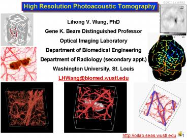
High Resolution Photoacoustic Tomography PowerPoint PPT Presentation
1 / 45
Title: High Resolution Photoacoustic Tomography
1
High Resolution Photoacoustic Tomography
- Lihong V. Wang, PhD
- Gene K. Beare Distinguished Professor
- Optical Imaging Laboratory
- Department of Biomedical Engineering
- Department of Radiology (secondary appt.)
- Washington University, St. Louis
- LHWang_at_biomed.wustl.edu
2
Credits to Lab Members
- CURRENT MEMBERS
- H. Fang
- A. Garcia-Uribe
- S. Hu
- X. Jin
- R. Kothapalli
- C. Kim
- G. Ku
- C. Li
- L. Li
- Y. Li
- K. Maslov
- M. Pramanik
- K. Song
- L. Song
- E. Stein
- M. Todorovic
- X. Xu
- X. Yang
- FORMER MEMBERS
- X. Xie, MS
- M. Xu, PhD
- Y. Xu, PhD
- G. Yao, PhD
- W. Yu, MS
- X. Zhao, MS
- FORMER MEMBERS
- J. Ai, PhD
- D. Feng, MS
- J. Hollmann
- S. Jiao, PhD
- J. Li, PhD
- M. Li, PhD
- G. Marquez, PhD
- M. Mehrubeoglu, PhD
- J. Oh, PhD
- Y. Pang, MS
- S. Sakadzic, PhD
- M. Sivaramakrishnan, MS
- E. Smith, MS
- H. Sun, PhD
- X. Wang, PhD
- Y. Wang, MS
3
Credits to Collaborators
- Texas AM University (Animal study)
- G. Stoica, DVM
- UT MD Anderson Cancer Center (Clinical study
contrast agents) - M. Duvic, MD
- B. Fornage, MD
- K. Hunt, MD
- C. Li, PhD
- V. Prieto, MD
- Nanospectra (Nanoshells)
- P. ONeal, PhD
- J. Schwartz, PhD
- D. Payne
- USC (high-frequency ultrasound array)
- K. Shung, PhD (R. Bitton, PhD candidate)
4
Outline
- Motivation and challenges
- Reconstruction-based photoacoustic tomography
- Confocal photoacoustic microscopy
- Deeply penetrating RF-based thermoacoustic
tomography - Summary
5
Motivation for Optical Imaging
- Safety Non-ionizing radiation photon energy is
2 eV. - Physics Related to the molecular conformation
of tissue. - Optics High intrinsic contrast
- Optical absorption oxyhemoglobin,
deoxyhemoglobin, melanin, and exogenous contrast
agents. - Optical scattering Size of cell nuclei.
- Optical polarization Collagen and muscle fibers.
- Physiology Functional imaging of physiological
parameters - Oxygen saturation of hemoglobin (related to
hyper-metabolism) - Total hemoglobin concentration (related to
angiogenesis) - Enlargement of cell nuclei
- Orientation of collagen
- Denaturation of collagen
- Blood flow (Doppler)
- Physiology Molecular imaging
- Integrin, VEGF, etc
- Reporter genes
- .
6
Challenges in Optical Imaging
OM, SNOM
1 mm
CFM, 2PM, SHM, etc.
Soft limit lt
OCT
DOT, UOT, PAT
Hard limit 10 d 5 cm
- OM Optical microscopy
- SNOM Scanning near-field optical microscopy
- CFM Confocal microscopy
- 2PM Two-photon microscopy
- SHM Second harmonic microscopy
- OCT Optical coherence tomography
- DOT Diffuse optical tomography
- UOT Ultrasound-modulated optical tomography
- PAT Photoacoustic tomography
Simulation software MCML available
from http//oilab.seas.wustl.edu
7
High Relative ResolutionDepth-to-Resolution
Ratio gt 100
8
Outline
- Motivation and challenges
- Reconstruction-based photoacoustic tomography
- Confocal photoacoustic microscopy
- Deeply penetrating RF-based thermoacoustic
tomography - Summary
9
Reconstruction-based Photoacoustic Tomography
Physical Review E 71, 016706 (2005). Phys. Rev.
Letters 92, 033902 (2004).
10
Photoacoustic Forward Solutionin an Infinite
Medium Plane Wave
11
Photoacoustic Forward Solutionin an Infinite
Medium Spherical Wave
12
Experimental Setup of Orthogonal-mode
Photoacoustic Tomography
13
Photoacoustic Inverse Solutionin an Infinite
Medium Time-domain Reconstruction
Beamforming
High-frequency contribution
Physical Review E 71, 016706 (2005). Physical
Review Letters 92, 033902 (2004).
14
Transcranial Functional Photoacoustic Imaging of
RatWhisker Stimulation In Vivo Hemodynamics
Left-whisker stimulation
Right-whisker stimulation
Nature Biotech. 21, 803 (2003).
15
Photoacoustic Angiography of Rat Brains In Vivo
(B) With ICG-PEG
- Without ICG-PEG
- Speckle free
- High resolution 60 mm
- High sensitivity fmol
(D) Open-skull photo
(C) B A
Optics Letters 8, 608 (2004).
16
Monitoring of Dynamic Optical Absorption of
Nanoshells
Nano Letters 4, 1689 (2004).
17
Functional and Molecular Photoacoustic
ImagingNude Mouse with a U87 Glioblastoma
Xenograft in the Brain
(b) Molecular imaging Tumor over-expression of
integrin
(a) Functional imaging Tumor hypoxia
Proc. of IEEE (accepted, 2007). Contrast agent
provided by Chun Lis Group (MD Anderson)
18
Photoacoustic Imaging usingHigh-frequency (30
MHz) Ultrasound Array
Transducer built by Kirk Shungs Group (USC)
19
Real-time Imaging of Nude Mouse Heart Beating
20
Deeply Penetrating Photoacoustic Tomographywith
NIR Excitation ICG Contrast
Optics Letters 30, 507 (2005).
21
Photoacoustic Imaging of the Breast (Fairway
Medical)
Alexander Oraevskys Group
22
Photoacoustic Imaging of the Breast(Fairway
Medical) Images
X-ray Mammogram
Tumors detected 29/30
23
Outline
- Motivation and challenges
- Reconstruction-based photoacoustic tomography
- Confocal photoacoustic microscopy
- Deeply penetrating RF-based thermoacoustic
tomography - Summary
24
Reflection-mode Photoacoustic Microscopy
Illustration
Sphere
25
Reflection-mode Dark-field ConfocalPhotoacoustic
Microscopy System
Optics Letters 30, 625 (2005). Nature Biotech.
24, 848 (2006).
26
System Parameters
- Laser
- Tunability 570-770 nm
- Repetition rate 10 Hz
- Pulse width 6.5 ns
- Optical fiber 0.6 mm diameter
- Energy per pulse 0.2 mJ
- Energy density at focus 6 mJ/cm2 lt20 mJ/cm2
(ANSI safety limit) - High-frequency ultrasound transducer
- Center frequency 50 MHz
- Nominal bandwidth 70 of 50 MHz
- NA 0.44
27
Imaging Depth and Resolution
- Imaging depth 3 mm
- Axial resolution 15 microns
- Depth/resolution 200 pixels
- Lateral resolution 45 microns
- Acquisition time 2 ms/A-scan
- No signal averaging
3 mm
B-scan of a black double-stranded cotton thread
embedded in rat
Optics Letters 30, 625 (2005).
28
Volumetric Imaging of Rat Microvasculature In Vivo
Maximum amplitude projection onto the skin
1 mm
Volume 10 mm x 8 mm x 3 mm
Optics Express 14, 9317 (2006).
29
Imaging of Skin Burn in Pigs
Acute thermal (175 oC, 20 s) burn in pig skin in
vivo. Postmortem imaging at 584-nm optical
wavelength.
Photograph
Photoacoustic image
B-scan image
Histology
J Biomed Optics 11, 054033 (2006).
30
Imaging of Hemoglobin Oxygen Saturation (SO2) In
Vivo
Total hemoglobin concentration
SO2 in segmented venules and arterioles
4
1
5
2
3
1 mm
Histology
Arterial microsphere perfusion
Nature Biotech. 24, 848 (2006).
31
Hemodynamics In Vivo(578, 584, 590, and 596 nm)
Total hemoglobin
Oxygen saturation
Arteries and veins
Change in oxygenation
Appl. Phys. Lett. 90, 053901 (2007).
32
Imaging of Melanoma In Vivo
Composite photoacoustic image acquired at 584 and
764 nm
B-scan image at 764 nm
Photograph
Histology
Movie
Surface rendering
Contrasts Vessel 13 Melanoma 69
Nature Biotech. 24, 848 (2006).
33
In Vivo Genetic ImagingGene Expression in
Gliosarcoma Tumor in Rat
- LacZ (gene)
- Beta-galactosidase (enzyme )
- X-gal (colorless substrate)
- Blue product
Image of blood vessels at 584-nm wavelength
Image of expression of LacZ reporter gene at
635-nm wavelength
Composite image
1 mm
J Biomed Optics 12(2), 020504 (2007).
34
Imaging of Human Palm In Vivo
Photo
Maximum amplitude projection onto the skin
B-scan image
Skin surface
Stratum
corneum
4
6
3
7
2
5
1
1 mm
Optical absorption
Nature Biotech. 24, 848 (2006).
35
Deep Reflection-Mode Photoacoustic Imaging
SystemSchematic
Prism
Concave lens
Spherical conical lens
Z
Optical condenser
US transducer
Y
Sample
X
(A) Schematic
(B) Light path
- Laser source
- Wavelength 804 nm
- Pulse width lt15 ns
- Repetition rate 10 Hz
- Transducer
- Frequency 5 MHz or 10 MHz
- Focal length 1
- Aperture 0.75 diameter
- f-number 1.33
36
Maximum Imaging Depth in Chicken Breast Tissue
- 804 nm optical wavelength
- Exposure lt31 mJ/cm2 (ANSI)
- 5 MHz transducer
- 30 times average
- SAFT
- Axial resolution 144 µm
- Transverse resolution 560 µm
- SNR
- 37 dB (17 mm)
- 24 dB (30 mm)
37
Noninvasive Photoacoustic Imaging of Rabbit In
Situ
(a) Axial view
(c) Invasive photograph
Kidney
Intestine
Vessel
5 mm
(b) Saggital view
V
I
12 mm
K
38
Modern High-resolution Optical Microscopy(Depth
/ Resolution gt 100)
- Optics Letters 30, 625 (2005).
- Nature Biotech. 24, 848 (2006).
- Nature Protocols 2, 797 (2007).
39
Outline
- Motivation and challenges
- Reconstruction-based photoacoustic tomography
- Confocal photoacoustic microscopy
- Deeply penetrating RF-based thermoacoustic
tomography - Summary
40
Experimental System for Thermoacoustic Tomography
Stepper motor
Water tank
Ultrasonic transducers
Waveguide
Microwave generator
Amplifiers and scope
41
Thermoacoustic Image of a Mastectomy Specimen
- 11 cm diam. X 9 cm thick
- 51 contrast
- Invasive lobular carcinoma
Tech. in Cancer Res. Treatment 4, 559 (2005).
42
Hemispherical Detection Geometry
Robert Krugers Group http//optosonics.com
43
Summary
- Physically combining ultrasonic and
electromagnetic waves (light RF) provides - improved spatial resolution compared with
optical/RF imaging, - new contrast mechanisms compared with ultrasound
imaging. - Spatial resolution is determined by the
ultrasonic parameters. - Spatial resolution is scalable with the
ultrasonic parameters. - Contrast is provided by the electromagnetic
properties. - Deep (cm) tissue imaging can be achieved.
- Speckle artifacts do not exist.
- Functional imaging can be accomplished with
endogenous contrast. - Molecular imaging can be accomplished with
exogenous contrast agents. - Non-ionizing radiation is used.
- Costs are comparable with those of ultrasound
systems.
44
Limitations and Future Directions
- Limitation of light penetration up to 5 cm (10
cm thickness) - Limitation of ultrasound penetration through
cavities and bones - Commercialization of photoacoustic microscopic
and macroscopic imaging technologies - Adaptation of commercial ultrasound technologies
- Development of reporter genes that produce
optically absorbing gene expression products for
molecular (genetic) imaging - Development of RF contrast agents for molecular
imaging
45
Chapters
- Introduction to biomedical optics
- Single scattering Rayleigh theory and Mie theory
- Monte Carlo modeling of photon transport
- Convolution for broad-beam responses
- Radiative transfer equation and diffusion theory
- Hybrid model of Monte Carlo method and diffusion
theory - Sensing of optical properties and spectroscopy
- Ballistic imaging and microscopy
- Optical coherence tomography
- Mueller optical coherence tomography
- Diffuse optical tomography
- Photoacoustic tomography
- Ultrasound-modulated optical tomography
Homework solutions available to instructors.
Taped presentations available on our web.

