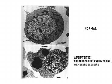
normal PowerPoint PPT Presentation
1 / 26
Title: normal
1
normal
Apoptotic Condensed nuclear material Membrane
blebbing
2
An Overview of the Apoptotic Cascade and Death
Domain Proteins The Death Domain (DD) is an 80aa
C-terminal domain shared among a large family of
transmembrane proteins such as FAS,TRADD, TNF-R1,
and RIP Thought to act as dimerization domain to
facilitate signaling pathways
3
(No Transcript)
4
The CASPASE Family of Proteases (cleaves after
Asp)
Activity of Initiator Caspases leads to the
Release of Active Effector Caspases
5
How do cells die?? A) Formation apoptosome starts
the cascade of caspases B) This leads to loss of
electron transport (and release of more
Cytochrome C) and build up of TOXIC free radicals
C) general loss of all cellular enzymes through
their digestion by Caspases
6
C. elegans and beyond.
FAS
A Separate Pathway from the Plasma Membrane
This Happens On the Mitochondrial Membrane
7
Apoptotic Pathways are Initiated on the
Mitochondrial Surface
Blk/Bax
mt
mt
Caspase9
Apaf-1
Bcl-2
8
Bcl/Bax/Bad Proteins influence the distribution
of Cytochrome C. In apoptotic cells, Cyto.C is
released into the cytosol to activate release of
Procaspase 9 by Apaf-1 This can be blocked by
over- expression of Bcl-2 and is promoted by
overexpression of Bax/Bad Association of Bad
with Bcl-2 is key event, this prevents the
anti-apoptotic activity of Bcl-2
CED4
CED9
23-50
9
Growth Factors Activate Kinase Cascades That
Target Bad Protein. Bad-P leaves the
mitochondrial membrane and is complexed
to 14-3-3 phosphoSer Scaffold proteins In this
situation, Bcl-2 is able to prevent the release
of Cyto. C from the mitochondria leaving the
Apaf-1/procaspase 9 complex INTACT So Apoptosis
is prevented, cell growth continues
23-50
10
Cancer
- A disease of multi-cellular organisms which have
lost some component of their cell cycle control
machinery or have otherwise acquired mutations
leading to the unrestricted growth on one cell
into a tumor.
11
Benign tumors arise with great frequency but
pose little risk because they are localized and
small
Figure 24-1
12
Malignant tumors generally invade surrounding
tissue and spread throughout the body
Alterations in cell-cell interactions and the
formation of new blood vessels are associated
with malignancy
Figure 24-2
13
Epidemiology of human cancers indicates that
development of cancer requires several mutations
Figure 24-5
14
Cloning of the RasD Oncogene the critical
observation was that DNA from tumor cells could
transform normal cultured cells into tumor cells
at (relatively) high frequencies DNA derived
from human tumor cells was used to transform
mouse cells and then a human specific Alu probe
(repetitive elment) was used to detect human DNA
that was incorporated into mouse genomes
24-4
15
The development of colon cancer is characterized
by a well-ordered series of mutations
Inherited mutations in tumor-suppressor
genes increase cancer risk
Figure 24-6
16
COOPERATIVITY- The OverExpression of Multiple
Oncogenes Increases Tumor Formation
Figure 24-7
17
Proto-oncogenes and tumor-suppressor genes the
seven types of proteins that participate in
controlling cell growth
Figure 24-9
18
Gain-of-Function Mutations Convert
Proto-Oncogenes Into Oncogenes
- Oncogenes were first identified in cancer-causing
retroviruses - The Rous sarcoma virus (RSV) contains a gene
(src) that is required for cancer-induction but
is not required for viral function - Normal cells contain a related gene that codes
for a protein-tyrosine kinase - The normal gene (c-src) is the proto-oncogene,
while the viral gene (v-src) is an oncogene that
codes for a constitutively active mutant
protein-tyrosine kinase - Many DNA viruses also contain oncogenes but these
have integral functions in viral replication
19
Virus-Encoded Activators of Growth-Factor
Receptors Act As Oncoproteins
Figure 24-14
20
Activating Mutations or Overexpression of
Growth-factor Receptors Can Transform Cells
Figure 24-15
21
Constitutively Active Signal-transduction
Proteins Are Encoded by Many Oncogenes
Figure 24-17
22
Passage from G1 to S phase is controlled by
proto-oncogenes and tumor-suppressor genes
Figure 24-19
23
Chromosomal Abnormalities Are Common in Human
Tumors
Figure 24-22
24
Mutations in p53 Abolish G1 Checkpoint Control
Some human carcinogens cause inactivating
mutations in the p53 gene and p53 activity is
also inhibited by certain proteins encoded by DNA
tumor viruses
Figure 24-21
25
Defects in DNA-Repair Systems Perpetuate
Mutations and Are Associated With Certain Cancers
26
Recent Topics..The Cutting Edge
Cell, Vol 117, 239-251, 16 April 2004 Cyclin
C/Cdk3 Promotes Rb-Dependent G0 Exit Shengjun Ren
and Barrett J. Rollins
Cell, Vol 117, 253-264, 16 April 2004 Negative
Regulation of dE2F1 by Cyclin-Dependent Kinases
Controls Cell Cycle Timing Tânia Reis and Bruce
A. Edgar
Molecular Cell, Vol 14, 81-93, 9 April
2004 Caspase Activation Inhibits Proteasome
Function during Apoptosis Xiao-Ming Sun, Michael
Butterworth, Marion MacFarlane, Wolfgang Dubiel,
Aaron Ciechanover, and Gerald M. Cohen
Molecular Cell, Vol 14, 105-116, 9 April
2004 Apoptotic Signaling in Response to a Single
Type of DNA Lesion, O6-Methylguanine Mark
J. Hickman and Leona D. Samson
Cancer Cell, Vol 5, 263-273, March
2004 Histologically normal human mammary
epithelia with silenced p16INK4a overexpress
COX-2, promoting a premalignant program Yongping
G. Crawford, Mona L. Gauthier, Anita Joubel,
Kristin Mantei, Krystyna Kozakiewicz 1, Cynthia
A. Afshari , and Thea D. Tlsty

