Radiographic Imaging Image Production - PowerPoint PPT Presentation
1 / 39
Title:
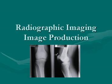
Radiographic Imaging Image Production
Description:
Attenuation ... The use of attenuating or absorbing material between the x-ray tube and the ... of the body that have the same attenuation of surrounding tissue ... – PowerPoint PPT presentation
Number of Views:2564
Avg rating:3.0/5.0
Title: Radiographic Imaging Image Production
1
Radiographic ImagingImage Production
2
Terms Related to Image Production
- Primary Radiation
- Refers to the x-ray beam after it exits the x-ray
tube and before it interacts with the patients
body - Remnant Radiation
- The remainder of radiation after it passes
through the patients body. - This is what produces the image on the
radiographic film - Secondary Scatter Radiation
- Radiation that may not be able to reach the film
but does not carry any useful information
3
Terms Related to Image Production
- Attenuation
- The process by which primary radiation is changed
or absorbed as it travels through the patient - Radiolucent
- Material that allow x-ray photons to pass through
easily (air) - Radiopaque
- Materials that do not allow x-ray photons to pass
through easily (bone)
4
Film / Screen RadiographyThe Imaging Chain
- Latent Image
- The image that is invisible on the radiographic
film until processing occurs - In order to release that image the film must be
developed.
5
Film / Screen RadiographyThe Imaging Chain
- Radiograph
- Image that is produced by x-ray photons on a
piece of radiographic film
6
Film / Screen RadiographyThe Imaging Chain
- Intensifying Screens
- Thin layers of cardboard or polyester coated with
layers of luminescent phosphor crystals that are
sensitive to x-rays. - In order to take full advantage of
intensification process, an intensifying screen
was placed in the front and back of the x-ray
film.
7
Film / Screen RadiographyThe Imaging Chain
- Double or Duplized emulsion film
- A special film that utilizes two intensifying
screens, one in the front and one in the back of
the film to enhance the intensification process.
8
Technical Exposure Factors
- Exposure Factors directly under the influence of
the radiographer - mAs
- kVp
- SID
9
mAs
- Milliampere seconds
- Controls the amount of radiation coming from the
x-ray tube and time the x-rays are being produced - Controls the quantity or number of x-ray photons
produced
10
kVp
- Kilovoltage peak or potential
- Measures the potential difference forcing the
current through the x-ray tube - It affects the energy or quality or power of the
x-ray photons
11
SID
- Source to image distance
- The distance between the point of x-ray emission
and the image receptor - Also known as focal film distance (FFD) or target
film distance (TFD)
12
Factors Affecting Radiographic Quality
- Density
- The overall blackening of the film
13
Variables That Affect Density
- Patient size and tissue composition
- The density of the tissues affect the visible
density on the radiographic film - The denser the tissue, the lighter the
corresponding film - mAs
- The chief controlling factor of exposure and
density - Increasing mA or time increases the radiographic
density
14
Variables That Affect Density
- kVp
- kVp affects density differently than mAs. In
order for there to be a significant increase in
density a 15 change in kVp must be made. - There is a peak or optimal kVp for each body part
- Distance
- Distance is inversely related to density
- A decrease of distance of the source of x-rays to
film increases the density and vice versa - Known as the Inverse Square Law
15
Factors Affecting Radiographic Quality
- Beam Modification
- Anything that changes the nature of the radiation
beam. - The beam may be modified before it enters the
patient (primary beam modification) or before it
interacts with the film (scatter control)
16
Factors Affecting Beam Modification
- Filtration
- The use of attenuating or absorbing material
between the x-ray tube and the patient that
filters out non-diagnostic, low energy, x-ray
photons. - Half value layer
- The amount of attenuating material that it takes
to reduce the primary x-ray beam to one half of
its original value. - Beam limitation devices
- Anything that will change the size of the primary
x-ray beam
17
Factors Affecting Radiographic Quality
- Grids
- A device that is designed to remove as many
scattered photons exiting the patient as possible
before they reach the film. - Consist of thin lead strips interspersed with
spacing material - Placed between the patient and the film to
intercept scattered photons leaving the patient.
18
Factors Affecting Grids
- Grid Ratio
- The ratio of the height of the lead strips to the
distance between them. - Grid ratios range from 51 to 161
- The higher the grid ratio the less density that
reaches the film
19
Factors Affecting Radiographic Quality
- Film / screen combinations
- Intensifying screens are fluorescent screens that
glow when exposed to x-radiation.. They are used
to enhance the radiation so that fewer x-ray
photons are used to create a radiographic image.
- The color of the glow of the intensifying screen
must match the color sensitivity of the film
(spectral matching)
20
Factors Affecting Radiographic Quality
- Relative speed of the film screen system
- The speed of an x-ray film system range from 50
to 1200. - The faster the speed of the system, the greater
the density on the radiographic film it creates
and the fewer x-ray photons it takes to create an
image.
21
Factors Affecting Radiographic Quality
- Processing
- Chemicals used to process or develop the
radiographic film may affect the density - Most common change in density is temperature
- Temperature to hot, increases radiographic
density - Temperature to cold, decreases radiographic
density
22
Contrast
- The visible difference between adjacent
radiographic densities. - The black and white and all shades of gray of the
x-ray film
23
Factors Affecting Radiographic Quality
- Patient Factors
- Because tissues in the body attenuate x-rays
differently, tissues with similar attenuation
will have similar density as well as contrast.
24
Factors Affecting Radiographic Contrast
- kVp
- The chief controlling factor of contrast
- The higher the kVp, the lower the contrast
- The lower the kVp, the higher the contrast
25
Factors Affecting Radiographic Contrast
- mAs
- A secondary factor for contrast. No change in
mAs can make up for inadequate penetration (kVp)
26
Factors Affecting Radiographic Contrast
- Beam Modification
- Anything that decreases scatter, increases
contrast.
27
Factors Affecting Radiographic Contrast
- Film / screen combination
- Imaging systems are complementary to the body
structures or areas of the body - In theory, the faster the system, the higher the
contrast
28
Factors Affecting Radiographic Contrast
- Contrast media
- Substances that attenuates the beam to a
different degree than the surrounding tissue - Used to enhance areas of the body that have the
same attenuation of surrounding tissue - Contrast media increases contrast on film
29
Factors Affecting Radiographic Contrast
- Processing
- Inadequate processing degrades the radiographic
contrast
30
Recorded Detail
- The distinct representation of an objects true
borders or edges - It is often called sharpness of detail,
definition or resolution
31
Factors Affecting Radiographic Recorded Detail
- Motion
- Voluntary motion
- Motion caused by the movement of the patient.
- Best controlled by good patient instructions
- Involuntary motion
- Motion caused by uncontrolled motion of the body
such as the heart beat or peristalsis - Best controlled by short exposure times
32
Factors Affecting Radiographic Recorded Detail
- Object unsharpness
- The inherent unsharpness of an object due to its
shape and location
33
Factors Affecting Radiographic Recorded Detail
- Focal spot size
- A small focal spot is used when fine detail is
needed - A large focal spot is used all other times
34
Factors Affecting Radiographic Recorded Detail
- Source to image distance (SID)
- As SID increases detail increase
- Penumbra
- A fuzzy border of an object that is obscure
- Umbra
- The true boarder
35
Factors Affecting Radiographic Recorded Detail
- Object to Image Distance (OID)
- The smaller the OID, the better the recorded
detail
36
Factors Affecting Radiographic Recorded Detail
- Material Unsharpness
- Faster systems produce greater unsharpness of
detail
37
Factors Affecting Radiographic Recorded Detail
- Distortion
- The misrepresentation of the true size or shape
of an object - Most commonly known as magnification
38
Types of Distortion
- Size distortion
- Magnification
- The best image is produced by the smallest OID
and the largest SID - Shape distortion
- The misrepresentation of the shape of a
radiographic image - Images in the direct path of the central ray are
the most accurately represented
39
THE END


![[PDF] Essentials of Radiographic Physics and Imaging 3rd Edition Kindle PowerPoint PPT Presentation](https://s3.amazonaws.com/images.powershow.com/10079275.th0.jpg?_=202407160511)
![get⚡[PDF]❤ Medical Imaging for the Health Care Provider: Practical Radiograph PowerPoint PPT Presentation](https://s3.amazonaws.com/images.powershow.com/10095504.th0.jpg?_=20240810027)