Protein Physics PowerPoint PPT Presentation
1 / 13
Title: Protein Physics
1
Protein Physics
- A. V. Finkelstein O. B. Ptitsyn
- LECTURE 15
Andrea Asztalos, 2008 February 15
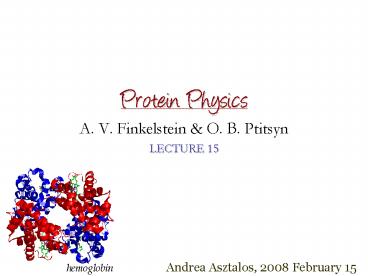
hemoglobin
2
Classification of Proteins andtheir Folds
1.Fibrous Proteins
2.Membrane Proteins
3.Globular Proteins
Dali/FSSP CATH
SCOP(structural classifications of proteins)
Orengo C. A. et al.,Structure, 5, 1093 (2005)
3
- Four major hierarchical levels in
- the CATH structural classification
- Families (gt40 sequence
- similarity) and superfamilies (lt30
- sequence similarity)
- Folds describe relative orientations
- of the secondary structures in 3D and
- the order in which they are connected
- At this level we observe similarities in
- proteins having no common ancestry
- or function.
- Architecture the overall shape of
- the protein
- Structural classes similar secondary
- structure compositions and packing
4
The CATHerine wheel
5
Unusual folds
Protein huristasin With no alpha and almost no
beta structure Special sequence with many
cysteines that form S-S bonds (shown as yellow
rods)
GFP (green fluorescent protein)
6
Open Questions Answers
- Why the majority of proteins fit a small set of
common folding patterns? - 80-20law 80 of proteins belong to only 20 of
observed folds (typical folds). Similarity of
protein tertiary structures is caused not only by
evolutionary divergence and not only by
functional convergence, but simply by
restrictions imposed on protein folds by some
physical regularities.
- Do we see the evolution of protein structures?
- 1). Do we see a microscopic evolution of
proteins? -gt a change - in protein structure would have an effect
upon the entire - organism.
- 2). Is there a macroscopic evolution of
proteins? -gt their - structure becomes more complex with
increasing complexity - of the organism.
7
- The hemoglobin from a lama binds oxygen more
strongly than hemoglobin from its animal
relatives living on the plain. It was showed that
microscopic changes in the hemoglobin structure
are responsible for this strengthening. - A review of protein structures shows that the
same folding patterns are observed both in
eukaryotes and prokaryotes, although the
distribution of the most popular folds in
eukaryotes is somewhat different from that in
prokaryotes.
Remark Fibrous proteins having the most simple
structure are typical for higher
organisms rather than prokaryotes. Remark The
proteins of eukaryotes are much more liable to
co- and post-translational
chemical modifications. Remark Eukaryotic
proteins are not only larger in size but also
contain a greater number of domains than
prokaryotic ones.
8
Determination of Protein Structures
- As of today, febr. 15th, 2008 the RCSB PDB has
48891 structures
- Different techniques used to study the structure
of proteins
Primary Structure obtained by biochemical
methods directly/indirectly
Quaternary Structure determined by electron
microscopy
Secondary/Tertiary Structure X-ray, NMR,
Circular dichroism (CD), UV, IR
Bernstein F.C., et al. J. Mol. Biol. 112, 535
(1977)
9
X-ray Crystallography of Proteins
- Braggs law
- Protein crystals are difficult to grow
- (50 solvent in large channels)
- Gravity obstacle to the formation of
- well-ordered crystals
Hanging-Drop technique
Equilibrium is reached by water diffusion
10
Reading out the structure
Each diffracted beam is defined by 3 properties
A intensity of the spot ? - set by the x-ray
source f lost in the experiment
The phase problem in X-ray crystallography
It is solved by Multiple Isomorphous
replacement (MIR)
- Introduces new X-ray scatterers
- in the crystal (heavy atoms)
- Positions of the heavy atoms gt
- the amplitude and phase of their
- contribution
- 3 amplitudes and one phase gt
- electron density map of the unit cell
Diffraction pattern from a retinol binding
protein crystal
- Resolution of diffraction data
- (1.7Å - 3Å - 5Å)
- Errors are removed by
- refinement R factor
- (0.0 1.5 -2.0 - 5.9)
11
NMR analysis of proteins in solution
- Used mainly for small molecules
- Applying radio-waves to excite the magnetic
moments of nuclei - aligned in a strong magnetic field
- Nuclei with non-zero nuclear
- spin ( 1H, 13C )
- Shielding effect of the surrounding
- electrons chemical shifts
NMR structure of BPTI
20 superimposed structures
- Obtain a list of distance constraints between
atoms
Wüthrich K., Science 243, 45 (1989) Nobel prize
2002
12
1D and 2D NMR spectroscopy
COSY (correlation spectroscopy) - gives peaks
between hydrogen atoms that are covalently
connected through one or two other atoms NOE
(nuclear Overhauser effect) - gives peaks
between pairs of hydrogen atoms that are
close together in space even if they are from
amino acid residues that are quite distant in
the primary sequence.
1H NMR spectra for bovine Pancreatic trypsin
inhibitor protein
2D NOE spectra of the protein BUSI IIA
13
- Structural Information on partially folded
polypeptides - Information on the frequencies of the rate
processes that mediate transitions
NMR structure of the bovine prion protein
Variation of its structure during 1ns (20
snapshots)
The globular domain maintains its main geometry,
whereas the tail undergoes large-scale changes
with time.

